


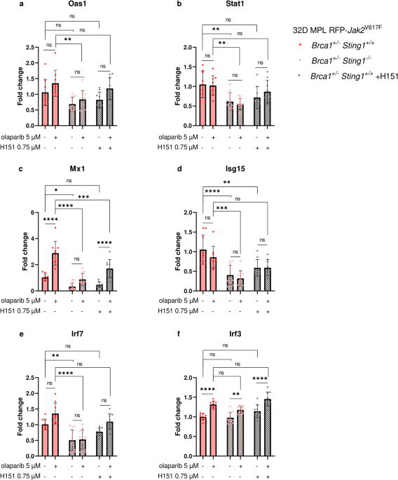
Our findings extend our previous observations of ISG upregulation in Brca1+/− 32D Jak2V617F cells upon olaparib treatment5. By introducing a genetic Sting1 knockout, we recapitulated the reduction in basal ISG expression previously observed with H-151, confirming a regulatory role of STING in ISG expression. Under basal conditions, analysis of Brca1+/+ cells demonstrated a comparable contribution of STING to ISG regulation, indicating that baseline STING activity is not restricted to the Brca1+/− background. Following olaparib treatment, significant induction of Oas1 and Mx1 remained confined to Brca1+/− cells, consistent with our previous findings that Brca1 haploinsufficiency enhances the interferon response to PARP inhibition. However, the persistence of olaparib induced ISG upregulation despite Sting1 loss or inhibition suggests that the heightened IFNα responsiveness and ISG induction in Brca1+/− Jak2V617F cells cannot be attributed to STING signaling alone and instead suggests the contribution of additional pathways activated in response to olaparib-induced DNA damage.
Among the analyzed ISGs, our data indicate heterogeneous responses to Sting1 loss, STING inhibition, and olaparib treatment. Basal expression was significantly reduced for nearly all genes except Oas1, suggesting that some ISGs rely more strongly on STING, whereas others may be maintained by STING-independent mechanisms. Furthermore, Stat1 and Isg15 did not show significant induction upon olaparib treatment. One possible explanation is that ISGs underly distinct expression kinetics following IFN-α or IFN-β release, as well as direct activation by IRF312,13, which may not have been fully captured in our experiments. Although we assessed different time points after olaparib treatment (Supplementary Fig. S18), additional time-resolved experiments would likely provide a more complete set of data.
Pharmacological STING inhibition with H-151 and genomic Sting1 knockout both reduced ISG expression, although the knockout produced a more pronounced effect. Under basal conditions, genetic Sting ablation significantly reduced ISG expression for most analyzed genes (5/6), whereas H151 treatment affected only Isg15. This difference is consistent with the distinct mechanisms of these approaches. H-151 covalently inhibits STING by preventing palmitoylation at Cys91, thereby blocking its activation14, whereas genomic deletion abolishes STING protein entirely. As a result, pharmacological inhibition is not expected to fully reproduce the effects of genetic Sting1 deficiency. Nevertheless, the observation that ISG expression are not entirely abolished, even in Sting1-deficient cells, suggests that basal ISG regulation is only partially dependent on STING and supports the notion that additional, STING-independent pathways contribute to maintaining basal ISG expression.
Despite clear differences in ISG expression, STING loss did not affect proliferation, viability, or apoptosis in 32D MPL RFP-Jak2V617F Brca1+/− cells. Prior studies in other cell systems have reported increased proliferation15, reduced apoptosis15,16 and resistance to radiation- and chemotherapy-induced cell death15,16,17 upon Sting1 knockout. This discrepancy may reflect cell type-specific dependencies on STING signaling or compensatory engagement of alternative pathways.
Because ISGs remained inducible despite STING loss, our data raise the possibility that alternative nucleic acid-sensing pathways compensate for absent STING signaling, as outlined in Fig. 5.
Fig. 5
Comparison of canonical cGAS-STING signaling and potential STING-independent pathways contributing to interferon type I signaling and ISG induction. (1) Genomic instability in MPN cells harboring Jak2V617F and Brca1-haploinsufficiency is driven by replication stress, defective homologous recombination, and PARP inhibition-induced DNA damage, resulting in accumulation and mislocalization of nucleic acids. (2) Mitochondrial RNA (mtRNA) is released into the cytosol as a consequence of mitochondrial dysfunction and metabolic stress and can activate RLR signaling. (3) Cytosolic dsDNA, arising from genomic instability and DNA damage (e.g., leakage of nuclear DNA into the cytosol), is sensed by cGAS, leading to production of the second messenger cGAMP, which binds and activates STING. (4) Cytosolic RNA is sensed by RIG-I, which signals through the mitochondrial adaptor MAVS, providing a parallel nucleic acid-sensing input that converges on downstream kinase activation. (5) Activated STING and MAVS converge on TBK1 (with auxiliary contribution from IKKε), resulting in its autophosphorylation and the activation of IRF3 and NF-κB. These transcription factors translocate to the nucleus and induce expression of type I interferons (e.g., IFNα) and NF-κB target genes (NFκBGs). Additionally, IRF3 can directly induce ISG expression by binding to IFN-stimulated responsive elements (ISREs) within the target gene promoters. (6) Secreted IFNα binds to the type I interferon receptor (IFNAR), leading to JAK1 (and TYK2) activation. (7) JAK1-dependent phosphorylation of STAT2 (and STAT1), together with IRF9, drives formation of the ISGF3 complex, which likewise targets ISREs to amplify ISG expression. Created with BioRender.com.
A prominent candidate is the retinoic acid-inducible gene I (RIG-I)-like receptors (RLRs) signaling pathway, originally known for its activation by sensing of viral RNA. RLRs also drive type I interferon production through TBK1 and IRF3 activation independently of STING18. Upon RNA binding, RIG-I or melanoma differentiation associated protein 5 (MDA5) signal via mitochondrial antiviral-signaling protein (MAVS), to activate inhibitor of nuclear factor kappa-B kinase epsilon (IKKε) and TBK1 directly, thereby bypassing STING and inducing IRF3/ IRF7- and nuclear factor kappa-light-chain-enhancer of activated B cells (NF-κB)-dependent ISG expression. Importantly, recent work has demonstrated that RLRs also recognize endogenous cytosolic RNA19,20, supporting the RLR axis as a plausible STING-independent mechanism of olaparib-induced ISG expression. Several mechanisms associated with DNA damage, replication stress and PARP inhibition can generate cytosolic RNA, including(i) mitochondrial RNA (mtRNA) leakage9,21,22, (ii) RIG-I-dependent ISG upregulation following PARP1 loss, where the specific RNA species responsible remain unidentified23, and (iii) replication stress- induced activation (e.g., long interspersed nuclear element-1 (LINE-1))24,25. Additionally, RNA-polymerase III (Pol III) can convert cytosolic dsDNA into dsRNA sensed by RIG-I, leading to an interferon type I response26,27, while genotoxic stress may promote cytosolic accumulation of small nuclear RNAs (snRNAs) acting as endogenous RIG-I agonists28,29. Collectively, these mechanisms suggest that olaparib may induce ISG expression through RLR-TBK1-IRF3 signaling even in the absence of STING. However, as the involvement of this pathway was not directly examined in the present study, its contribution remains speculative and should be addressed in future studies, for example through genetic ablation of RIG-I or MAVS. Another potential STING-independent pathway involves DNA-dependent activator of interferon regulatory factors (DAI/ZBP1), which senses cytosolic dsDNA and directly activates TBK1 to induce IRF3-dependent type I interferon signaling30,31. Despite cGAS-STING being widely considered the dominant cytosolic DNA sensor in most mammalian cell types32, DAI/ZBP1 may become particularly relevant when STING is absent.
Given that both RLR and DAI converge on TBK1, our results suggest that TBK1 may act as the central kinase governing IRF3 and NF-κB activation in the STING knockout setting. This may contribute to the persistent ISG expression despite the loss of STING and raises the possibility that TBK1 represents a key dependency node in this signaling context, suggesting that TBK1 deletion would provide critical mechanistic insight into IFN-mediated responses to PARP inhibition in this model.
Our results show a reduced, but not completely abolished, type I interferon response in Brca1-haploinsufficient Jak2V617F Sting1−/− cells, indicating that the innate immune response to genomic instability relies on parallel signaling pathways rather than solely on the cGAS-STING axis. In the context of MPN, where chronic replication stress leads to persistent nucleic acid damage, this plasticity enables compensatory ISG expression. Based on these findings, we hypothesize that downstream convergence points such as TBK1 may play an important role in modulating inflammation and sensitivity to PARP inhibitors, highlighting the need to define pathway hierarchies to identify therapeutic targets. Beyond the RLR–MAVS and DAI pathways discussed above, several other innate immune sensors, including AIM2, Toll-like receptors (TLRs), including TLR3/7/9, and DDX family members (e.g., DDX41 and DDX1), likely contribute to the redundancy of type I interferon signaling33. Defining the contribution of these pathways will be important for future studies aimed at establishing the hierarchy and interplay of innate immune signaling in the response to PARP inhibition in BRCA1 haploinsufficient MPN.
