


Human specimens
Brain tissue samples of five epilepsy patients were collected during therapeutic surgery in 2014 (Department of Neurosurgery, Charité-Universitätsmedizin Berlin, Germany). Patient characteristics: gender – 3/2 (f/m); age – 41.6 ± 15.3 years. All human experiments were conducted in accordance with relevant guidelines and regulations (including institutional, national, and international ethical standards). The study received formal ethical approval by the Ethical Committee of Charité-Universitätsmedizin Berlin (application number: EA4/065/13). Written informed consent was obtained from all participants prior to sample collection and analysis.
Animal work
Mice
Mice were housed in a 12/12-hour light-dark cycle with food and water provided ad libitum. Male mice (weight: 20–38 g; age: 8–52 weeks) were used for experiments. All experiments were performed according to the German Law for Animal Protection and the National Institute of Health Guidelines for Care and Use of Laboratory Animals. For animal experiments, we followed the ARRIVE guidelines. Animal studies were approved by the local ethics committee on animal research, LaGeSo Berlin (Permit Number: T0159/09, G152/09, G0376/11 and G0281/14). C57BL6/J mice (gender: male, age: 10–18 weeks, weight: 25–34 g, wildtype and as recipients for bone marrow; gender: male, age: 8–28 weeks, weight: 20–35 g as donor for bone marrow) were purchased from Charles River (Sulzfeld, Germany). Heterozygous C57BL6/J-TgN(beta-act-EGFP) mice (gender: male, age: 8–52 weeks, weight: 20–38 g as donor for bone marrow) and heterozygous Cx3cr1+/−-GFP (B6.129P2(Cg)-Cx3cr1tm1Litt/J) mice (gender: male, age: 10–18 weeks, weight: 25–34 g as recipient for bone marrow; gender: male, age: 10–48 weeks, weight: 22–38 g as donor for bone marrow) were purchased from Charles River and bred in-house (FEM, Berlin, Germany). Genotyping was performed for every transgenic animal. The recipient animals were matched according to sex, age and weight.
Animals were randomly assigned to groups and analyses were conducted blinded. The animal experiments were repeated two to three times to achieve a total of 5–6 animals per group.
Generation of bone marrow chimeras
Total body irradiated (TBI) chimeras were generated by standard procedure48,49. Mice (C57BL6/J) were anesthetized (90 mg/kg ketamine, Ketavet, Pfizer; 10 mg/kg xylazine, Rompun, Bayer HealthCare; intraperitoneal injection) and lethally irradiated (11.5 Gy) with cesium 137 (Gammacell® 40 Exactor from MDS Nordian, Deutsches Rheuma Forschungszentrum, Berlin, Germany). For HPI, animals (C57BL6/J or Cx3cr1+/−-GFP) were placed into a lead shield construction followed by irradiation17. Within 12 h, freshly collected 1.7-2.0 × 10⁷ bone marrow cells of either C57BL6/J-TgN(beta-act-EGFP) or C57BL6/J were injected intravenously (200µL/animal). After 8 weeks, reconstitution levels were examined by flow cytometry of blood samples as described before17,19. Only animals with a reconstitution level of above 75% of CD45+CD11b+ cells were further analyzed (reconstitution level: 75–95%).
Stereotactic tumor cell implantation
Eight weeks after the generation of bone marrow chimeras, we injected GL261 tumor cells intracranially. Anaesthetized mice (90 mg/kg ketamine; 10 mg/kg xylazine; intraperitoneal injection) were fixed in a stereotactic frame (Stoelting). Bepanthen eye cream (5% Dexpanthenol, Bayer) was used to cover the eyes during surgery. To prevent pain, the scalp was treated with lidocaine (Xylocain pump spray, Aspen). A midline incision was done on the scalp, a hole was drilled by a canula and tumor cells were injected 1 mm anterior and 2 mm lateral to the bregma. Here, a microliter Hamilton syringe (1µL; Carl Roth GmbH, Karlsruhe, Germany) was inserted into 4 mm depth, returned to 3 mm, and cells were slowly released (2.0 × 10⁴ cells/µL). After surgery, Paracetamol (300 mg/kg, Paracetamol-ratiopharm solution, Ratiopharm) was added to the drinking water for pain management (up to 72 h). Animals were monitored at least twice daily for the first 72 h for postoperative signs of distress. The endpoint of the study was day 21 after tumor cell implantation.
Magnetic resonance imaging (MRI)
Magnetic resonance imaging was performed on day 21 after GL261 tumor cell inoculation. Tumor-bearing mice were anesthetized by 1.5–2.0% isoflurane (Forene, Abbot, Wiesbaden, Germany) delivered in a O2/N2O mixture (30%/70%) and placed into a 7-Tesla rodent MRI scanner (Pharmascan 70/16, Bruker BioSpin, Rheinstetten, Germany). Brains were observed with T2-weighted two-dimensional turbo spin-echo sequences by Paravision 4.0 software (Bruker BioSpin). Calculation of tumor volumes was carried out with the program Analyze 5.0 (AnalyzeDirect, Lenexa, KS). Tumor volumes were as follows. Wildtype animals (WT, n = 5): 9.83 ± 5.20 mm3; total body irradiated chimeras (TBI, n = 5): 21.52 ± 10.00 mm3; head protected irradiated animals (HPI, n = 6): 9.60 ± 4.75 mm3. As expected, the tumor volumes of WT and HPI animals were comparable, while the tumor sizes of TBI mice doubled17.
Isolation of bone marrow cells
Naïve animals were anesthetized with isoflurane (Isofluran CP, Cp-Pharma) and sacrificed by cervical dislocation. Bone marrow cells were harvested by flushing femurs of C57BL6/J or Cx3cr1+/−-GFP mice with Ca2+-, Mg2+-free phosphate-buffered saline (PBS; PAA Laboratories). Suspensions were centrifuged and passed through a pre-separation filter (30 μm; Miltenyi Biotec, Bergisch Gladbach, Germany) to obtain single-cell suspensions. Viable cells were diluted in PBS to a concentration of 1.7–2.0 × 10⁷ cells/200 µL.
Isolation of microglia from adult brains
Naïve mice were anaesthetized (90 mg/kg ketamine; 10 mg/kg xylazine; intraperitoneal injection) and perfused intracardially with PBS. Brains were taken and processed under sterile conditions. Brains were homogenized by using the Neural Tissue Dissociation Kit (P) (Miltenyi Biotec) and the gentleMACS™ Octo Dissociator with Heaters (Miltenyi Biotec). Isolation of microglia was performed by addition of CD11b MicroBeads (Miltenyi Biotec) and two subsequently used columns (LS column, MS column; Miltenyi Biotec). Purity of microglia was analyzed by FACS (BD FACS CantoII) following staining with CD11b-PE (Biolegend) and CD45-APC (BD Biosciences) antibodies. Purity of eluted cells was approx. 95% CD45+CD11b+ cells.
Cell culture
Cultivation of GL261 cells for implantation
GL261 cells (passage 14) were grown in high-glucose Dulbecco’s modified Eagle’s medium (DMEM) containing stable glutamine and sodium pyruvate (PAA Laboratories) supplemented with 10% fetal calf serum (PAA Laboratories), 100 units of penicillin/ml and 100 µg/mL of streptomycin at 37 °C and 5% CO2 atmosphere. The GL261 cell line, an established murine glioblastoma model, was obtained from the German Collection of Microorganisms and Cell Cultures (DSMZ, ACC802), Germany. Cells were cultured for three days until a confluence of 80% was reached. Cells were detached using 0.5% trypsin/EDTA (Life Technologies, Darmstadt, Germany), washed with PBS and diluted to a concentration of 2 × 10⁴ cells in 1 µL.
Preparation of tumor-conditioned medium
GL261 cells were cultured as described above but with 5% FCS. After three days in culture (confluence approx. 80%), supernatant was collected and centrifuged two times at 4000 rpm to exclude cells and debris.
Differentiation and cultivation of macrophages
Bone marrow cells were seeded in a T75 flask (Sarstedt) using RPMI 1640 medium (Gibco, ThermoFisher Scientific) containing stable glutamine with 10% fetal calf serum, 100 units of penicillin/ml and 100 µg/ml of streptomycin. Additionally, M-CSF (20 ng/mL; Miltenyi Biotec) was added and medium change was performed every second or third day for eight days. Macrophages attached to the bottom. On day eight of culture, macrophages were detached by a cell scraper, washed and plated in 8-well chamber slides (x-well cell culture chamber, 8 wells, on PCA slide; Sarstedt). 1.2 × 10⁵ cells/well were plated in RPMI medium including supplements for two days.
Plating of primary microglia
Primary isolated microglia were seeded into 8-well chamber slides (x-well cell culture chamber, 8 wells, on PCA slide). Cells were cultured for three days in high-glucose Dulbecco’s modified Eagle’s medium (DMEM) containing stable glutamine and sodium pyruvate supplemented with 10% fetal calf serum, 100 units of penicillin/ml and 100 µg/ml of streptomycin at 37 °C and 5% CO2 atmosphere. 2.5 × 10⁵ cells/well were plated in DMEM medium including supplements for three days.
Cultivation and plating of BV2 cells
BV2 cells (passages 5–6), an established immortalized murine microglial cell line, were used. The cell line was kindly provided by PD Dr. Marina Jendrach (Department of Neuropathology, Charité – Universitätsmedizin Berlin, Germany). Cells were cultured in high-glucose Dulbecco’s modified Eagle’s medium (DMEM) containing stable glutamine and sodium pyruvate supplemented with 5% fetal calf serum, 100 units of penicillin/ml and 100 µg/ml of streptomycin at 37 °C and 5% CO2 atmosphere. Cells were grown for three days. Cells were detached using 0.5% trypsin/EDTA. Cells were counted and 7,500 cells were plated for each well of an 8 well-chamber slide (x-well cell culture chamber, 8 wells, on lumox® slide, Sarstedt). DMEM with 5% FCS or tumor-conditioned medium (TCM) was used. After three days of culture, cells were fixed and stained.
Immunofluorescence staining
Processing, immunofluorescence staining and analyses of human tissue samples
Directly after surgery, human brain tissues were fixed in 4% PFA for 24 h and dehydrated in rising sucrose concentrations (10%/20%/30%; Roth, Karlsruhe, Germany). The brain tissue samples were carefully frozen with liquid nitrogen. Frozen sections were prepared at 10 μm thickness.
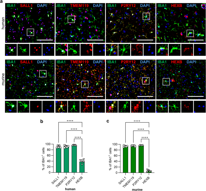
All sections were treated with Autofluorescence Eliminator Reagent (Millipore, Burlington, MA, USA) following the instructions of the manufacturer. Afterwards, human brain sections were permeabilized by 0.1% Triton X-100/PBS (20 min, room temperature; SALL1, P2RY12, HEXB) or directly blocked with 1% Casein/PBS (30 min, RT; TMEM119). Sections were stained with primary antibodies for 2 h at RT. To identify microglia/macrophages, IBA1 staining (goat anti-IBA1, Abcam, Cambridge, UK, ab5076, 1:200) was performed. Additionally, the following antibodies to detect specific markers were used: rabbit anti-SALL1 (1:100; PA5-62057, ThermoFisher Scientific), rabbit anti-TMEM119 (1:100; ab185337, Abcam), rabbit anti-P2RY12 (1:100; NBP2-33870, Novus Biologicals), rabbit anti-HEXB (1:100; PA5-101082, ThermoFisher Scientific). After washing section 3 × 5 min in 0.5% Casein/PBS, secondary antibodies were applied (AF647-conjugated anti-goat IgG; Cy3-conjugated anti-rabbit IgG; Dianova, Hamburg, Germany, each 1:200). After incubation for 1.5 h at RT, sections were washed 2 × 5 min in PBS and water. DAPI-containing mounting medium (Immunoselect Antifading Mounting Medium DAPI, Dianova) was used to stain nuclei.
Images were acquired using a Zeiss Axio Observer Z1 fluorescence microscope (Zeiss MicroImaging GmbH, Jena, Germany) at room temperature. Approximately 24 images for each patient at three different brain tissue areas were taken. ImageJ 1.54j (NIH, Bethesda, MD, USA) was used to analyze images.
Processing of murine brains, and immunofluorescence staining and analyses of sections
For immunohistochemically studies, on day 21 of tumor growth, glioma-bearing mice were anaesthetized (90 mg/kg ketamine; 10 mg/kg xylazine; intraperitoneal injection) and perfused intracardially with 4% paraformaldehyde (PFA). Brains were fixed in 4% PFA and subsequently dehydrated by sucrose solutions in rising concentrations (10%/20%/30%). Frozen brains were embedded in gelatine and sliced by a cryostat (Microm HM 560, Microm International GmbH) in coronal sections of 10 μm thickness.
For staining of TMEM119, sections were directly blocked with 1% Casein/PBS (30 min, RT). For staining of SALL1, sections were fixed with methanol (10 min, -20 °C) and blocked, subsequently. For P2RY12 and HEXB staining, sections were treated with Antigen Retrieval Reagent-Universal (VCTS023, R&D Systems; heating up to 90 °C, 2–5 min) and blocking was performed with 0.1% Triton X-100 in 1% Casein/PBS (30 min, RT). Primary antibodies were used to detect microglia/macrophages (IBA1), and the different markers: goat anti-IBA1 (1:200; ab5076, Abcam), rabbit anti-SALL1 (1:100; ab31526, Abcam), rabbit anti-TMEM119 (1:50; ab209064, Abcam), rat anti-P2RY12 (1:100; 848002, Biolegend), rabbit anti-HEXB (1:100; PA5-101082, ThermoFisher Scientific). Following 2 h of incubation at room temperature, sections were washed with 0.5% Casein (4 × 5 min). Secondary antibodies were applied for 1.5 h: AF647-conjugated anti-goat IgG; Cy3-conjugated anti-rabbit IgG, Cy3-conjugated anti-rat IgG (1:200; Dianova). Sections were washed 3 × 5 min in PBS and water. DAPI-containing mounting medium (Dianova) stained nuclei.
Images were acquired using Zeiss Axio Observer Z1 fluorescence microscope at RT. Up to 15 images for each brain region of three different sections were taken (animals: 4–6 in total per group). Images were analyzed by ImageJ 1.54j (NIH).
Negative controls were performed using secondary antibodies alone. For each condition, two to three brain sections from two different TBI mice were included and processed according to the same staining protocols as the experimental samples. Imaging (three regions per section: contralateral, peritumoral, and intratumoral), as well as image processing and analysis, were performed using the same settings and workflow as for the corresponding immunostainings.
Fixation and staining of cultured cells (primary microglia, macrophages and BV2 cells)
Primary microglia, macrophages and BV2 cells were fixed with 4% PFA (RT, 20 min). For SALL1 and TMEM119 blocking was performed with 1% Casein/PBS. For P2RY12 and HEXB blocking was performed with 0.1% Triton X-100 (SigmaAldrich) in 1% Casein/PBS (RT, 30 min) The following primary antibodies were used for staining: rabbit anti-SALL1 (1:100; Abcam), rabbit anti-TMEM119 (1:50; Abcam), rat anti-P2RY12 (1:100; Biolegend), rabbit anti-HEXB (1:100; ThermoFisher Scientific), goat anti-IBA1 (1:100; Abcam) or Phalloidin (1:200; Alexa Fluor™ 488 Phalloidin, A12379, Invitrogen). Incubation of primary antibodies was 2 h at RT. Following washing, secondary antibodies were applied (dilution for each antibody was 1:200): Cy3-conjugated anti-rabbit IgG, Cy3-conjugated anti-rat IgG, AF647-conjugated anti-goat IgG (Dianova). DAPI-containing mounting medium was used to stain nuclei.
Images were acquired using Zeiss Axio Observer Z1 fluorescence microscope at RT. Approx. 8 images were taken for each well (4–11 wells/marker). Images were analyzed by ImageJ 1.54j (NIH).
Negative controls for primary microglia/macrophages and BV2 cells were performed using secondary antibodies alone. For each condition, one to two wells were included, and 5–8 images were acquired per well. Imaging and subsequent image processing were performed using the same settings and workflow as for the corresponding stained samples.
Statistics
Statistical analyses were performed using GraphPad Prism (GraphPad Software 10). Depending on the experimental setup, comparisons between two groups were conducted using unpaired Student’s t-test, whereas comparisons among multiple groups were analyzed using one-way analysis of variance (ANOVA) followed by Bonferroni post hoc correction. Data are presented as mean ± standard deviation. A p-value of < 0.05 was considered statistically significant.
