


We conducted a retrospective, multicenter study of patients who underwent DHC for MCI with HT between 2014 and 2024 at five German university hospitals. The study was conducted in accordance with the Declaration of Helsinki and approved by the Institutional Review Board of Rostock University Medical Center (approval number A2025-0195), which waived the requirement for informed consent because of the retrospective nature of the study. The first and last author had full access to all the data in the study and take responsibility for its integrity and the data analysis.
Patient population
Eligible patients were adults diagnosed with MCI who underwent DHC and had radiologically confirmed HT prior to surgery. The diagnosis of MCI required both clinical and radiological criteria. Radiologically, MCI was defined as hypodensity on computed tomography (CT) and/or CT-perfusion mismatch involving at least 50% of the MCA territory. Clinically, MCI was defined as progressive neurological deterioration with a decrease of at least two points on the Glasgow Coma Scale (GCS). HT was defined as the conversion of a bland infarction into a hemorrhagic area and was objectivized according to the Heidelberg Bleeding Classification22.
The decision to perform DHC was based on interdisciplinary assessment by a senior neurologist and a senior neurosurgeon, integrating the overall clinical and radiological presentation. According to institutional protocols, HT was not considered a contraindication to DHC. Postoperatively, all patients were admitted to interdisciplinary neurological intensive care units, where they were treated according to German national protocols and guidelines.
Demographic data (age, sex), vascular risk factors (hypertension, diabetes mellitus, coronary artery disease, atrial fibrillation, hyperlipidemia, smoking status, chronic obstructive pulmonary disease), and prior medication use (anticoagulants, antiplatelet agents) were recorded. History of prior stroke was also documented. Baseline neurological status was assessed using the GCS and National Institutes of Health Stroke Scale (NIHSS) at admission. The presence of anisocoria prior to surgery was documented as a marker of impending herniation.
Radiological variables
Preoperative imaging variables included infarct side, infarct volume, hemorrhage volume, Heidelberg bleeding classification, and the hemorrhage-to-infarct volume ratio. Volumes were calculated with the region of interest function in the Brainlab® software. Postoperative imaging was assessed for hematoma expansion, HT, hematoma volume, and Heidelberg bleeding classification.
Interventions
Reperfusion therapies were recorded, including IVtPA and MT, along with treatment-to-event intervals, and time from symptom onset to surgery. All patients underwent DHC according to institutional standards. The interval from ictus to surgery was recorded in hours. Additional surgical procedures, including infarct debridement and hematoma evacuation, were documented.
Outcome measures
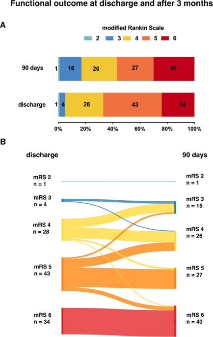
The primary endpoints were functional outcome and mortality 90-day follow-up. Functional outcome was assessed using the modified Rankin Scale (mRS). Both variables were recorded at discharge and at the 3-month follow-up.
Statistical analysis
Continuous variables are presented as mean ± SD or median (interquartile range [IQR]), as appropriate, and categorical variables as counts and percentages. Patients were stratified according to 90-day functional outcome. Group comparisons were performed using the Mann–Whitney U test for continuous variables and the χ² or Fisher exact test for categorical variables, as appropriate.
Given the highly skewed 90-day mRS distribution and low counts in the lower mRS levels, mRS was analyzed as an ordered categorical outcome grouped into 2–3, 4, and 5–6. Proportional odds regression was designated the primary analysis to retain the ordinal information of the mRS. Binary logistic regression (mRS 2–4 vs. 5–6) was performed as a sensitivity analysis. This threshold was chosen because very few patients achieved functional independence, making the conventional definition of functional independence as mRS ≤ 2 uninformative in this severely affected population.
Covariates were selected a priori based on biological plausibility and the primary study objective and included age (per 10 years), treatment modality, hemorrhage volume (per 10 mL), and infarct volume (per 50 mL). Hemorrhage and infarct volumes were modeled as continuous measures of structural tissue burden. Hemorrhage volume was preferred over the Heidelberg Bleeding Classification because it provides a quantitative measure of hemorrhagic burden, while Heidelberg subtypes represent distinct hemorrhage patterns and were partly sparsely represented. Age was retained irrespective of univariable significance given its established prognostic role after DHC. Additional candidate variables were assessed in univariable regression analyses (Supplemental Material). Sex and NIHSS were not associated with outcome, while comorbidities such as diabetes and coronary artery disease were not included as core adjustment variables. Given the limited sample size, the primary multivariable model was deliberately kept parsimonious to reduce the risk of overfitting. Analyses were conducted using complete-case data. Odds ratios (ORs) are reported with 95% confidence intervals (CIs). Collinearity among predictors was assessed using Spearman correlation coefficients and variance inflation factors (VIFs), with VIF < 2 indicating no relevant multicollinearity. The proportional odds assumption was evaluated using the test of parallel lines. All tests were two-sided, and p values < 0.05 were considered statistically significant. Statistical analyses were performed using SPSS version 31.0 (IBM Corp., Armonk, NY, USA).
