


Discovery of Kobe3708 as a prototype RAS/RAF binding inhibitor by in silico screening
Previously, we performed a docking-based screen focused on the surface structure of RAS·GTP to develop compounds that bind directly to RAS, and identified Kobe0065 family compounds that displayed blocking activity against RAS/RAF binding and exhibited antitumour activity against RAS-driven cancer cell xenografts33. In an effort to expand our search for more potent small molecule inhibitors of RAS/RAF binding with modes of action distinct from those identified in our previous research33, we conducted in silico screening of a commercial library containing 2.5 million compounds using a ligand-based, computer-assisted active learning system34 rather than a docking-based virtual screening approach. The details of this active learning-based screening procedure are described in the Methods section entitled “In silico screening”. Notably, this screening did not incorporate structural information from the target proteins RAS or RAF, and virtual hit compounds with structural similarity to previous RAS inhibitors33 were excluded during the sequential screening process to ensure the selection of a structurally diverse set of compounds.
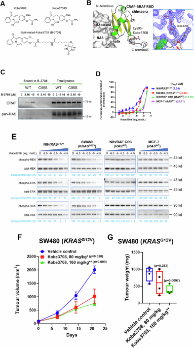
The several selected compounds were then evaluated by cell growth inhibition assays using NIH3T3 cells stably expressing HRASG12V (NIH/RASG12V) and the CR3 kinase domain of CRAF (NIH/RAF CR3: constitutively active RAF due to RBD deletion)33,35 to confirm target specificity and lack of RAF kinase inhibition. One compound, Kobe3708, inhibited the growth of NIH/RASG12V but not NIH/RAF CR3 cells, whereas sorafenib, an ATP-competitive pan-RAF kinase inhibitor used as a positive control, inhibited the growth of cells expressing both recombinant proteins (Fig. 1A and Supplementary Table 1). In addition, Kobe3708, but not sorafenib, potently inhibited the binding of HRASG12V preloaded with GppNHp (HRASG12V·GTP) to the CRAF RBD with an IC50 value of 7.62 μM (Fig. 2B and Supplementary Table 1). Collectively, the stepwise screening identified the potential of Kobe3708 to inhibit RAS/RAF binding. Therefore, we decided to examine Kobe3708 as a prototype compound in greater detail.
Fig. 1: Discovery of Kobe3708, a prototype RAS/RAF-binding inhibitor.
A Structure of Kobe3708, Kobe3708S, and biotinylated-Kobe3708 (B-3708). B Crystal structure of CRAF-BRAF RBD chimaera/Kobe3708 complex. The overall crystal structure of the complex is shown, considering the three-dimensional position of the RAS/RAF-binding interface using HRAS/CRAF RBD model (PDB: 4G0N). HRAS and CRAF-BRAF RBD chimaera are shown as ribbon and surface models, respectively. Strands, helices, and loops in the CRAF-BRAF RBD chimaera are shown in yellow, green, and gray, respectively. The side chains of Cys95 and Kobe3708 are presented as a ball-and-stick models. The binding site of Kobe3708 is located away from the RAS/RAF-binding interface. The magnified image of the covalent binding of Kobe3708 with the side chain of Cys95 is shown in the right panel. The 2mFo-DFc electron density map contoured at 1.5 σ from the X-ray crystallographic data is shown as a blue mesh, where the electron densities of the side chain of Cys95 and the carbon atom neighbouring the oxindole ring in Kobe3708 (red arrow) overlap. C CRAF bound to B-3708. NIH3T3 stably expressing CRAFWT or CRAFC95S were treated with B-3708 at the concentrations indicated for 3 h at 37 °C. Cell lysates were then mixed with streptavidin agarose for 1 h at 4 °C to immobilize the proteins bound to the biotinylated compound, separated by SDS-PAGE, and detected with anti-CRAF and anti-pan-RAS antibodies. The results shown are representative of three independent experiments. Uncropped western blot images with molecular weight markers are provided in the Source Data file. D Inhibition of anchorage-dependent cell growth by Kobe3708 in NIH/RASG12V, SW480 (KRASG12V), NIH/RAF CR3 and MCF-7 (RASWT) cells. Cells were treated with Kobe3708 at the indicated concentration in 0.5% FBS-containing medium for 72 h. After compound treatment, cell counting reagent SF was added and the number of viable cells was determined by directly measuring the absorbance of the formazan produced. Biological replicates consisted of independently cultured and treated cells and were measured in triplicate or quadruplicate technical wells. Technical replicates were averaged and treated as a single biological replicate for statistical analysis. Data are presented as mean ± SEM. IC50 values were calculated using GraphPad Prism 9. The number of biological replicates (n) for each cell line was as follows: NIH/RASG12V, n = 8; SW480 (KRASG12V), n = 7; NIH/RAF CR3, n = 10; and MCF-7 (RASWT), n = 4. E Inhibition of downstream molecules of RAS signalling by Kobe3708. Lysates were prepared from cells treated with the indicated concentration of Kobe3708 under the same assay conditions as in C. Phosphorylated (phospho)-MEK, phospho-ERK, phospho-p90RSK (RSK), total MEK, total ERK, and total RSK were detected by western blotting with the respective antibodies. The values of phospho/total MEK, ERK, and RSK in compound-treated cells relative to those in vehicle-treated cells were calculated by pixel count. The results shown are representative of three independent experiments. Uncropped western blot images with molecular weight markers are provided in the Source Data file. F Antitumour effects of Kobe3708 in SW480 xenograft model. Female athymic nude mice (n = 4 animals/group) were subcutaneously injected with 5 × 10⁶ cells into the right flank. When the tumour reached 84.73 ± 6.36 mm3, the compounds were intraperitoneally administered for 5 consecutive days per week for a total of 21 days at the indicated dosages, and tumour volume was continuously monitored. Data are presented as mean ± SEM. Unpaired t-test was used for statistical analysis: *, p < 0.05 and **, p < 0.01 compared to the vehicle control. The exact p values are shown in the figure. G Tumour weights after treatment with the indicated compounds or vehicle control. Box plots show the median (centre line), 25th and 75th percentiles (box limits), and minimum and maximum values (whiskers). The number of biologically independent mice (n) in each group was 4. One-way ANOVA was used for statistical analysis: *p < 0.05 and **p < 0.01 versus vehicle control. The exact p values are shown in the figure.
Fig. 2: Development and profiling of Kobe3708 derivatives RK440 and RK447.
A Structures of RK440 and RK447. B ELISAs to detect the inhibition of HRASG12V·GTP binding to CRAF RBD by the compounds. The amount of RAS bound to RAF was quantified by measuring the colour development of the substrate at OD450 nm. The IC50 values were calculated using GraphPad Prism 9. The number of replicates (n) for each compound was as follows: Kobe3708, n = 7; RK440, n = 7; and RK447 (RASWT), n = 6. Data are presented as mean ± SEM. C In vitro sensitivity of RAS-driven cancer cell lines. Cells were treated with the compounds in 0.5% FBS-containing medium for 72 h. After compound treatment, cell counting reagent SF was added and the number of viable cells was determined by directly measuring the absorbance of the formazan produced. Biological replicates consisted of independently cultured and treated cells and were measured in triplicate or quadruplicate technical wells. Technical replicates were averaged and treated as a single biological replicate for statistical analysis. Data are presented as mean ± SEM. IC50 values were calculated using GraphPad Prism 9. Cancer cell lines and their RAS mutation status are shown under the X-axis. The number of biological replicates (n) for each cell line was as follows: SHP-77, n = 5; MOLT-4, n = 4; CCRF-CEM, n = 6; HL-60, n = 13; SW620, n = 8; THP-1, n = 5; Hs578T, n = 4; PANC-1, n = 4; DLD-1, n = 5; HCT116, n = 4; MIA PaCa-2, n = 4; A375, n = 4; 786-O, n = 4; COLO205, n = 4; BxPC-3, n = 4; and MCF-7, n = 4. D Immunoprecipitation assay and western blot analysis to detect RAF bound to RAS and subsequent MEK/ERK activation in HL-60 cells harbouring NRASQ61L (left) and SHP-77 cells harbouring KRASG12V (right). HL-60 and SHP-77 cells were treated with vehicle (DMSO) or the compounds at the indicated concentration in 0.5% FBS-containing medium for 6 h. BRAF and CRAF co-immunoprecipitated with an anti-pan-RAS antibody, and total BRAF and CRAF were detected by western blotting with the respective antibodies. RAS, phosphorylated (phospho)-MEK, phospho-ERK, total MEK, and total ERK were detected using their respective antibodies. The values of bound/total BRAF, CRAF, phospho/total MEK, and ERK in compound-treated cells relative to those in vehicle-treated cells were calculated by pixel count. The results shown are representative of three independent experiments. Uncropped western blot images with molecular weight markers are provided in the Source Data file.
Structural analyses revealed that covalent binding between Kobe3708 and the CRAF RBD allosterically disrupts RAS/RAF binding
To determine whether RAS or the CRAF RBD was the target of Kobe3708, we conducted binding experiments for Kobe3708 with HRAS (HRAST35S) and the CRAF RBD using 15N heteronuclear single quantum coherence nuclear magnetic resonance (HSQC NMR) spectroscopy. The HRAS NMR spectra in the presence and absence of Kobe3708 overlapped very well (Supplementary Fig. 1A), whereas several residues in the CRAF RBD exhibited a significant peak shift in the presence of Kobe3708 (Supplementary Fig. 1B). These results indicate that Kobe3708 specifically binds to the CRAF RBD but not to RAS.
To investigate the effect of Kobe3708 binding on the structure of the CRAF RBD, we analysed the signal perturbation changes in signal intensity with the addition of Kobe3708. The results indicated that the signal intensity of the β2-strand (principal RAS binding interface) in the CRAF RBD was slightly perturbed by Kobe3708 treatment (Supplementary Fig. 1C). In contrast, signal perturbations were pronounced at the C-terminus of the α-helix located in the region adjacent to the β2-strand (Supplementary Fig. 1C). In addition, some residues including cysteine exhibited time-dependent signal changes, such as splitting and decay, in the 15N HSQC NMR spectra of the CRAF RBD with the addition of Kobe3708 (Supplementary Fig. 1D), suggesting the covalent binding of Kobe3708 to the CRAF RBD. These results imply that Kobe3708 induces conformational changes around the RAS binding interface by covalently binding to the CRAF RBD.
To clarify the binding mode of Kobe3708 and its mechanism of inhibiting the RAS/RAF interaction, we used X-ray crystallography to determine the structure of the complex between the CRAF RBD and Kobe3708. Although we first attempted to make a crystal of the wild-type CRAF RBD/Kobe3708 complex, we could not get any crystals. Then, we shifted to use a CRAF-BRAF RBD chimaera because its biochemical activities, such as RAS/RAF binding and Kobe3708-induced RAS binding inhibition, were equivalent to those of the wild-type CRAF RBD (Supplementary Fig. 1E, F) and because it possesses favourable properties for crystallization via RBD dimer formation, leading to stable crystal packing. The overall crystal structure of the CRAF-BRAF RBD chimaera exhibited a ubiquitin-like β-grasp fold (Fig. 1B), similar to those of wild-type BRAF and CRAF RBDs (PDB ID: 3NY5, 4G0N). The structure of the complex revealed that Kobe3708 binds to Cys95 which is located far from the RAS binding interface of RAF. Furthermore, the uninterrupted electron density between the sulfur atom of the side chain in Cys95 and the carbon atom neighbouring the oxindole ring of Kobe3708 confirmed the authentic covalent binding of Kobe3708 to the CRAF RBD (Fig. 1B and Supplementary Table 2). Indeed, in pull-down assays utilizing GST-RAF RBD and RAS, an upward shift in the electrophoretic bands of wild-type CRAF RBD and CRAF-BRAF RBD chimaera was observed in the presence of Kobe3708, reflecting the covalent binding of Kobe3708 to the CRAF RBD, and that is accompanied by RAS binding inhibition (Supplementary Fig. 1F and yellow asterisks). The importance of covalent binding was further confirmed by a compound lacking the Michael acceptor (Kobe3708S), which lost a binding to RAF in ELISA (Fig. 1A and Supplementary Table 1). The comparison between the Kobe3708-bound and apo-forms revealed substantial changes in the dihedral angles of C-terminus of the α-helix at the RAS binding interface, while those in the β2-strand appeared to be negligible presumably due to restriction by the crystal packing (Supplementary Fig. 1G). Collectively, these data suggest that Kobe3708 covalently binds to CRAF Cys95 and may interfere with RAS/RAF binding via not direct PPI inhibition but allosteric effects over the RAS/RAF binding interface.
Cys95 is a key residue in the covalent binding for Kobe3708 to exert inhibition of the full-length CRAFWT/RAS binding
To evaluate the importance of Cys95 in the CRAF RBD as a target residue for covalent binding of Kobe3708, we first performed a pull-down assay with biotinylated-Kobe3708 (B-3708, Fig. 1A) in NIH3T3 cells stably expressing either full-length CRAFWT or the CRAFC95S mutant. Streptavidin-collected complexes revealed dose-dependent binding of Kobe3708 to CRAFWT, but not to CRAFC95S (Fig. 1C), indicating that Cys95 is critical for compound binding. A similar pull-down assay in cancer cells (SW620 and SHP-77) harboring KRASG12V demonstrated that Kobe3708 binding to endogenous CRAF disrupted the RAS/RAF interaction (Supplementary Fig. 1O).
We next conducted an in vitro RAS/RAF binding assay using lysates from NIH3T3 cells expressing either CRAFWT or CRAFC95S, together with purified recombinant RAS. Kobe3708 induced an evident upward mobility shift of CRAFWT, reflecting covalent binding, which was accompanied by inhibition of RAS/RAF binding (Supplementary Fig. 1H). In contrast, CRAFC95S showed neither a mobility shift nor RAS/RAF binding inhibition in the presence of the compound. At higher concentrations (50 µM), CRAFWT displayed an additional mobility shift relative to 20 µM, suggesting that Kobe3708 may additionally bind to cysteine residues other than Cys95 at substantially higher concentrations (Supplementary Fig. 1H), possibly facilitated by conformational changes in the RBD induced by initial compound binding.
Collectively, these results support our model that Cys95 serves as the primary target of Kobe3708 in the CRAF RBD and that covalent binding to this residue is crucial for inhibition of the RAS/RAF interaction in cells.
Kobe3708 inhibits cell proliferation and activation of downstream RAS/RAF signalling in cancer cells with active RAS mutations
Next, cancer cells with or without active RAS mutations were treated with increasing concentrations of Kobe3708 to evaluate its potential as a therapeutic agent against mutant RAS-driven cancers. First, the effect of Kobe3708 on serum-independent growth was assessed in anchorage-dependent cell growth assays with low serum (0.5%)-containing media; Kobe3708 inhibited the growth of cancer cells with active RAS mutations such as NIH/RASG12V and human colorectal cancer SW480 (carrying the active KRASG12V mutation) cells in a dose-dependent manner, with IC50 values of 3.04 and 4.22 µM, respectively (Fig. 1D). On the other hand, the compound showed less sensitive growth inhibition against cancer cells without active RAS mutations, such as NIH/RAF CR3 and human breast cancer MCF-7 cells, with IC50 values of 14.70 and 20.71 µM, respectively (Fig. 1D). Kobe3708 was further profiled in 66 cancer cell lines, including 21 with RAS mutations and 45 with wild-type RAS. Kobe3708 demonstrated lower IC50 values in the RAS-mutated cancer cell lines compared to those with wild-type RAS, in both low serum and normal serum (10%) conditions; the IC50 values were 2.14 µM for RAS mutants and 3.70 µM for wild-type RAS in low serum media, and 4.66 µM for RAS mutants and 6.04 µM for wild-type RAS in normal serum media (Supplementary Fig. 1I). These findings demonstrate that Kobe3708 exerts anti-proliferative effects on a broad spectrum of RAS-mutated cancers including N, H, and KRAS variants, as well as on some RAS-wild type cancers which may rely on RAS/RAF signalling. This effect may be attributed to its ability to target RAF_RBD, which recognizes GTP-bound RAS irrespective of RAS mutations.
Interestingly, a volcano plot reporting type II ANOVA performed on a subset of cancer genes36,37 showed that RAS mutations, especially in NRAS, were significantly correlated with susceptibility to Kobe3708 (p = 0.00568; Supplementary Fig. 1J), suggesting a potential drug effect of Kobe3708 on cancers driven by mutant RAS, including NRAS.
We then evaluated the inhibitory effect of Kobe3708 on downstream molecules of RAS/RAF signalling in these cells and found that Kobe3708 treatment induced dose-dependent reductions in the levels of phosphorylated MEK, ERK, and p90RSK (RSK) in NIH/RASG12V and SW480 cells with active RAS mutations (Fig. 1E), while negligible reduction was observed in NIH/RAF CR3 or MCF-7 cells without active RAS mutations (Fig. 1E). Collectively, Kobe3708 exerted anti-cancer cell growth effects in vitro with a broad range of RAS variants, presumably through its inhibitory capacity on RAS/RAF binding and subsequent RAF/MEK/ERK signaling.
Kobe3708 inhibits mutant RAS-driven tumour growth by preventing RAS/RAF-mediated ERK activation
Kobe3708 did not cause more than 20% inhibition except for one target (Serotonin 5-HT2B, 49% inhibition at 1 µM) in a panel of 50 targets commonly used in safety profiling38, suggesting low cytotoxicity (Supplementary Fig. 1K). Last, we assessed the antitumour activity of Kobe3708 in vivo using SW480 xenograft tumour-bearing nude mice. Five consecutive days of treatment per week with 80 or 160 mg/kg Kobe3708 were well tolerated and resulted in a dose-dependent inhibition of tumour growth (Supplementary Fig. 1L and Fig. 1F, G). Immunostaining of tumour sections collected 21 days after compound administration showed that Kobe3708 decreased phosphorylated ERK levels in a dose-dependent manner, suggesting that Kobe3708 hinders tumour growth by inhibiting RAS/RAF-mediated ERK activation (Supplementary Fig. 1M).
Synthetic development of Kobe3708 derivatives and in vitro SAR studies to yield two more potent compounds, RK440 and RK447, with different chemical core structures
To further enhance the in vitro activity of Kobe3708, which inhibited the binding between HRASG12V·GTP and the CRAF RBD in ELISA with an IC50 value of 7.62 μM (Figs. 1A and 2B), structure-activity relationship (SAR) studies were conducted as follows (Supplementary Fig. 2A): Introduction of a morpholine to the carboxamide group of Kobe3708 was tolerated in terms of ELISA activity (RK311). Replacement of the methoxyphenyl group of RK311 with a quinolyl group led to a 5-fold increase in the binding affinity (RK418). It was speculated that the electron-deficient nature of the quinoline group contributed to the increased binding affinity to the CRAF RBD. We also found that the morpholine amide and the quinolyl groups of RK418 were interchangeable with a tetrahydropyranyl and a benzothiazolyl group, respectively, without causing activity loss; the resultant RK440 and RK447 exhibited potent HRAS/CRAF binding inhibition in ELISA with IC50 values of 1.41 and 1.15 μM, respectively (Fig. 2A, B and Supplementary Fig. 2A).
We next conducted ELISA to compare the RAS/RAF binding inhibition capacity of three compounds across wild-type and active RAS mutations (G12V and G12D) in H, K, and NRAS, with RAF RBDs of the three RAF isoforms (A, B, and CRAF). The results showed that all three compounds effectively blocked RAS binding to the RAF isoforms; Kobe3708, RK440, and RK447 inhibited RAS/RAF binding with IC50 values ranging from 4.89 to 19.16 µM, 1.41 to 12.55 µM, and 1.15 to 7.52 µM, respectively (Supplementary Fig. 2C), confirming the enhanced inhibitory capacities of the derivatives. Notably, RK440 and RK447 showed the similar inhibitory profiling of Kobe3708 as shown in the cancer cell panel (Supplementary Fig. 1I), suggesting a broad spectrum of RAS-mutated cancers as well as some RAS-wild type cancers.
RK440 and RK447 inhibit RAS-driven cancer cell growth, RAS/RAF binding, and the subsequent activation of downstream signalling
We assessed the cellular activity of Kobe3708 and its derivatives using anchorage-dependent cell growth assays in several cancer cell lines. Kobe3708, RK440 and RK447 effectively inhibited the growth of RAS-driven cancer cells with a wide variety of RAS mutations, as follows: CCRF-CEM (KRASG12D), PANC-1 (KRASG12D), SHP-77 (KRASG12V), SW620 (KRASG12V), HL-60 (NRASQ61L), Hs578T (HRASG12D), MOLT-4 (NRASG12C), THP-1 (NRASG12D), MIA PaCa-2 (KRASG12C), DLD-1 (KRASG13D), and HCT116 (KRASG13D) cells, with IC50 values ranging from 0.74 to 4.73 µM (Fig. 2C). In contrast, cells without RAS mutations, such as melanoma A375, renal cancer 786-O, colon cancer COLO205, pancreatic cancer BxPC-3, and breast cancer MCF-7 cells, showed less sensitivities with IC50 values ranging from 8.64 to 22.85 µM (Fig. 2C). In addition, these compounds were less effective with LD50 values ranging from 8.67 to >31.6 µM in other cancer cell lines without RAS mutations (Supplementary Table 3) and showed less sensitive growth inhibition against noncancerous mouse fibroblast-like L929 and mouse fibroblast BALB/c-3T3 cells harbouring wild-type RAS, with IC50 values ranging from 10.67 to 20.40 µM (Supplementary Fig. 2D). The specificity of the compounds for RAS was further evaluated by volcano plot analysis. A volcano plot reporting type II ANOVA36,37 showed a correlation between NRAS mutations and sensitivity to RK440 (p = 0.010; Supplementary Fig. 2E) as well as Kobe3708 (Supplementary Fig. 1J). Furthermore, in patient-derived colorectal cancer organoids, both RK440 and RK447 exhibited significant (p = 0.0035 – 0.0305) or trending suppression of cell growth at a concentration of 1 μM in organoids with NRASQ61L or KRASG12D mutations. However, neither compound displayed significant inhibition in organoid with wild-type RAS (Supplementary Fig. 2F). Collectively, Kobe3708 and its derivatives, RK440 and RK447, have a potency in inhibiting the growth of multiple RAS-variant cancers, with higher sensitivity against active RAS cancers compared to RAS wild-type, while showing less inhibitory effects on non-transformed cells.
Based on these results, we next evaluated the inhibitory effects of the compounds on downstream molecules in the RAS/RAF signalling pathway at the cellular level. To this end, we first attempted to use the top five cancer cell lines in the Fig. 2C: SHP-77 (human small-cell lung cancer with the KRASG12V mutation), MOLT-4 (human acute lymphoblastic leukemia with theNRASG12C mutation), CCRF-CEM (human acute lymphoblastic leukemia with the KRASG12D mutation), HL-60 (human acute promyelocytic leukaemia with the NRASQ61L mutation) and SW620 (human colorectal cancer with the clinically prevalent KRASG12V mutation) cells to the compounds. However, the basal levels of total and/or phosphorylated MEK/ERK were very low in cultured MOLT-4 and CCRF-CEM cells, and the remaining three cell lines were determined through the employment of western blot analysis. The results demonstrated that the compounds reduced the fraction of RAF bound to RAS in HL-60 (NRASQ61L), SHP-77 (KRASG12V), and SW620 (KRASG12V) in a dose-dependent manner, as well as the phosphorylation levels of the downstream molecules MEK and ERK (Fig. 2D and Supplementary Fig. 2G). In summary, Kobe3708 and its derivatives RK440 and RK447 may inhibit the growth of a broad spectrum of RAS-driven cancer cells by preventing RAS/RAF interactions and subsequent downstream signalling.
Kobe3708 and its derivatives RK440 and RK447 suppress tumour growth and inhibit RAS/MAPK signalling in in vivo models of RAS-driven cancers
Based on their potent cellular activity and favourable pharmacokinetic properties, such as Cmax (Kobe3708: 0.288, RK440: 3.69, and RK447: 1.24) and AUC values (Kobe3708: 6.95, RK440: 18.9, and RK447: 6.36) (Supplementary Fig. 3A, B), the antitumour activities of RK440, RK447, and Kobe3708 were assessed using a nude mouse xenograft model. Among the top five cancer cell lines with in vitro high sensitivity to the compounds in Fig. 2C, we selected two cell lines: HL60 (NRASQ61L) and SHP-77 (KRASG12V), because the compounds have been found to affect the RAS signalling pathway in these cells (Fig. 2D) and were therefore considered suitable for xenograft assays. We first assessed the antitumour activity of the compounds with HL-60 cells (NRASQ61L). After administration for five consecutive days per week, these compounds showed a dose-dependent suppression of tumour volume and weight. Kobe3708 at 90 mg/kg was found to inhibit tumour growth, while RK440 and RK447 showed more potent activity than Kobe3708 even at doses as low as 30 mg/kg (Fig. 3A, B). Based on body weight analysis, the treatments were well tolerated except for 60 mg/kg RK440, which resulted in 7.87% weight loss in the mice on Day 21 (Supplementary Fig. 3C).
Fig. 3: Antitumour activities of compounds against RAS-driven tumour xenograft models.
A, C Tumour volumes of HL-60 with the active NRASQ61L (A) and SHP-77 with the active KRASG12V (C) xenograft tumours after treatment with the indicated compounds or the vehicle control. Female athymic nude mice (n = 4 animals/group) were subcutaneously injected with HL-60 cells (5 × 10⁶ cells) or SHP-77 cells (1 × 10⁷ cells) into the right flank. When the tumours reached 192.28 ± 7.38 mm3 and 141.24 ± 5.01 mm3, respectively, the compounds were administered intraperitoneally for 5 consecutive days per week for a total of 21 days at the indicated dosages, and tumour volume was continuously monitored. Data are presented as mean ± SEM. The unpaired t-test was used for statistical analysis: * p < 0.05, ** p < 0.01, and *** p < 0.005 compared to the vehicle control. The exact p values are as follows: A vs. 30 mk/kg Kobe3708, p = 0.17; 90 mk/kg Kobe3708, p = 0.0097; 30 mk/kg RK440, p = 0.0053; 60 mk/kg RK440, p = 0.0031; 30 mk/kg RK447 p = 0.0013; and 75 mk/kg RK447 p = 0.0020, C vs. 30 mk/kg Kobe3708, p = 0.23; 90 mk/kg Kobe3708, p = 0.285; 30 mk/kg RK440, p = 0.10; 60 mk/kg RK440, p = 0.025; 30 mk/kg RK447 p = 0.045; and 75 mk/kg RK447 p = 0.037. B, D Tumour weights after treatment with the indicated compounds or vehicle control. Box plots show the median (centre line), 25th and 75th percentiles (box limits), and minimum and maximum values (whiskers). The number of biologically independent mice (n) in each group was 4. One-way ANOVA was used for statistical analysis: *p < 0.05, **p < 0.01, and ***p < 0.005 versus vehicle control. The exact p values are as follows: A vs. 30 mk/kg Kobe3708, p = 0.60; 90 mk/kg Kobe3708, p = 0.064; 30 mk/kg RK440, p = 0.040; 60 mk/kg RK440, p = 0.0033; 30 mk/kg RK447 p = 0.0043; and 75 mk/kg RK447 p = 0.0048. C vs. 30 mk/kg Kobe3708, p = 16; 90 mk/kg Kobe3708, p = 0.018; 30 mk/kg RK440, p = 0.079; 60 mk/kg RK440, p = 0.0089; 30 mk/kg RK447 p = 0.18; and 75 mk/kg RK447 p = 0.033. E Inhibition of ERK phosphorylation in HL-60 and SHP-77 tumours. After intraperitoneal administration of the indicated doses of the compounds to mice bearing xenograft tumours for 2 h, the tumours were surgically removed and dissected to prepare the tumour extracts. The expression of phosphorylated (phospho) and total ERK in the tumours was detected by western blotting with the respective antibodies. The values of phospho/total ERK relative to those in vehicle-treated tumours were calculated using pixel count. The number of biological replicates (n) was 4. Uncropped western blot images with molecular weight markers are provided in the Source Data file.
We next assessed the antitumour activity of the compounds in a xenograft model with SHP-77 (KRASG12V) cells. The effects of administering Kobe3708, RK440, and RK447 were similar in both xenograft models (Fig. 3C, D). The treatments were also well tolerated except for 60 mg/kg RK440, which resulted in 19.48% weight loss in the mice on Day 21 (Supplementary Fig. 3D). Furthermore, dose-dependent inhibition of ERK phosphorylation was observed in tumours at 2 h (Fig. 3E) and 21 days (Supplementary Fig. 3E) after compound administration, strongly suggesting that the compounds inhibited tumour growth by blocking ERK activation. Therefore, based on their cellular activity, we suggest that Kobe3708 and its derivatives RK440 and RK447 may inhibit the growth of diverse RAS-driven xenograft tumours.
RK440 binding induces negative allosteric modulation of RAF RBD conformation, thereby preventing RAS/RAF interaction
To elucidate the molecular mechanism by which Kobe3708 derivatives inhibit RAS/RAF binding at the atomic level, we first attempted to solve and analyse the X-ray crystal structures of the CRAF-BRAF RBD chimaera in complex with RK440 and RK447. However, unlike Kobe3708, we were unable to obtain crystals of the complexes; therefore, we focused on NMR analysis. In the case of RK447, the spectral quality of the obtained protein complex was very poor and unfavourable for data analysis. Therefore, we conducted a series of CRAF RBD WT measurements using RK440. Almost all signals in the 15N HSQC spectrum of the CRAF RBD in complex with RK440 were successfully assigned, with the exception of Asn56, Gln92, and Cys95 (Supplementary Fig. 4A). Backbone amide assignments covered 78 of 78 non-proline residues in the RAF1 residues 51–131 region of CRAF RBD_apo (100.0%) and 75 of 78 non-proline residues in the CRAF RBD/RK440 complex (96.2%). The electrophilic structural properties of RK440 and the pattern of missing signals from Cys95 and Gln92 were similar to those observed in the Kobe3708 spectrum (Fig. 4A and Supplementary Figs. 1B, 2A and 4A). These data suggest that RK440 covalently binds to Cys95 and elicits structural changes in the adjacent region, including Gln92 and Cys96.
Fig. 4: Allosteric conformational changes in CRAF RBD upon RK440 binding were revealed by NMR.
A 15N HSQC NMR spectra of CRAF RBD in the presence (red) and absence (black) of RK440 at 25 °C. Expanded 1H-15N resonance of Cys95 in the 7.60-8.12 ppm region (left panel). This signal vanished upon RK440 binding, as highlighted by the dashed square indicating the target residue. Expanded 1H-15N resonance of Cys96 in the 7.95-8.20 ppm region (right panel). Shifts in the signals of the residues are represented by gradient arrows from black to red. B Chemical shift perturbation (CSP) analysis of CRAF RBD backbone amide group upon RK440 binding (upper panel). The amino acid residues with significant CSPs, with a Δδ higher than the average of 0.019, are presented in orange. Some signals in the spectrum acquired after RK440 binding could not be assigned because compound binding to Cys95 resulted in broadening of the signals around this residue, which therefore fell below the threshold values. Chemical shift mapping of the CRAF RBD (lower panel). The residues that exhibited CSPs in the presence of RK440 (upper panel) are presented as orange ribbons with individual labels superimposed on the HRAS/CRAF RBD complex (PDB: 4G0N). C Superimposition of CRAF RBD_apo (pink) and the complex (in the presence of RK440) (light blue). These models were superimposed to minimize the RMSDs of the main chains of the residues relative to the known crystal structure (PDB: 4G0N). RK440 target residue Cys95 is shown in yellow. Compound binding to Cys95 caused displacement of the β2-strand and tilting of the α-helix toward RAS, as indicated by the arrows.
Chemical shift perturbation (CSP) analysis of the backbone amide group in the presence of RK440 was conducted using the 15N HSQC spectra. The perturbed residues were mapped onto a crystal structure model (PDB ID: 4G0N, Fig. 4B). These results indicate that RK440 binding induced changes in not only the residues near Cys95 (residues 94-99, 127-131) but also those of the direct RAS binding interface: the changes in the residues in the β2-strand and its connected N-terminal loop (residues 63-71) and α-helix (residues 78-89), which are far from the compound binding site, suggested allosteric modulation of the CRAF RBD conformation. (Fig. 4B).
To obtain more precise and detailed information on the conformational changes that occur upon compound binding, we solved the solution structures of CRAF RBD alone (termed CRAF RBD_apo) and in complex with RK440 (termed CRAF RBD_complex) using NMR spectroscopy. Using this approach, we obtained a set of 20 structures with the lowest target function that converged well, with mean backbone root-mean-square deviation (RMSD) values of 0.185 ± 0.080 (CRAF RBD_apo) and 0.265 ± 0.077 (CRAF RBD_complex) (Supplementary Fig. 4C, D and Supplementary Table 4). The overall structure of CRAF RBD_apo was similar to the previously solved solution structure (PDB: 1RFA) and the crystal structure in complex with RAS (PDB ID: 4G0N), with backbone RMSDs of 1.241 Å and 1.296 Å, respectively. Superimposition of the representative CRAF RBD_apo structure with that of the CRAF RBD_complex revealed that RK440 binding to Cys95 caused pronounced tilting of the α-helix and the following loop (Fig. 4C). Along with these structural changes, the spatially adjacent β2-strand, which is also a principal RAS binding interface, was slightly shifted toward RAS (Fig. 4C). These findings indicated that the compound binding induces an unfavourable conformation causing steric hindrance against RAS. Notably, these regions include Asn64, Gln66 and Arg89, whose interactions with RAS Switch I residues (Glu37 and Asp38) are essential for RAS/RAF complex stabilization (PDB ID: 4G0N)12, suggesting that the positional changes of these key residues could contribute to impairment of RAS recognition.
In order to investigate the intra-molecular events allowing these conformational changes upon the compound binding, we compared all the 20 structures for CRAF RBD_apo and CRAF RBD_complex by focusing on the region around the compound target residue, Cys95. In CRAF RBD_apo, the region of interest was converged well and adopted a short helix-like structure that was stabilized by the hydrogen bonding interactions with Cys95 and Cys96 centred around Gln92 and those between Pro93 and His79 (Figs. 5A and 5C_left). On the other hand, in CRAF RBD_complex, Gln92, Pro93, and Cys95 moved away toward bulk solvent, that could be attributed to occupation of SH group in Cys95 by the covalent binding of RK440 (Figs. 5B and 5C_right). Also, the RK440 binding-induced structural changes presumed to induce the positional change of His79 that interacts with Pro93 in CRAF RBD_apo, and the adjacent Trp114 (Fig. 5B). The outward deviation of Gln92, Pro93, and Cys95 was accompanied by destabilization of the short helix-like structure, which is stabilized by the interactions between these key residues found in CRAF RBD_apo, resulting in a prominent elongation of the connecting loop between the α-helix and the β3-strand (Fig. 5C). These changes would induce a prominent conformational change causing steric hindrance against RAS i.e., tilting in the C-terminus of the α-helix and the displacement of the adjacent β2-strand toward RAS (Figs. 4C and 5C). The structural differences were also detected as prominent changes in torsion angles, in which ϕ angles for Gln92 and Asn64, and ψ angles for Leu91, Pro93, Cys95, and Pro63 were substantially different between CRAF RBD_apo and CRAF RBD_complex (Supplementary Fig. 4E). Thus, detailed NMR analysis in ensemble structures demonstrated that the RK440 binding to Cys95 allosterically induces the conformational change at the RAS binding interface to cause the steric hindrance against RAS (Fig. 5A–C and Supplementary Fig. 5A, B), that is, negative allosteric modulation, thereby effectively impairing the RAS/RAF interaction.
Fig. 5: Allosteric changes at atomic level in the RAS-binding interface induces steric hindrance against RAS.
A, B Ensemble structures for CRAF RBD_apo (A) and CRAF RBD_complex (B) at the region around compound target residue, Cys95. Apo and complex structures are coloured in pink and cyan, respectively. Shown are the key residues discussed in the main text, and the representative structure and the other structures in the ensemble are represented by stick and line models, respectively. The inter-residue interactions found in CRAF RBD_apo are indicated by dashed yellow lines. In (B), the key residues in the representative CRAF RBD_apo structure are shown as semi-transparent stick models coloured in pink, and the positional shifts of each residue induced by compound binding are indicated by black arrows. C Schematic diagram of molecular mechanism of impairing RAS recognition by RK440 binding. In the left panel, the inter-residue interactions discussed in the main text are indicated by dashed orange lines. In the right panel, the conformational changes induced by compound binding are indicated by red allows. D Mechanism of RAS signal inhibition by negative allosteric modulators in pan-RAS cancers and RAS-driven acquired drug resistance. Kobe3708 and its derivatives bind not to a direct RAS-binding interface in RBD, but to a unique site in RBD, and disrupt RBD conformation allosterically, thereby preventing RAS/RAF interaction and downstream signalling in multiple RAS-driven cancers (upper panel). Furthermore, the compounds effectively suppressed signalling in BRAFV600E-cancer cells with RAS-driven acquired resistance to RAF kinase inhibitors by blocking the RAS/RAF interaction and downstream signalling (lower panel). KD: kinase domain; comp: Kobe3708 and its derivatives. E CRAF RBD/RK440 binding mode. The model was generated via covalent docking simulations using the NMR structure obtained in this study. The main chain of CRAF RBD is shown in blue. The CRAF RBD side chain residues Glu94, Cys95, Ala 97 and Leu131 are presented as stick models. RK440 is shown in the ball and sick representation.
Kobe3708 and its derivatives prevent BRAF inhibitor-induced paradoxical RAS signalling activation and exhibit antitumour activity against BRAF inhibitor-resistant melanoma
Finally, we focused on drug-resistant melanoma to investigate the efficacy of Kobe3708 and its derivatives, RK440 and RK447, as antitumour agents. Approximately 70% of melanomas have the constitutively active BRAF mutation V600E39. The therapeutic effects of BRAF inhibitors (ATP-competitive BRAF-specific kinase inhibitors), such as PLX4032, are transient in such tumours. In almost all cases, after several months of a good initial response, the tumour becomes dominated by drug-resistant cells in which BRAF inhibitors promote wild-type RAS/RAF binding-mediated RAF dimer formation in the presence of BRAF inhibitors, leading to paradoxical RAS signal activation10,11,12,13. Thus, based on information regarding the in vitro activity and mode of action of Kobe3708 and its two derivatives as PPI inhibitors, we hypothesized that these compounds may have an effect on melanoma cells that have acquired drug resistance to BRAF-specific inhibitors. To this end, we generated the PLX4032-resistant melanoma cell lines A375R and HT-144R from the parental A375 and HT-144 cell lines (both carrying BRAFV600E), respectively, by continuously adding 1 µM PLX-4032 to the culture medium for more than 3 months. The generated PLX4032-resistant melanoma cell line A375R showed increased amounts of BRAF and CRAF bound to RAS compared to the parental A375 cells, suggesting a possible reactivation of RAS/MAPK signalling (Supplementary Fig. 6A). Treatment with 1 µM PLX4032 inhibited the growth of A375 cells but had no effect on A375R cells (Fig. 6A). Monotherapy with Kobe3708 and its derivatives RK440 and RK447 did not inhibit the growth of either A375 or A375R cells. In contrast, Kobe3708, RK440, and RK447 in combination with PLX4032 potently and dose-dependently inhibited the growth of A375R cells. In particular, the combination of RK447 and PLX4032 markedly inhibited the growth of A375R cells (Fig. 6A), suggesting its potential as an antitumour agent for drug-resistant melanoma. Similar results were obtained when the same experiments were performed using HT-144R cells (Supplementary Fig. 6B).
Fig. 6: Antitumour activities of compounds against RAF kinase inhibitor-resistant melanoma.
A Cellular activity of compounds against human melanoma A375 and PLX4032-resistant A375 (A375R) cells. A375 or A375R cells were treated with the compounds at the indicated concentration in the presence or absence of 1 µM PLX4032 for 72 h in 0.5% FBS-containing medium. After compound treatment, cell counting reagent SF was added and the number of viable cells was determined by directly measuring the absorbance of the formazan produced. Biological replicates consisted of independently cultured and treated cells and were measured in triplicate or quadruplicate technical wells. Technical replicates were averaged and treated as a single biological replicate for statistical analysis. Data are presented as mean ± SEM. The number of biological replicates (n) for each condition was 4. Statistical analysis was performed using an unpaired t-test: *p < 0.05, **p < 0.01, and ***p < 0.005 versus vehicle control. The exact p values are shown in the figure. B Immunoprecipitation assay and western blot analysis to detect RAF binding to RAS and subsequent MEK/ERK/RSK activation in A375R cells. A375R cells were treated with vehicle (DMSO) or the compounds at the indicated concentration in 0.5% FBS-containing medium for 6 h. BRAF and CRAF co-immunoprecipitated with an anti-pan-RAS antibody, and total BRAF and CRAF were detected by western blotting with the respective antibodies. RAS, phosphorylated (phospho) and total MEK, ERK, and RSK were detected using their respective antibodies. The values of bound/total BRAF or CRAF and phospho/total MEK, ERK, and RSK in compound-treated cells relative to those in vehicle-treated cells were calculated by pixel count. The results shown are representative of three independent experiments. Uncropped western blot images with molecular weight markers are provided in the Source Data file. C Antitumour effect of Kobe3708, RK440 and RK447 in combination with 1 µM PLX4032 in the A375R xenograft model. Female athymic nude mice (n = 4 animals/group) were implanted with A375R cells (5 × 10⁶ cells) into the right flank. When the tumours reached 98.57 ± 5.87 mm3, the compounds were intraperitoneally administered for 5 consecutive days per week for a total of 21 days at the indicated dosages, and tumour volume was continuously monitored. Unpaired t-test was used to analyse the significance of tumour volume compared to the vehicle-treated group: * p < 0.05, compared to the vehicle control. Data are presented as mean ± SEM. The exact p values are shown in the figure. D Dissected tumour weights at day 21 after treatment with the indicated compounds or the vehicle control. Box plots show the median (centre line), 25th and 75th percentiles (box limits), and minimum and maximum values (whiskers). The number of biologically independent mice (n) in each group was 4. One-way ANOVA was used for statistical analysis: *p < 0.05 versus vehicle control. The exact p values are shown in the figure. E Inhibition of ERK phosphorylation in PLX4032-resistant A375 (A375R) cells. After intraperitoneal administration of the indicated doses of the compounds in the presence of 30 mpk PLX4032 to mice bearing xenograft tumours for 2 h, the tumours were surgically removed and dissected to prepare the tumour extracts. The expression of phosphorylated (phospho) and total ERK in the tumours was detected by western blotting with the respective antibodies. The values of phospho/total ERK relative to those in vehicle-treated tumours were calculated using pixel count. The number of biological replicates (n) was 4. Uncropped western blot images with molecular weight markers are provided in the Source Data file.
To evaluate the effects of the compounds on the intracellular RAS/RAF signalling activities, we performed immunoprecipitation with anti-pan-RAS antibody and western blotting to detect RAF binding to RAS and the phosphorylation levels of downstream molecules, respectively. Upon compound treatment, PLX4032-resistant A375R cells showed a dose-dependent decrease in the levels of BRAF and CRAF bound to RAS, which was not observed in the parental A375 cells (Supplementary Fig. 6A), accompanied by decreased phosphorylation of MEK, ERK, and p90RSK (Fig. 6B). Taken together, these results suggested that the compounds inhibited A375R cell growth by blocking RAS/RAF binding and subsequent downstream signalling.
Next, we evaluated the in vivo effects of these compounds in combination with PLX4032 in an A375R tumour xenograft model. When the compounds were administered for 5 consecutive days per week for 21 days, 75 mg/kg RK447 reduced A375R cell-derived tumour growth when combined with 30 mg/kg PLX4032 (Fig. 6C, D). Of note, the nude mice bearing A375R xenografts showed body weight loss, even in the vehicle-treated group (Supplementary Fig. 6C), which may have been due to cancer cachexia40. Furthermore, dose-dependent inhibition of ERK phosphorylation was observed in tumours at 2 h (Fig. 6E) and 21 days (Supplementary Fig. 6D) after compound administration, suggesting that the combination therapy inhibits RAS-dependent signalling, including ERK activation, thereby inhibiting A375R tumour growth. Together, these data suggest that Kobe3708 and its derivatives have the potential to inhibit the growth not only of cancers with a variety of RAS mutations but also those with acquired resistance to ATP-competitive BRAF kinase inhibitors.
