


The prognosis of PDAC remains poor, largely due to difficulty in early diagnosis14. Risk factors include family history, diabetes, chronic pancreatitis, intraductal papillary mucinous neoplasm (IPMN), smoking, and alcohol, but most PDAC arises without clear risk factors15. PDAC often does not exhibit specific symptoms in the early stages, and by the time symptoms appear, many patients are already inoperable. Various imaging modalities, including CT, MRI, and EUS, are used to detect early-stage PDAC, which is still operable. However, stage 0 PDAC, in particular, often lacks tumor mass formation. In such cases, only indirect findings, such as changes in the main pancreatic duct or focal pancreatic atrophy, provide important diagnostic clues16. In Japan, EUS-FNA has excellent diagnostic accuracy for the pathological diagnosis of pancreatic solid lesions, but its performance decreases to 83.2% for tumors < 20 mm in diameter17. When no mass is detected, SPACE is often performed with ENPD in cases where PDAC is suspected, with a reported diagnostic accuracy of approximately 74.3%18. Consequently, some early-stage PDACs remain pathologically unconfirmed preoperatively, creating an unmet need for highly sensitive complementary biomarkers.
Traditional serum tumor markers are simple and minimally invasive. CA19-9 is widely used as a traditional tumor marker for PDAC, but as was the case in our study, its sensitivity is insufficient for early-stage disease. In previous studies of early-stage PDAC, serum CA19-9 elevation has been reported in only approximately 27–50% of patients6,7, although the exact assay thresholds may vary across studies. Furthermore, individuals who genetically express non-sialylated Lewis blood group antigens do not express CA19-9 at all19. Other serum markers such as CEA, DUPAN-2, and Span-1 show even lower diagnostic rates6,20.
Recently, several blood-based and urine-based biomarkers for PDAC have been investigated. However, the definition of “early-stage PDAC” varies among studies, and many reports have evaluated stage I–II or resectable PDAC rather than stage 0 disease. Apolipoprotein A2 isoforms have shown sensitivities of 47.4% for stage I PDAC and 16.7% for stage 0 PDAC20,21, and a next-generation sequencing-based microRNA (miRNA) model combined with CA19-9 demonstrated a sensitivity of approximately 60% for stage 0 or I PDAC22. A urinary exosomal miRNA assay also demonstrated a sensitivity of around 90% for stage I or IIA PDAC23. Other approaches, including thrombospondin-2 (THBS2), PAM4-based immunoassay, and rapid magnetic nanosensor assay have shown encouraging diagnostic performance for stage I–II, or resectable PDAC24,25,26. In contrast, circulating tumor DNA (ctDNA)-based approaches may have inherent limitations for detecting very early-stage PDAC because ctDNA shedding is strongly influenced by tumor burden. For example, Jiang et al. reported that ctDNA was detected in only 53.8% of patients with stage I PDAC, whereas the detection rate increased with disease stage27. Thus, although several circulating biomarkers have shown potential for stage I or resectable PDAC, robust evidence for detecting stage 0 disease remains limited.
In this single-center, prospective pilot diagnostic accuracy study, we evaluated a whole-blood mRNA-based diagnostic system in a cohort of patients with stage 0 or I PDAC. The key finding is that the mRNA-based system detected 9 of 10 early-stage PDAC patients, corresponding to a sensitivity of 90.0%, whereas CA19-9 identified only one patient (10.0%). The AUC of the mRNA-based system (0.84) also exceeded that of CA19-9 (0.66), highlighting its superior overall diagnostic discrimination in this setting. On the other hand, CA19-9 showed superior specificity compared to the mRNA-based system (97.1% vs. 70.2%). These findings indicate that the mRNA-based system and CA19-9 provide complementary diagnostic information. The mRNA-based system consistently demonstrated superior sensitivity across matched conditions, whereas CA19-9 provided high specificity. The combined mRNA/CA19-9 score improved specificity at the expense of sensitivity, suggesting that different clinical contexts may require different diagnostic strategies.
Compared with the previous study by Sakai et al.8, which reported an overall sensitivity of 73.6% and specificity of 64.7%, with a sensitivity of 78.6% in the stage I–II subset, the present study showed a sensitivity of 90.0% and specificity of 70.2% in patients with stage 0 or I PDAC. Although this comparison should be interpreted with caution because of differences in study design, patient population, and the small number of early-stage cases in the present cohort, these findings may suggest that the mRNA-based system retains, or may even show higher, sensitivity in very early-stage PDAC. Further validation in larger, independent cohorts is warranted to determine its diagnostic performance across different disease stages.
To our knowledge, this is the first study to assess this mRNA-based system specifically in carcinoma in situ, providing rare data on biomarker performance at the earliest stages of PDAC. While direct cross-study comparisons should be made cautiously, our findings suggest that peripheral blood gene expression profiling may complement conventional tumor markers, particularly in patients with minimal tumor burden. This mRNA-based system is blood-based, minimally invasive, and can be performed using standard RT-PCR platforms.
Our previous study has demonstrated a correlation between peripheral blood gene expression changes and systemic inflammatory responses in PDAC11. The 56 genes comprising this mRNA-based system are largely immune-related, consistent with the notion that PDAC is involved in both local and systemic immune responses8,11. The results of this study support the possibility that even small PDAC lesions may induce local inflammation and immune alterations, which is reflected in peripheral blood transcriptome signatures. Our previous studies have suggested that monocytes/macrophages and CD4 + T cells play an important role in shaping the tumor microenvironment in early-stage PDAC, and capturing these immune responses in peripheral blood provides a diagnostic mechanism using this mRNA system11. This approach differs from conventional protein biomarkers, such as CA19-9, which are highly dependent on tumor burden and secreted protein levels28.
In the additional stage II–IV subset, CA19-9 and the combined score showed higher overall positivity rates than the mRNA-based system, particularly in stage III–IV disease. In contrast, the mRNA-based system showed relatively consistent positivity across stage 0–I and stage II–IV disease, supporting the possibility that this assay may capture host-response signatures that are not solely dependent on tumor burden. However, because mRNA data were available only in a subset of the entire PDAC cohort, these findings should be interpreted cautiously and do not eliminate the possibility of selection bias.
Moreover, because this immune-related whole-blood mRNA signature may reflect systemic host responses rather than PDAC-specific tumor-derived signals, its disease specificity remains uncertain. This panel has not been systematically evaluated in other malignancies or inflammatory conditions, and therefore cross-reactivity with non-PDAC cancers or non-malignant inflammatory diseases cannot be excluded. Furthermore, because this algorithm was developed as a weighted diagnostic classifier rather than a mechanistic feature-importance model, the biological contribution of each individual gene remains to be clarified in future studies.
From a clinical perspective, the moderate specificity (70.2%) of the mRNA-based system represents an important limitation, particularly if used as a stand-alone diagnostic test. In screening or population-based settings, false positives could lead to unnecessary anxiety and subsequent excessive tests. It is also unclear whether some mRNA-positive HCs in this study harbor subclinical pancreatic malignancy or simply reflect background variation. Although HCs had no imaging or laboratory findings suggestive of PDAC or related pancreatic diseases, systematic evaluation for occult malignancies in other organs, chronic inflammatory conditions, or other immune-modifying disorders was incomplete, and longitudinal follow-up data for mRNA-positive HCs were unavailable. Therefore, mRNA positivity among HCs may reflect non-specific systemic immune activation rather than preclinical PDAC. Future studies including disease-control cohorts and longitudinal follow-up of mRNA-positive individuals are required to clarify the disease specificity and predictive value of this assay.
The PDAC group had a higher prevalence of family history of PDAC than HCs, raising the possibility that hereditary predisposition could influence the performance of the mRNA-based system. However, the results of exploratory analyses do not suggest a clear dependence of mRNA positivity on family history alone. Nevertheless, the number of participants with a family history was very small, and germline mutation status was not systematically assessed. Therefore, whether hereditary predisposition or germline variants influence the performance of this assay remains unclear and should be evaluated in future studies involving genetically defined high-risk cohorts.
At present, the most appropriate role for this assay may be as a complementary tool in well-defined clinical scenarios rather than as a general screening test. Potential applications include:
-
(i)
individuals with strong PDAC risk factors (e.g. familial PDAC, hereditary pancreatitis, hereditary pancreatic cancer syndrome, long-standing new-onset diabetes, chronic pancreatitis)15,20,29;
-
(ii)
patients with indirect imaging findings (e.g. main pancreatic duct changes, focal atrophy) but no detectable mass16; and.
-
(iii)
cases in which EUS-FNA or SPACE fail to provide a definitive pathological diagnosis17,18.
In such situations, a highly sensitive blood-based test could guide intensified surveillance, additional imaging, or surgical decision-making. However, this study should be interpreted as a case–control diagnostic accuracy study in a highly selected cohort, rather than a screening or early detection study. Establishing a diagnostic algorithm combining this mRNA-based system with laboratory and imaging tests may improve diagnostic stratification of selected patients with suspected resectable-stage PDAC.
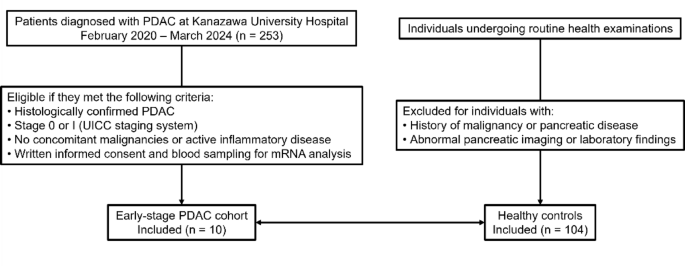
This study has several limitations. First, this was a single-center case–control diagnostic accuracy study with a small number of stage 0–I PDAC cases, resulting in wide CIs for estimates of sensitivity and AUC. Although the observed sensitivity was high, these findings should be interpreted as preliminary and hypothesis-generating rather than definitive evidence of diagnostic utility. Second, no formal a priori sample size calculation was performed because of the extreme rarity of PDAC detected at stage 0 or I, even in high-volume centers. Accordingly, all eligible stage 0–I patients diagnosed over a four-year period were prospectively enrolled, and the study was designed as an exploratory pilot investigation. Third, the use of HCs rather than clinically relevant disease controls, such as patients with chronic pancreatitis, IPMN, pancreatic cystic lesions, or individuals at high risk because of genetic predisposition or family history, substantially limits the generalizability of the findings to real-world diagnostic settings. In particular, because the mRNA-based system reflects immune-related whole-blood gene expression, false-positive results may arise from non-PDAC malignancies, inflammatory conditions, or other systemic immune alterations. This mRNA panel has not been systematically evaluated in other malignancies or inflammatory diseases; therefore, cross-reactivity with non-PDAC cancers or non-malignant inflammatory conditions cannot be excluded, and disease specificity remains insufficiently established. In addition, although HCs had no findings suggestive of PDAC or related pancreatic diseases on health-checkup evaluations, systematic screening for occult malignancies in other organs or chronic inflammatory conditions was incomplete, and longitudinal follow-up data for mRNA-positive HCs were not available. Fourth, this study represents an internal evaluation of a previously developed diagnostic system rather than an independent external validation study. Although the diagnostic algorithm and cut-off were predefined and not re-optimized in the present cohort, validation in independent multicenter cohorts is required. Fifth, our algorithm was developed as a weighted diagnostic classifier rather than a mechanistic feature-importance model. Therefore, the relative biological contribution of each gene remains to be clarified in future studies. Sixth, potential confounding factors, including subclinical inflammation, comorbidities, and concomitant medications, may influence peripheral blood gene expression, although no clear associations beyond age were detected in this small sample. Finally, this study did not evaluate cost-effectiveness or logistical issues related to implementing RT-PCR-based assays in routine practice. Future studies should include larger numbers of early-stage PDAC cases, appropriate disease-control cohorts, longitudinal follow-up of mRNA-positive controls, and external validation of the diagnostic algorithm before clinical implementation.
In conclusion, this pilot prospective study demonstrates that the mRNA-based diagnostic system shows substantially higher sensitivity than CA19-9 for the detection of early-stage PDAC, including carcinoma in situ. Although limited by sample size and moderate specificity, these findings provide preliminary evidence that peripheral blood transcriptomic signatures may capture early tumor-associated immune alterations not detectable by conventional tumor markers. Future multicenter studies with adequately powered cohorts, disease control groups, and refined diagnostic algorithms are warranted to confirm diagnostic accuracy, optimize specificity, and define the most appropriate clinical contexts for implementation. Ultimately, integration of this mRNA-based system into existing diagnostic strategies may improve diagnostic evaluation of selected patients with suspected early-stage PDAC and ultimately contribute to improved patient outcomes.
