


Schaum, N. et al. Single-cell transcriptomics of 20 mouse organs creates a Tabula Muris. Nature 562, 367–372 (2018).
Google Scholar
Baysoy, A., Bai, Z., Satija, R. & Fan, R. The technological landscape and applications of single-cell multi-omics. Nat. Rev. Mol. Cell Biol. 24, 695–713 (2023).
Google Scholar
The Tabula Sapiens Consortium. The Tabula Sapiens: a multiple-organ, single-cell transcriptomic atlas of humans. Science 376, eabl4896 (2022).
Google Scholar
Moffitt, J. R., Lundberg, E. & Heyn, H. The emerging landscape of spatial profiling technologies. Nat. Rev. Genet. 23, 741–759 (2022).
Google Scholar
Hickey, J. W. et al. Spatial mapping of protein composition and tissue organization: a primer for multiplexed antibody-based imaging. Nat. Methods 19, 284–295 (2022). This is a thorough review of multiplexed antibody-based imaging approaches with a focus on experimental design, computational analysis and practical guidelines.
Google Scholar
Bollhagen, A. & Bodenmiller, B. Highly multiplexed tissue imaging in precision oncology and translational cancer research. Cancer Discov. 14, 2071–2088 (2024).
Google Scholar
de Souza, N., Zhao, S. & Bodenmiller, B. Multiplex protein imaging in tumour biology. Nat. Rev. Cancer 24, 171–191 (2024).
Google Scholar
Method of the Year 2024: spatial proteomics. Nat. Methods 21, 2195–2196 (2024).
Gerdes, M. J. et al. Highly multiplexed single-cell analysis of formalin-fixed, paraffin-embedded cancer tissue. Proc. Natl Acad. Sci. USA 110, 11982–11987 (2013).
Google Scholar
Gut, G., Herrmann, M. D. & Pelkmans, L. Multiplexed protein maps link subcellular organization to cellular states. Science 361, eaar7042 (2018).
Google Scholar
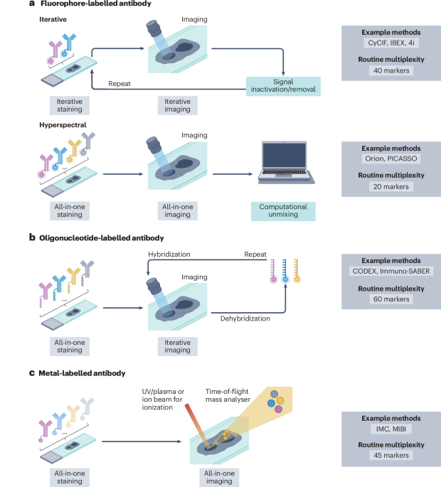
Lin, J.-R. et al. Highly multiplexed immunofluorescence imaging of human tissues and tumors using t-CyCIF and conventional optical microscopes. eLife 7, e31657 (2018).
Google Scholar
Radtke, A. J. et al. IBEX: a versatile multiplex optical imaging approach for deep phenotyping and spatial analysis of cells in complex tissues. Proc. Natl Acad. Sci. USA 117, 33455–33465 (2020).
Google Scholar
Lin, J. R. et al. High-plex immunofluorescence imaging and traditional histology of the same tissue section for discovering image-based biomarkers. Nat. Cancer 4, 1036–1052 (2023).
Google Scholar
Seo, J. et al. PICASSO allows ultra-multiplexed fluorescence imaging of spatially overlapping proteins without reference spectra measurements. Nat. Commun. 13, 2475 (2022).
Google Scholar
Goltsev, Y. et al. Deep profiling of mouse splenic architecture with CODEX multiplexed imaging. Cell 174, 968–981.e15 (2018).
Google Scholar
Black, S. et al. CODEX multiplexed tissue imaging with DNA-conjugated antibodies. Nat. Protoc. 16, 3802–3835 (2021).
Google Scholar
Saka, S. K. et al. Immuno-SABER enables highly multiplexed and amplified protein imaging in tissues. Nat. Biotechnol. 37, 1080–1090 (2019).
Google Scholar
Angelo, M. et al. Multiplexed ion beam imaging of human breast tumors. Nat. Med. 20, 436–442 (2014).
Google Scholar
Giesen, C. et al. Highly multiplexed imaging of tumor tissues with subcellular resolution by mass cytometry. Nat. Methods 11, 417–422 (2014). This landmark article introduces IMC.
Google Scholar
Windhager, J. et al. An end-to-end workflow for multiplexed image processing and analysis. Nat. Protoc. 18, 3565–3613 (2023). This article describes an extendable and customizable end-to-end workflow for multiplexed tissue image processing and analysis, specifically developed for IMC.
Google Scholar
Jackson, H. W. et al. The single-cell pathology landscape of breast cancer. Nature 578, 615–620 (2020). This study pioneers the use of IMC in a large breast cancer cohort and shows that spatial single-cell phenotyping can improve patient stratification.
Google Scholar
Rendeiro, A. F. et al. The spatial landscape of lung pathology during COVID-19 progression. Nature 593, 564–569 (2021). This study uses IMC to map COVID-19 lung disease progression, showing that SARS-CoV-2 mainly infects alveolar epithelial cells and triggers local hyperinflammation.
Google Scholar
Damond, N. et al. A map of human type 1 diabetes progression by imaging mass cytometry. Cell Metab. 29, 755–768.e5 (2019).
Google Scholar
Wang, Y. J. et al. Multiplexed in situ imaging mass cytometry analysis of the human endocrine pancreas and immune system in type 1 diabetes. Cell Metab. 29, 769–783.e4 (2019).
Google Scholar
Karimi, E. et al. Single-cell spatial immune landscapes of primary and metastatic brain tumours. Nature 614, 555–563 (2023).
Google Scholar
Sorin, M. et al. Single-cell spatial landscapes of the lung tumour immune microenvironment. Nature 614, 548–554 (2023). This article maps the tumour microenvironment of non-small-cell lung cancer using IMC and links its spatial organization to clinical outcomes.
Google Scholar
Wang, X. Q. et al. Spatial predictors of immunotherapy response in triple-negative breast cancer. Nature 621, 868–876 (2023). This study uses IMC in a randomized clinical trial for biomarker discovery and identifies spatial predictors of immunotherapy response in triple-negative breast cancer.
Google Scholar
Meyer, L. et al. A stratification system for breast cancer based on basoluminal tumor cells and spatial tumor architecture. Cancer Cell 43, 1637–1655.e9 (2025).
Google Scholar
Danenberg, E. et al. Breast tumor microenvironment structures are associated with genomic features and clinical outcome. Nat. Genet. 54, 660–669 (2022).
Google Scholar
Bandura, D. R. et al. Mass cytometry: technique for real time single cell multitarget immunoassay based on inductively coupled plasma time-of-flight mass spectrometry. Anal. Chem. 81, 6813–6822 (2009).
Google Scholar
Hyperion XTi Imaging System with and Without Slide Loader User Guide FLDM-00672. Standard BioTools https://www.standardbio.com/support/instrument-support/hyperion-xti-support (2023).
Han, G., Spitzer, M. H., Bendall, S. C., Fantl, W. J. & Nolan, G. P. Metal-isotope-tagged monoclonal antibodies for high-dimensional mass cytometry. Nat. Protoc. 13, 2121–2148 (2018).
Google Scholar
Lou, X. et al. Polymer-based elemental tags for sensitive bioassays. Angew. Chem. Int. Ed. 46, 6111–6114 (2007).
Google Scholar
Casanova, R. et al. Standardization of suspension and imaging mass cytometry single-cell readouts for clinical decision making. Cytom. A 107, 390–403 (2025).
Google Scholar
Han, G. et al. Atomic mass tag of bismuth-209 for increasing the immunoassay multiplexing capacity of mass cytometry. Cytom. A 91, 1150–1163 (2017).
Google Scholar
Hartmann, F. J. et al. in Mass Cytometry. Methods in Molecular Biology Vol. 1989 (eds McGuire, H. & Ashhurst, T.) 55–81 (Humana, 2019).
Mei, H. E., Leipold, M. D. & Maecker, H. T. Platinum-conjugated antibodies for application in mass cytometry. Cytom. A 89, 292–300 (2016).
Google Scholar
Elaldi, R. et al. High dimensional imaging mass cytometry panel to visualize the tumor immune microenvironment contexture. Front. Immunol. 12, 666233 (2021).
Google Scholar
Schulz, D. et al. Simultaneous multiplexed imaging of mRNA and proteins with subcellular resolution in breast cancer tissue samples by mass cytometry. Cell Syst. 6, 25–36.e5 (2018). This article shows that IMC can detect RNA transcripts and proteins simultaneously in the same tissue section.
Google Scholar
Wang, F. et al. RNAscope: a novel in situ RNA analysis platform for formalin-fixed, paraffin-embedded tissues. J. Mol. Diagn. 14, 22–29 (2012).
Google Scholar
Schulz, D. Oligonucleotide-polymer conjugation for imaging mass cytometry v2. Preprint at https://doi.org/10.17504/protocols.io.dm6gpjz91gzp/v2 (2023).
Bodenmiller, B. Multiplexed epitope-based tissue imaging for discovery and healthcare applications. Cell Syst. 2, 225–238 (2016).
Google Scholar
Meyer, L. et al. A spatial multi-omic portrait of survival outcome for clear cell renal cell carcinoma. Preprint at medRxiv https://doi.org/10.64898/2026.03.02.26347390 (2026).
Kuett, L. et al. Distant metastases of breast cancer resemble primary tumors in cancer cell composition but differ in immune cell phenotypes. Cancer Res. 85, 15–31 (2025).
Google Scholar
Steenbuck, N. et al. Imaging mass cytometry reveals functional and immunological changes during type 1 diabetes progression in human pancreata. Nat. Metab. https://doi.org/10.1038/s42255-026-01559-z (2026).
Google Scholar
Matos, T. R., Liu, H. & Ritz, J. Research techniques made simple: experimental methodology for single-cell mass cytometry. J. Investig. Dermatol. 137, e31–e38 (2017).
Google Scholar
Chevrier, S. et al. Compensation of signal spillover in suspension and imaging mass cytometry. Cell Syst. 6, 612–620.e5 (2018).
Google Scholar
Takahashi, C. et al. Mass cytometry panel optimization through the designed distribution of signal interference. Cytom. A 91, 39–47 (2017).
Google Scholar
Lun, X. K. et al. Influence of node abundance on signaling network state and dynamics analyzed by mass cytometry. Nat. Biotechnol. 35, 164–172 (2017).
Google Scholar
Uhlen, M. et al. A proposal for validation of antibodies. Nat. Methods 13, 823–827 (2016).
Google Scholar
Bordeaux, J. et al. Antibody validation. Biotechniques 48, 197–209 (2010).
Google Scholar
Røgenes, H. et al. Development of 42 marker panel for in-depth study of cancer associated fibroblast niches in breast cancer using imaging mass cytometry. Front. Immunol. 15, 1325191 (2024).
Google Scholar
Thul, P. J. & Lindskog, C. The human protein atlas: a spatial map of the human proteome. Protein Sci. 27, 233–244 (2018).
Google Scholar
Bouzekri, A., Esch, A. & Ornatsky, O. Multidimensional profiling of drug-treated cells by imaging mass cytometry. FEBS Open Bio 9, 1652–1669 (2019).
Google Scholar
Lotsberg, M. L. et al. Intrinsic differences in spatiotemporal organization and stromal cell interactions between isogenic lung cancer cells of epithelial and mesenchymal phenotypes revealed by high-dimensional single-cell analysis of heterotypic 3D spheroid models. Front. Oncol. 12, 818437 (2022).
Google Scholar
Zanotelli, V. R. et al. A quantitative analysis of the interplay of environment, neighborhood, and cell state in 3D spheroids. Mol. Syst. Biol. 16, MSB20209798 (2020).
Google Scholar
Taube, J. M. et al. The society for immunotherapy in cancer statement on best practices for multiplex immunohistochemistry (IHC) and immunofluorescence (IF) staining and validation. J. ImmunoTher. Cancer 8, e000155 (2020).
Google Scholar
Eling, N. et al. Multi-modal image analysis for large-scale cancer tissue studies within IMMUcan. Cell Rep. Methods 5, 101170 (2025). This study describes the multi-modal image analysis framework used in the IMMUcan project, which generated a large manually curated reference dataset for spatial single-cell phenotyping.
Google Scholar
Bost, P., Schulz, D., Engler, S., Wasserfall, C. & Bodenmiller, B. Optimizing multiplexed imaging experimental design through tissue spatial segregation estimation. Nat. Methods 20, 418–423 (2023).
Google Scholar
Baker, E. A. G., Schapiro, D., Dumitrascu, B., Vickovic, S. & Regev, A. In silico tissue generation and power analysis for spatial omics. Nat. Methods 20, 424–431 (2023).
Google Scholar
Ravi, V. M. et al. Spatially resolved multi-omics deciphers bidirectional tumor-host interdependence in glioblastoma. Cancer Cell 40, 639–655.e13 (2022).
Google Scholar
Yuan, M. et al. Smart spatial omics (S2-omics) optimizes region of interest selection to capture molecular heterogeneity in diverse tissues. Nat. Cell Biol. 27, 2225–2238 (2025).
Google Scholar
Fustinoni, S. et al. Exposure and management of the health risk for the use of formaldehyde and xylene in a large pathology laboratory. Ann. Work Expo. Health 65, 805–818 (2021).
Google Scholar
Grady, C. et al. Broad consent for research with biological samples: workshop conclusions. Am. J. Bioeth. 15, 34–42 (2015).
Google Scholar
Schapiro, D. et al. MCMICRO: a scalable, modular image-processing pipeline for multiplexed tissue imaging. Nat. Methods 19, 311–315 (2022).
Google Scholar
McQuin, C. et al. CellProfiler 3.0: next-generation image processing for biology. PLoS Biol. 16, e2005970 (2018).
Google Scholar
Berg, S. et al. ilastik: interactive machine learning for (bio)image analysis. Nat. Methods 16, 1226–1232 (2019).
Google Scholar
Baranski, A. et al. MAUI (MBI Analysis User Interface)—an image processing pipeline for multiplexed mass based imaging. PLoS Comput. Biol. 17, e1008887 (2021).
Google Scholar
Milosevic, V. Different approaches to Imaging Mass Cytometry data analysis. Bioinform. Adv. 3, vbad046 (2023).
Google Scholar
Meyer-Bender, M. et al. Spatialproteomics: an interoperable toolbox for analyzing highly multiplexed fluorescence image data. Nat. Methods 23, 1564–1573 (2026).
Google Scholar
Guazzini, M., Reisach, A. G., Weichwald, S. & Seiler, C. spillR: spillover compensation in mass cytometry data. Bioinformatics 40, btae337 (2024).
Google Scholar
Lu, P. et al. IMC-Denoise: a content aware denoising pipeline to enhance Imaging Mass Cytometry. Nat. Commun. 14, 1601 (2023).
Google Scholar
Ijsselsteijn, M. E., Somarakis, A., Lelieveldt, B. P. F., Höllt, T. & de Miranda, N. F. C. C. Semi-automated background removal limits data loss and normalizes imaging mass cytometry data. Cytom. A 99, 1187–1197 (2021).
Google Scholar
Stringer, C., Wang, T., Michaelos, M. & Pachitariu, M. Cellpose: a generalist algorithm for cellular segmentation. Nat. Methods 18, 100–106 (2021). This article introduces Cellpose, a common deep learning model for cell segmentation in IMC workflows.
Google Scholar
Greenwald, N. F. et al. Whole-cell segmentation of tissue images with human-level performance using large-scale data annotation and deep learning. Nat. Biotechnol. 40, 555–565 (2022).
Google Scholar
Pachitariu, M., Rariden, M. & Stringer, C. Cellpose-SAM: superhuman generalization for cellular segmentation. Preprint at bioRxiv https://doi.org/10.1101/2025.04.28.651001 (2025).
Marks, M. et al. CellSAM: a foundation model for cell segmentation. Nat. Methods 22, 2585–2593 (2025).
Google Scholar
Goldsborough, T. et al. A novel channel invariant architecture for the segmentation of cells and nuclei in multiplexed images using InstanSeg. Preprint at bioRxiv https://doi.org/10.1101/2024.09.04.611150 (2024).
Cords, L. et al. Cancer-associated fibroblast phenotypes are associated with patient outcome in non-small cell lung cancer. Cancer Cell 42, 396–412.e5 (2024).
Google Scholar
van Maldegem, F. et al. Characterisation of tumour microenvironment remodelling following oncogene inhibition in preclinical studies with imaging mass cytometry. Nat. Commun. 12, 5906 (2021).
Google Scholar
Rumberger, J. L. et al. Automated classification of cellular expression in multiplexed imaging data with Nimbus. Nat. Methods 22, 2161–2170 (2025).
Google Scholar
Bai, Y. et al. Adjacent cell marker lateral spillover compensation and reinforcement for multiplexed images. Front. Immunol. 12, 652631 (2021).
Google Scholar
Lee, Y. et al. Segmentation aware probabilistic phenotyping of single-cell spatial protein expression data. Nat. Commun. 16, 389 (2025).
Google Scholar
Liu, C. C. et al. Robust phenotyping of highly multiplexed tissue imaging data using pixel-level clustering. Nat. Commun. 14, 4618 (2023).
Google Scholar
Gerber, R. et al. Spatially-smoothed quantification improves cell typing in imaging mass cytometry datasets. Preprint at bioRxiv https://doi.org/10.64898/2025.12.16.693927 (2025).
Gentleman, R. C. et al. Bioconductor: open software development for computational biology and bioinformatics. Genome Biol. 5, R80 (2004).
Google Scholar
Eling, N., Damond, N., Hoch, T. & Bodenmiller, B. cytomapper: an R/Bioconductor package for visualization of highly multiplexed imaging data. Bioinformatics 36, 5706–5708 (2020).
Google Scholar
Righelli, D. et al. SpatialExperiment: infrastructure for spatially-resolved transcriptomics data in R using Bioconductor. Bioinformatics 38, 3128–3131 (2022).
Google Scholar
Chen, J. G. et al. Giotto Suite: a multiscale and technology-agnostic spatial multiomics analysis ecosystem. Nat. Methods 22, 2052–2064 (2025).
Google Scholar
Sofroniew, N. et al. napari: a multi-dimensional image viewer for Python. Zenodo https://doi.org/10.5281/zenodo.13863809 (2024).
Bankhead, P. et al. QuPath: open source software for digital pathology image analysis. Sci. Rep. 7, 16878 (2017).
Google Scholar
Schindelin, J. et al. Fiji: an open-source platform for biological-image analysis. Nat. Methods 9, 676–682 (2012).
Google Scholar
Meyer, L., Eling, N. & Bodenmiller, B. cytoviewer: an R/Bioconductor package for interactive visualization and exploration of highly multiplexed imaging data. BMC Bioinform. 25, 9 (2024).
Google Scholar
McInnes, L., Healy, J., Saul, N. & Großberger, L. UMAP: uniform manifold approximation and projection. J. Open Source Softw. 3, 861 (2018).
Google Scholar
Haghverdi, L., Lun, A. T. L., Morgan, M. D. & Marioni, J. C. Batch effects in single-cell RNA-sequencing data are corrected by matching mutual nearest neighbors. Nat. Biotechnol. 36, 421–427 (2018).
Google Scholar
Korsunsky, I. et al. Fast, sensitive and accurate integration of single-cell data with Harmony. Nat. Methods 16, 1289–1296 (2019).
Google Scholar
Levine, J. H. et al. Data-driven phenotypic dissection of AML reveals progenitor-like cells that correlate with prognosis. Cell 162, 184–197 (2015).
Google Scholar
Van Gassen, S. et al. FlowSOM: using self-organizing maps for visualization and interpretation of cytometry data. Cytom. A 87, 636–645 (2015).
Google Scholar
Hao, Y. et al. Dictionary learning for integrative, multimodal and scalable single-cell analysis. Nat. Biotechnol. 42, 293–304 (2024).
Google Scholar
Wolf, F. A., Angerer, P. & Theis, F. J. SCANPY: large-scale single-cell gene expression data analysis. Genome Biol. 19, 15 (2018).
Google Scholar
Bussi, Y. et al. CellTune: an integrative software for accurate cell classification in spatial proteomics. Nat. Methods https://doi.org/10.1038/s41592-026-03162-2 (2026).
Hoch, T. et al. Multiplexed imaging mass cytometry of the chemokine milieus in melanoma characterizes features of the response to immunotherapy. Sci. Immunol. 7, eabk1692 (2022).
Google Scholar
Shaban, M. et al. MAPS: pathologist-level cell type annotation from tissue images through machine learning. Nat. Commun. 15, 28 (2024).
Google Scholar
Amitay, Y. et al. CellSighter: a neural network to classify cells in highly multiplexed images. Nat. Commun. 14, 4302 (2023).
Google Scholar
Brbić, M. et al. Annotation of spatially resolved single-cell data with STELLAR. Nat. Methods 19, 1411–1418 (2022).
Google Scholar
Sun, H. et al. Flexible and robust cell-type annotation for highly multiplexed tissue images. Cell Syst. 16, 101374 (2025).
Google Scholar
Cheng, C. et al. AICellType: a large language model-based platform for accurate cell type annotation. Brief. Bioinform. 27, bbag151 (2026).
Google Scholar
Geuenich, M. J. et al. Automated assignment of cell identity from single-cell multiplexed imaging and proteomic data. Cell Syst. 12, 1173–1186.e5 (2021).
Google Scholar
Zhang, W. et al. Identification of cell types in multiplexed in situ images by combining protein expression and spatial information using CELESTA. Nat. Methods 19, 759–769 (2022).
Google Scholar
Huynh, K. L. A. et al. Deconvolution of cell types and states in spatial multiomics utilizing TACIT. Nat. Commun. 16, 3747 (2025).
Google Scholar
Robinson, M. D., McCarthy, D. J. & Smyth, G. K. edgeR: a Bioconductor package for differential expression analysis of digital gene expression data. Bioinformatics 26, 139–140 (2010).
Google Scholar
Amezquita, R. A. et al. Orchestrating single-cell analysis with Bioconductor. Nat. Methods 17, 137–145 (2020). This study describes common workflows for single-cell analysis within the Bioconductor framework and provides a rich online resource.
Google Scholar
Ripley, B. D. The second-order analysis of stationary point processes. J. Appl. Probab. 13, 255–266 (1976).
Google Scholar
Canete, N. P. et al. spicyR: spatial analysis of in situ cytometry data in R. Bioinformatics 38, 3099–3105 (2022).
Google Scholar
Ameen, F., Robertson, N., Lin, D. M., Ghazanfar, S. & Patrick, E. Kontextual reframes analysis of spatial omics data and reveals consistent cell relationships across images. Cell Rep. Methods 5, 101175 (2025).
Google Scholar
Benimam, M. M. et al. Statistical analysis of spatial patterns in tumor microenvironment images. Nat. Commun. 16, 3090 (2025).
Google Scholar
dos Santos Peixoto, R. et al. Characterizing cell-type spatial relationships across length scales in spatially resolved omics data. Nat. Commun. 16, 350 (2025).
Google Scholar
Palla, G. et al. Squidpy: a scalable framework for spatial omics analysis. Nat. Methods 19, 171–178 (2022).
Google Scholar
Nirmal, A. J. & Sorger, P. K. SCIMAP: a python toolkit for integrated spatial analysis of multiplexed imaging data. J. Open Source Softw. 9, 6604 (2024).
Google Scholar
Schiller, C., Ibarra-Arellano, M. A., Bestak, K., Tanevski, J. & Schapiro, D. Comparison and optimization of cellular neighbor preference methods for quantitative tissue analysis. Nat. Commun. 17, 3514 (2026).
Google Scholar
Schürch, C. M. et al. Coordinated cellular neighborhoods orchestrate antitumoral immunity at the colorectal cancer invasive front. Cell 182, 1341–1359.e19 (2020). This article introduces the concept of cellular neighbourhoods and shows that coordinated spatial immune-cell organization at the colorectal cancer invasive front is associated with patient survival.
Google Scholar
Chen, Z., Soifer, I., Hilton, H., Keren, L. & Jojic, V. Modeling multiplexed images with spatial-LDA reveals novel tissue microenvironments. J. Comput. Biol. 27, 1204–1218 (2020).
Google Scholar
Kim, J. et al. Unsupervised discovery of tissue architecture in multiplexed imaging. Nat. Methods 19, 1653–1661 (2022).
Google Scholar
Patrick, E. et al. Spatial analysis for highly multiplexed imaging data to identify tissue microenvironments. Cytom. A 103, 593–599 (2023).
Google Scholar
Ranek, J. S. et al. The automated computational workflow QUICHE reveals structural definitions of antitumor responses in triple-negative breast cancer. Nat. Cancer https://doi.org/10.1038/s43018-026-01122-5 (2026).
Google Scholar
Varrone, M., Tavernari, D., Santamaria-Martínez, A., Walsh, L. A. & Ciriello, G. CellCharter reveals spatial cell niches associated with tissue remodeling and cell plasticity. Nat. Genet. 56, 74–84 (2024).
Google Scholar
Możejko, M. et al. Cellohood: multi-granular discovery of cellular neighborhoods with a permutation-invariant set transformer auto-encoder. Preprint at bioRxiv https://doi.org/10.1101/2025.11.10.687184 (2025).
Wu, Z. et al. Graph deep learning for the characterization of tumour microenvironments from spatial protein profiles in tissue specimens. Nat. Biomed. Eng. 6, 1435–1448 (2022).
Google Scholar
Hu, Y. et al. Unsupervised and supervised discovery of tissue cellular neighborhoods from cell phenotypes. Nat. Methods 21, 267–278 (2024).
Google Scholar
Zhao, E. et al. Spatial transcriptomics at subspot resolution with BayesSpace. Nat. Biotechnol. 39, 1375–1384 (2021).
Google Scholar
Dong, K. & Zhang, S. Deciphering spatial domains from spatially resolved transcriptomics with an adaptive graph attention auto-encoder. Nat. Commun. 13, 1739 (2022).
Google Scholar
Crowell, H. L. et al. Orchestrating spatial transcriptomics analysis with Bioconductor. Preprint at bioRxiv https://doi.org/10.1101/2025.11.20.688607 (2026).
Flint, L. E. et al. Characterization of an aggregated three-dimensional cell culture model by multimodal mass spectrometry imaging. Anal. Chem. 92, 12538–12547 (2020).
Google Scholar
Flint, L. E. et al. Comparison of osteosarcoma aggregated tumour models with human tissue by multimodal mass spectrometry imaging. Metabolites 11, 506 (2021).
Google Scholar
Berg, H. F. et al. Patient-derived organoids reflect the genetic profile of endometrial tumors and predict patient prognosis. Commun. Med. 1, 20 (2021).
Google Scholar
Bollhagen, A. et al. High-resolution imaging mass cytometry to map subcellular structures. Nat. Methods 22, 2601–2608 (2025). This study improves the resolution of IMC to below 350 nm using laser oversampling and pixel deconvolution, enabling detailed mapping of subcellular structures.
Google Scholar
Zabransky, D. J. et al. Profiling of syngeneic mouse HCC tumor models as a framework to understand anti–PD-1 sensitive tumor microenvironments. Hepatology 77, 1566–1579 (2023).
Google Scholar
Peran, I. et al. Cadherin 11 promotes immunosuppression and extracellular matrix deposition to support growth of pancreatic tumors and resistance to gemcitabine in mice. Gastroenterology 160, 1359–1372.e13 (2021).
Google Scholar
Snyder, M. P. et al. The human body at cellular resolution: the NIH Human Biomolecular Atlas Program. Nature 574, 187–192 (2019).
Google Scholar
Curtis, C. et al. The genomic and transcriptomic architecture of 2,000 breast tumours reveals novel subgroups. Nature 486, 346–352 (2012).
Google Scholar
Ali, H. R. et al. Imaging mass cytometry and multiplatform genomics define the phenogenomic landscape of breast cancer. Nat. Cancer 1, 163–175 (2020).
Google Scholar
Enfield, K. S. S. et al. Spatial architecture of myeloid and T cells orchestrates immune evasion and clinical outcome in lung cancer. Cancer Discov. 14, 1018–1047 (2024).
Google Scholar
Voglis, S. et al. Spatially-resolved single-cell imaging of melanoma brain metastases identifies localized immune patterns predictive of immune checkpoint blockade response. Neuro-Oncol. https://doi.org/10.1093/neuonc/noag128 (2026).
Zhang, H. et al. Spatially resolved C1QC+ macrophage-CD4+ T cell niche in colorectal cancer microenvironment: implications for immunotherapy response. Cell Discov. 11, 60 (2025).
Google Scholar
Li, M. et al. Spatial proteomics of immune microenvironment in nonalcoholic steatohepatitis-associated hepatocellular carcinoma. Hepatology 79, 560–574 (2024).
Google Scholar
Hong, L. et al. Spatial single-cell proteomics landscape decodes the tumor microenvironmental ecosystem of intrahepatic cholangiocarcinoma. Hepatology 83, 57–74 (2026).
Google Scholar
Louis Sam Titus, A. S. C. et al. Molecular architecture of proliferative lupus nephritis as elucidated using 50-plex imaging mass cytometry proteomics. Clin. Immunol. 254, 109713 (2023).
Google Scholar
Ramaglia, V. et al. Multiplexed imaging of immune cells in staged multiple sclerosis lesions by mass cytometry. eLife 8, e48051 (2019).
Google Scholar
Kondo, A. et al. Highly multiplexed image analysis of intestinal tissue sections in patients with inflammatory bowel disease. Gastroenterology 161, 1940–1952 (2021).
Google Scholar
Gianni, L. et al. Pathologic complete response (pCR) to neoadjuvant treatment with or without atezolizumab in triple-negative, early high-risk and locally advanced breast cancer: NeoTRIP Michelangelo randomized study. Ann. Oncol. 33, 534–543 (2022).
Google Scholar
Ludford, K. et al. Neoadjuvant pembrolizumab in localized microsatellite instability high/deficient mismatch repair solid tumors. J. Clin. Oncol. 41, 2181–2190 (2023).
Google Scholar
Irmisch, A. et al. The Tumor Profiler Study: integrated, multi-omic, functional tumor profiling for clinical decision support. Cancer Cell 39, 288–293 (2021).
Google Scholar
Miglino, N. et al. Feasibility of multiomics tumor profiling for guiding treatment of melanoma. Nat. Med. 31, 2430–2441 (2025). This article reports on the results from the melanoma arm of the Tumor Profiler trial, in which multiomics profiling improved treatment decisions and IMC was highlighted as a key contributing technology.
Google Scholar
Tornaas, S. et al. Development of a high dimensional imaging mass cytometry panel to investigate spatial organization of tissue microenvironment in formalin-fixed archival clinical tissues. Heliyon 10, e31191 (2024).
Google Scholar
Schulz, A. R. et al. Stabilizing antibody cocktails for mass cytometry. Cytom. A 95, 910–916 (2019).
Google Scholar
O’Hara, D. M. et al. Recommendations for the validation of flow cytometric testing during drug development: II assays. J. Immunol. Methods 363, 120–134 (2011).
Google Scholar
Wood, B. et al. Validation of cell-based fluorescence assays: practice guidelines from the ICSH and ICCS – part V – assay performance criteria. Cytom. B Clin. Cytom. 84, 315–323 (2013).
Google Scholar
Wilkinson, M. D. et al. Comment: the FAIR guiding principles for scientific data management and stewardship. Sci. Data 3, 160018 (2016).
Google Scholar
Cleary, B. et al. Compressed sensing for highly efficient imaging transcriptomics. Nat. Biotechnol. 39, 936–942 (2021).
Google Scholar
Hosogane, T., Santana, L. S., Eling, N., Moch, H. & Bodenmiller, B. Compressed sensing expands the multiplexity of imaging mass cytometry. Nat. Commun. 16, 11671 (2025).
Google Scholar
Ben-Uri, R. et al. High-dimensional imaging using combinatorial channel multiplexing and deep learning. Nat. Biotechnol. https://doi.org/10.1038/s41587-025-02585-0 (2026).
Tietscher, S. et al. A comprehensive single-cell map of T cell exhaustion-associated immune environments in human breast cancer. Nat. Commun. 14, 98 (2023).
Google Scholar
Lun, X. K. et al. Signal amplification by cyclic extension enables high-sensitivity single-cell mass cytometry. Nat. Biotechnol. 43, 811–821 (2025).
Google Scholar
Hosogane, T., Casanova, R. & Bodenmiller, B. DNA-barcoded signal amplification for imaging mass cytometry enables sensitive and highly multiplexed tissue imaging. Nat. Methods 20, 1304–1309 (2023).
Google Scholar
Dicks, S. et al. GPU-accelerated single-cell analysis at scale with rapids-singlecell. Preprint at https://doi.org/10.48550/arXiv.2603.02402 (2026).
Moore, J. et al. OME-Zarr: a cloud-optimized bioimaging file format with international community support. Histochem. Cell Biol. 160, 223–251 (2023).
Google Scholar
Lüthi, J. et al. Fractal: towards FAIR bioimage analysis at scale with OME-Zarr-native workflows. Preprint at bioRxiv https://doi.org/10.64898/2026.03.05.709921 (2026).
Bell, A. T. F. et al. PanIN and CAF transitions in pancreatic carcinogenesis revealed with spatial data integration. Cell Syst. 15, 753–769.e5 (2024).
Google Scholar
Griner, J., Gerber, R., Robinson, M. D., Krieg, C. & Guglietta, S. Resolving sialylated N-glycans and immune cell landscapes using a unified same-section IMC–MSI workflow. Preprint at bioRxiv https://doi.org/10.64898/2026.02.21.707206 (2026).
Baars, M. J. D. et al. MATISSE: a method for improved single cell segmentation in imaging mass cytometry. BMC Biol. 19, 99 (2021).
Google Scholar
Kim, E. N. et al. Dual-modality imaging of immunofluorescence and imaging mass cytometry for whole-slide imaging and accurate segmentation. Cell Rep. Methods 3, 100595 (2023).
Google Scholar
Nunes, J. B. et al. Integration of mass cytometry and mass spectrometry imaging for spatially resolved single-cell metabolic profiling. Nat. Methods 21, 1796–1800 (2024).
Google Scholar
Chen, W. et al. A visual–omics foundation model to bridge histopathology with spatial transcriptomics. Nat. Methods 22, 1568–1582 (2025).
Google Scholar
Enninful, A. et al. Integration of imaging-based and sequencing-based spatial omics mapping on the same tissue section via DBiTplus. Nat. Methods https://doi.org/10.1038/s41592-025-02948-0 (2026).
He, S. et al. Starfysh integrates spatial transcriptomic and histologic data to reveal heterogeneous tumor–immune hubs. Nat. Biotechnol. 43, 223–235 (2025).
Google Scholar
Liu, Y. et al. High-parameter spatial multi-omics through histology-anchored integration. Nat. Methods https://doi.org/10.1038/s41592-025-02926-6 (2026).
Li, Z. et al. AI-enabled virtual spatial proteomics from histopathology for interpretable biomarker discovery in lung cancer. Nat. Med. https://doi.org/10.1038/s41591-025-04060-4 (2026).
Google Scholar
Valanarasu, J. M. J. et al. Multimodal AI generates virtual population for tumor microenvironment modeling. Cell 189, 386–400.e19 (2026).
Google Scholar
Bai, B. et al. Deep learning-enabled virtual histological staining of biological samples. Light Sci. Appl. 12, 57 (2023).
Google Scholar
Shaban, M. et al. A foundation model for spatial proteomics. Preprint at https://doi.org/10.48550/arXiv.2506.03373 (2025).
Wenckstern, J. et al. The Virtual Tissues foundation model resolves spatial proteomics across scales. Nature https://doi.org/10.1038/s41586-026-10884-y (2026).
Uchal, D. et al. ImmuVis: hyperconvolutional foundation model for imaging mass cytometry. Preprint at https://doi.org/10.48550/arXiv.2602.04585 (2026).
Kuett, L. et al. Three-dimensional imaging mass cytometry for highly multiplexed molecular and cellular mapping of tissues and the tumor microenvironment. Nat. Cancer 3, 122–133 (2022). This study extends IMC to 3D analysis by reconstructing multiplexed tissue volumes from serial sections, revealing breast cancer tissue architecture not visible in two dimensions.
Google Scholar
Fischer, R. S., Wu, Y., Kanchanawong, P., Shroff, H. & Waterman, C. M. Microscopy in 3D: a biologist’s toolbox. Trends Cell Biol. 21, 682–691 (2011).
Google Scholar
Chen, S. et al. Three-dimensional mapping in multi-samples with large-scale imaging and multiplexed post staining. Commun. Biol. 6, 148 (2023).
Google Scholar
Yapp, C. et al. Highly multiplexed 3D profiling of cell states and immune niches in human tumors. Nat. Methods 22, 2180–2193 (2025).
Google Scholar
Allam, M. et al. Spatially variant immune infiltration scoring in human cancer tissues. NPJ Precis. Oncol. 6, 60 (2022).
Google Scholar
Parkinson, J. E. et al. Extracellular matrix phenotyping by imaging mass cytometry defines distinct cellular matrix environments associated with allergic airway inflammation. Mol. Syst. Biol. https://doi.org/10.1038/s44320-026-00234-5 (2026).
Google Scholar
Somer, J., Mannor, S. & Alon, U. Temporal tissue dynamics from a spatial snapshot. Nature 650, 490–499 (2026).
Google Scholar
