


Brain tumours are a clinically important cause of neurological disorders in dogs, although they occur infrequently2. Brain tumours also have a low frequency in humans, and dogs could represent a valuable translational model for human brain tumours, as spontaneous canine brain tumours share histopathological and genetic characteristics with their human counterparts8.
There had been no published data on the incidence or prevalence of canine brain tumours in Australia. In this study, meningioma was the most common primary brain tumour, representing 60.6% of diagnoses. This aligns with studies from Asia and North America, where meningioma accounted for 45–51% of canine brain tumours2,6,9,52, although Hayes et al.53. found glial tumours more common than meningioma. The discrepancy is likely attributable to differences in tumour classification methodology; our study classified each tumour type separately, whereas Hayes et al.53, grouped several tumour types under glial tumours. Overall, the literature consistently identifies meningioma as the predominant primary canine brain tumour54,55, likely reflecting its origin from arachnoid cap cells of the meninges and its extra-axial location, which typically produces clearer margins on imaging; this makes meningiomas easier to detect than many intra-axial tumours55,56.
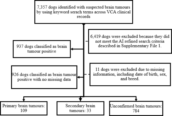
In the current study, secondary brain tumours accounted for 3.6% of all cases, a lower proportion than the 14.5% reported by Kishimoto et al.9. Pituitary macroadenoma was the most frequently identified secondary tumour (36.4%). Although the pituitary gland lies within the cranial cavity, pituitary neoplasms are often classified as secondary brain tumours because they originate from endocrine rather than neural tissue. Snyder et al.10 also reported pituitary tumours as the predominant secondary brain tumours. In contrast, Okonji et al.57 identified hemangiosarcoma, carcinoma, and melanoma as the most common metastatic tumours. The variation in reported frequency among studies likely reflects differences in data sources used, dissimilar sampling approaches and tumour classification systems. Studies restricted to specific tumour types or based on data from referral-centre populations may also influence the observed frequency of brain tumours.
In the current study, 84.6% of cases were classified as unconfirmed brain tumours, representing dogs with a high clinical suspicion of brain tumours rather than with a definitive diagnosis. This high proportion reflects the fact that dogs presenting with severe or rapidly progressive central neurological signs often show rapid clinical deterioration and have a poor prognosis and therefore a definite diagnosis is not obtained. In fact, many suspected end-stage brain tumour cases are euthanised before advanced imaging or histopathological examination can be performed58,59. Consequently, suspected tumour cases based on clinical signs from veterinary clinic records might underrepresent true tumour incidence (primary and secondary brain tumours) because definitive confirmation requires biopsy or post-mortem examination, which is not always feasible in clinical practices8. On the other hand, absence of diagnostic confirmation requires cautious interpretation of suspected cancer cases based on clinical signs8,59. Therefore, in contrast to previous studies6,9,10,53, which included only confirmed primary and secondary tumours, the present study incorporates both confirmed tumours and cases with a high clinical suspicion of brain tumours, representing a key methodological difference in case selection that should be considered when comparing results between different studies. However, the consistency of patterns between confirmed and unconfirmed cases suggests that inclusion of unconfirmed cases is unlikely to materially influence the overall conclusions.
In this study, the overall incidence rate of brain tumours was 8.7 per 100,000 dog-years at risk, which is lower than the 14.5 per 100,000 dogs at risk reported by Vandevelde60,61, for central nervous system tumours. Within primary tumours, meningiomas had the highest incidence at 0.62 per 100,000 dog-years, followed by glioma (0.25; 95% CI: 0.16–0.35). These findings are consistent with those reported by Dhein et al.27, who reported a meningioma incidence of 1.2 (95% CI: 1.0-1.5) in Swiss dogs based on population-level pathology data. The current results also correspond with human data from Rohringer et al.62, who reported crude incidence rates of 2.3 per 100,000 for all meningiomas and 0.17 per 100,000 for malignant meningiomas in Manitoba, Canada. Variations in incidence rates likely reflecting variations in study designs, case ascertainment, and population denominators. However, overall, the findings suggest a broadly similar pattern in the relative frequency and predominance of meningioma across dogs and humans. This cross-species consistency in relative meningioma predominance underscores the value of dogs as a translational model for human meningioma epidemiology and therapeutic research.
Across age groups, brain tumour incidence was highest in dogs aged 10–12 years (12.7 per 100,000 dog-years), followed by those ≥ 13 years (11.2) and 7–9 years (8.3) old, consistent with previous studies identifying older age as a major risk factor in dogs2,9,52,53. The higher incidence in elderly dogs likely reflects biological ageing process, including the accumulation of somatic mutations and age-related immune decline, which reduce the ability to eliminate abnormal cells63,64. In humans, the incidence of brain and central nervous system cancers is estimated at 1.3 per 100,000 persons for children aged 0–14 years65, and paediatric brain and central nervous system cancers rank second globally for both incidence and mortality within this age group66. Brain cancer incidence also increases with age in humans and in Australia, incidence rates among those aged 80 years and older were 24 cases per 100,000 people in 202167. While some dog breeds show elevated risk, most human brain tumours have no recognised inherited predisposition at the population level68, underscoring species differences in susceptibility.
Among breed categories, utility breeds (such as boxers, bullmastiffs, schnauzers) had the highest incidence of brain tumours (14.3 per 100,000 dog-years), followed by terriers (10.8), non-sporting breeds (such as bulldogs, French bulldogs, poodles, chow chows, dalmatians) (8.9), and crossbreeds (8.2). These findings are consistent with previous studies reporting that boxers and terriers are frequently affected by glial tumours53, while higher rates of brain tumours have been reported in golden retrievers, schnauzers, bulldogs, and boxers9. Song et al.2 also noted increased prevalence of primary brain tumours in crossbreeds, golden retrievers, schnauzers, boxers, and bulldogs. Variation in incidence across breeds likely reflects genetic predisposition, breed-specific anatomy, and inherited risk factors that elevate susceptibility in certain pedigree populations. The current findings support that underlying heritable determinants play a role in tumour development. This pattern is consistent with evidence that some breeds have a disproportionately higher risk for specific tumour types, indicating substantial genetic contribution to susceptibility69.
Spatial analysis revealed geographic variation in primary and secondary brain tumour occurrence across Australia. New South Wales had the highest incidence rate 2.3 per 100,000 dog-years), followed by the ACT (2.2), Victoria (1.4), and QLD (1.2). In contrast, cases in Queensland were predominantly concentrated in coastal regions. These spatial differences may reflect geographic variations in dog populations. In addition, the uneven representation of clinics within the VetCompass Australia network may have influenced the spatial distribution of recorded brain tumour cases. Consequently, observed geographic hotspots should be interpreted cautiously, as they may reflect differences in dog population structures and veterinary access by dog owners, as well as variations in clinic density rather than true brain tumour risk13,27.
The temporal trend analysis of primary and secondary brain tumour incidence revealed considerable variation over the study period. Incidence increased from 0.6 cases per 100,000 dog-years at risk in 2001 to 3.9 cases per 100,000 dog-years at risk in 2015. This upward trend may partly reflect improvements in diagnostic technologies, clinical awareness, and reporting practices rather than a true increase in disease occurrence. Over the same period, the number of participating veterinary clinics within the VCA program increased, potentially enhancing case detection and ascertainment. Advances in diagnostic imaging and clinical have improved the differentiation of neoplastic from non-neoplastic brain lesions and facilitated more accurate tumour classification and grading70,71,72,73. Consequently, lesions that may previously have remained undetected or misclassified are now more likely to be diagnosed as brain tumours. Improved data recording and increasing completeness of records in the VCA database may also have contributed to the observed trend. Therefore, the increase in incidence should be interpreted cautiously, as it may partly reflect improved case ascertainment rather than a true increase in disease occurrence.
Kulldorff’s spatial scan analysis identified a significant cluster of primary and secondary brain tumour cases in NSW (RR = 5.8, p = 0.001), together with two significant clusters of unconfirmed brain tumour cases. These findings suggest geographic variations in brain tumour occurrence; however, the clusters should be interpreted carefully given potential influences of dog population distribution, clinic participation in VCA and diagnostic access by dog owners.
The cluster analysis identified a grouping of glioma and lymphosarcoma cases with particular breeds, including French bulldogs and boerboels. Within this cluster, 93.1% of dogs were diagnosed with glioma. French Bulldogs comprised 13.8% of the cluster, while Australian Bulldogs, Pugs, Rottweilers, and Beagles each accounted for 6.9%. These findings suggest a greater representation of brachycephalic breeds within the glioma-associated cluster, consistent with previous studies reporting increased glioma occurrence in short-skulled breeds74,75. Secondary brain adenocarcinoma clustered with Australian cattle dogs and Irish terriers, while glioblastoma clustered with Moodle dogs, a Maltese-poodle crossbreed. In cluster 5, English springer spaniels were grouped with nasal lymphoma (v-test ≥ 2, p < 0.05). As the MCA/HCPC approach is exploratory, these findings should be interpreted as patterns of co-occurrence rather than direct measures of breed-specific risk. Nevertheless, previous studies have reported increased susceptibility to certain cancers among some pedigree breeds, suggesting that genetic and morphological factors may contribute to observed brain tumour patterns69.
The limited treatment options for many brain tumours, particularly those unsuitable for surgery, likely contribute to the survival differences observed across tumour types. Median survival times for glioma, astrocytoma, and pituitary macroadenomas in this study were shorter than those reported in studies involving advanced interventions such as surgical debulking or radiotherapy, which generally result in longer survival12,76,77. In contrast, the median survival for meningioma was higher than those reported by Heidner et al.78 and Rossmeisl et al.58, but remained lower than the 386 days reported by Forward et al.54 following surgical resection. These difference highlights how treatment modalities influence outcomes, and multiple studies have demonstrated significantly improved survival following radiotherapy for canine brain tumour cases79,80,81.
In addition to treatment effects, biological factors may also contribute to the observed variation in reported survival times. Smaller breeds typically living longer than larger breeds, and this interaction between body size, morphology, and longevity may partly explain extended survival in some cohorts82. When examining survival across tumour types, glioma showed the highest mortality risk compared with meningioma, consistent with findings by Jose-Lopez et al.76 and Rossmeisl et al.58, who noted poor prognoses in dogs with glioma. Overall, survival estimates should be interpreted with caution, as they may be influenced by treatment availability, access to specialist care, owner decision-making, and determining whether dogs are euthanised at or shortly after diagnosis.
Among the animal-level risk factor assessed for survival following tumour diagnosis, neutered dogs exhibited a slightly higher risk of death than intact dogs (HR = 1.1; 95% CI: 0.5–2.3; p > 0.05), contrasting with Rossmeisl et al.58, who reported reduced primary brain tumour risk in spayed females. Such discrepancies likely reflect differences in study designs, treatment pathways, and the distribution of sex and neuter status across populations. Overall, canine brain tumours generally carry a poor prognosis57,78, often resulting in euthanasia as clinical signs progress78. Although neuter status has not been directly linked to primary brain tumour risk, it has been associated with increased risk of other cancers, including hemangiosarcoma, lymphoma, osteosarcoma and mast cell tumours, which may influence broader survival patterns in dogs83.
In the analysis of animal-level risk factors for tumour incidence, male dogs had a higher incidence of both confirmed and unconfirmed brain tumours than females, a finding that contrast with Kishimoto et al.9, who reported similar prevalence between sexes in Japan. This discrepancy may reflect differences in study design, population characteristics, or the influence of reproductive status on disease occurrence8. Among breed categories, utility breeds (e.g., boxer, bullmastiff, akita, schnauzer, rottweiler) had an incidence rate 3.9 times higher than that of crossbreed dogs (p ≤ 0.001), while terrier breeds showed a 1.8-fold higher (p < 0.05). These findings are consistent with previous studies reporting increased occurrence of brain tumours in breeds such as boxers, French bulldogs, rat terriers, and Boston terriers2.
Age showed a strong positive association with brain tumour incidence in confirmed cases. Compared with dogs aged 1–3 years, those aged 4–6 years had IRRs of 3.0, while IRRs increased to 6.4, 7.0, and 4.1 for dogs aged 7–9, 10–12 and ≥ 13 years, respectively (p ≤ 0.001). This age-related pattern is consistent with previous studies reporting a higher occurrence of brain tumour in middle-aged and older dogs2,8,9. Differences across studies may reflect variation in tumour types, study populations, and their underlying demographic characteristics. Biologically, the strong age effect may reflect the cumulative impact of genetic and environmental damage, reduced DNA repair capacity, declining immune surveillance, and prolonged exposure to carcinogenic factor in older dogs84.
In the current study, a GPT-4o model accessed through the OpenAI Batch API was used to process a large dataset of VCA clinical records. This AI-assisted approach enabled structured extraction of tumour classification and related variables from unstructured diagnostic text, demonstrating the potential of large language models to support scalable and efficient retrospective research. However, careful validation remains essential to minimise misclassification and ensure data reliability when using AI approaches and had been implemented in this study.
This study has several limitations. VCA dogs were used as the denominator population because no comprehensive state- or SA2-level dog population registry exists in Australia, therefore, findings may not be fully representative of the wider Australian dog population. In addition, differences in diagnostic access, referral patterns, and clinic participation may have influenced the observed incidence, spatial patterns, and temporal trends.
The findings of this study have practical implications for both veterinarians and pet owners. A clear understanding of canine brain tumour incidence, spatio-temporal patterns, tumour type-breed-cluster, time to death, and risk factors for both, incidence of brain tumours and survival following diagnosis can support earlier recognition of brain tumours, enhance informed decision-making by clinicians, and the application of appropriate management strategies. Importantly, the ability to identify areas with higher incidence and spatial clustering can guide targeted clinical attention and resource allocation, ultimately improving pet welfare.
In conclusion, this study provides the first nationwide description of the incidence, spatio-temporal patterns, and survival outcomes of canine brain tumours in Australia using VCA data from 1994 to 2024. Future research should build upon the VCA registry to further investigate these patterns and refine prognostic models. Establishing a standardized, population-based canine registry would enhance the accuracy of representativeness, and generalizability of epidemiological analyses across Australia.
