


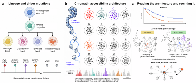
In a recent study published in Nature, Ochi et al. used ATAC-seq to profile bone marrow or peripheral blood tumor cells from 1563 newly diagnosed cases of acute myeloid leukemia (AML), resolving the disease into 16 chromatin-accessibility subgroups (the eCHROMA study) with independent prognostic and pharmacological value, most of them invisible to current genetic classification.1 This chromatin-based framework raises a sharper question: how much of that architecture can actually be engineered by drugs designed to rewrite it?
For three decades, AML classification has rested almost entirely on genetics. Driver mutations and structural variants underpin AML subtyping,2 informing the World Health Organization (WHO), International Consensus Classification (ICC), and European LeukemiaNet (ELN) systems that guide diagnosis, risk stratification and drug selection. Yet clinical heterogeneity and treatment resistance persist even within genetically defined categories, evidencing that mutation catalogs alone do not capture the full biology of the disease.
The central result is a stratification of AML into 16 chromatin-accessibility subgroups (A to P), built from 176,853 recurrent ATAC peaks. What makes this more than a descriptive exercise is what the subgroups are not. Exhaustive decision-tree analysis using all known driver lesions and whole-genome sequencing in a 213-case subset could uniquely define only three subgroups by their canonical fusions (PML::RARA, RUNX1::RUNX1T1, CBFB::MYH11) and partially a fourth by CEBPA bZIP mutations. The remaining twelve show reproducible, clinically distinct chromatin states that current genomic tools cannot predict, even at whole-genome resolution. That is the paper’s strongest empirical claim, and it is a well-supported one.
Importantly, the authors are careful not to overclaim independence from genotype. Many subgroups still map, imperfectly, onto known genetic lesions, but the real advance lies in subdividing genetically defined categories into biologically and clinically distinct entities. NPM1-mutated and KMT2A-rearranged AML, each treated as near-monolithic in current classifications, jointly resolve into four HOX-driven subgroups with distinct maturation blocks and mutation co-occurrence patterns, a heterogeneity within NPM1-mutated AML also reported independently at the transcriptomic level.3 TP53-mutated AML, similarly, splits into three subgroups nominally distinguished by differentiation trajectory (erythroid, primitive, and a third) and by outcome. The underlying deconvolution data are dispersed enough to blur the boundaries between all three, however, and none of the three subgroup assignments is checked against variant allele frequency or karyotype complexity, both plausible confounders of a deconvolution-based reading. This refinement, rather than wholesale rejection of genetics, is where the clinical value concentrates.
Mechanistically, each subgroup is anchored in a distinct gene-regulatory architecture. The authors apply two independent, previously validated computational tools, transcription-factor network inference and super-enhancer-based regulatory mapping, to their own data. This identifies subgroup-specific master regulators: HOXA family factors and E2F3 in the HOX-driven subgroups, BCL11A and IRF factors in the RUNX1-mutated subgroup, and SPI1 and C/EBP factors in the monocytic subgroups. Of 1718 non-redundant super-enhancer loci identified across the cohort, 387 were unique to a single subgroup, and 833 were shared by only a few, a substrate for subgroup-specific dependency that goes beyond a correlative chromatin signature. Single-cell multiomics on 281,167 cells from 36 patients confirms that this identity is established early and preserved through the leukemic hierarchy, including in leukemic stem cell (LSC)-enriched populations, arguing that the epigenomic fingerprint is not merely a bystander mark of differentiation state but a stable property of the malignant clone.
The clinical implications are concrete, if modest in some respects. Incorporating the ATAC subgroup into ELN risk stratification improved the concordance index by 0.036 to 0.039 in the Swedish and Japanese cohorts, respectively, an increase the authors and prior methodological work treat as clinically meaningful, though it should be read as a refinement rather than a replacement of ELN staging. More striking is the drug-sensitivity data. Subgroup K, defined by RUNX1 mutation and a maturation block at the common lymphoid progenitor stage, showed pronounced sensitivity to ABL inhibitors that was absent in RUNX1-mutated samples from other subgroups, a context-dependent vulnerability that genotype alone would have missed. Subgroups C, F, and H showed sensitivity to MEK1/2 inhibitors regardless of RAS-pathway mutation status, consistent with epigenetic activation of the pathway. For clinical translation, the authors trained expression-based classifiers using only 30 genes that predict high-risk ATAC subgroups with accuracies between 0.77 and 0.95, depending on ELN category. This yields a parsimonious and, in principle, transferable signature. Three gaps stand between this signature and clinical use. It still needs prospective validation across ethnically diverse populations and heterogeneous laboratory platforms. Its stability under therapeutic pressure remains unknown, which bears directly on its potential for measurable residual disease (MRD) monitoring. Regulatory and cost-effectiveness hurdles also stand in the way of adding a new assay to routine diagnostic workflows.
One extrapolation deserves a more cautious framing than the paper itself offers. Leukemic identity, including in LSC-enriched fractions, is chromatin-stable across the differentiation hierarchy. This stability is suggestive of MRD monitoring, since a subgroup-specific accessibility or expression signature could in principle be more robust to clonal drift than a single mutation-based marker. This is not purely speculative. An independently derived chromatin signature, based on accessibility at transposable elements rather than driver mutations, was recently shown to stratify AML relapse risk across three cohorts.4 Whether the ATAC subgroups identified here retain that stability under treatment and relapse, rather than shifting, remains untested, as the present cohort was not designed to track chromatin accessibility longitudinally.
The paper’s forward-looking discussion, reasonably, gestures toward a new generation of epigenetic therapies that might reprogram these chromatin landscapes to relieve differentiation blocks. That optimism deserves a mechanistic check. Rai and colleagues show,5 across CRISPR screens, that catalytically dead HDAC mutants and a chemically modified SAHA analog stripped of HDAC-inhibitory activity, that global histone hyperacetylation is neither necessary nor sufficient for the anticancer effects of HDAC inhibitors. A compound that never engages HDAC catalytic activity retains full antitumor efficacy in vivo; conversely, matching a drug’s histone acetylation footprint does not recapitulate its transcriptional or cytotoxic effects. The lesson generalizes beyond HDAC biology. Producing a defined chromatin-accessibility state pharmacologically will not automatically deliver the transcriptional program that state is associated with in an observational atlas. eCHROMA’s own transcription-factor and super-enhancer networks point to a more precise target. Each subgroup’s architecture identifies specific regulatory circuitry as load-bearing, not the bulk accessibility mark itself. That logic is not merely aspirational. A phase 2 trial of the menin inhibitor revumenib is testing this genotype-agnostic principle in HOX-driven leukemias spanning several genetic lesions. The intended long-term biomarker of eligibility is HOX/MEIS1 expression itself, not the underlying mutation (NCT06229912). Chromatin state is a legitimate and underused axis for classifying and stratifying AML, but engineering that axis therapeutically will require targeting the regulatory logic each subgroup encodes, not simply moving the epigenetic needle (Fig. 1).
Fig. 1
Chromatin accessibility resolves acute myeloid leukemia into subgroups that genotype alone does not predict, with consequences that diverge for prognosis and for therapy. a Current classification of acute myeloid leukemia (AML), codified in the WHO, ICC, and ELN systems that guide diagnosis, risk stratification, and drug selection, rests on recurrent driver mutations and fusions mapped onto the myeloid differentiation hierarchy from hematopoietic stem cell to mature blast. Four representative lesions, PML::RARA, RUNX1::RUNX1T1, CBFB::MYH11, and CEBPA-bZIP, each defines a single, non-overlapping disease category. Others, including NPM1 and TP53 mutations, are each associated with multiple distinct clinical and biological presentations, a heterogeneity that genetics alone leaves unresolved. b Applying ATAC-seq to 1563 newly diagnosed AML cases resolves chromatin accessibility, summarized here from 176,853 recurrent accessible regions, into sixteen reproducible subgroups (A-P). Each subgroup is anchored by a distinct transcription factor and super-enhancer network rather than by a single mutation. Colored rings mark the four subgroups, A, B, C, and I, that correspond one-to-one with a driver lesion shown in panel a; the remaining twelve subgroups, shown with a grey ring, are not predicted by known genetics, a result that holds even after whole-genome sequencing of a subset of cases. c Two consequences follow from this second, chromatin-defined layer of classification. Top, incorporating chromatin subgroup into ELN risk stratification improves the prognostic concordance index by 0.036 to 0.039 across independent cohorts (schematic survival curves), and identifies drug sensitivities that genotype alone does not: ABL-inhibitor (dasatinib shown) sensitivity restricted to the RUNX1-mutated subgroup K, and MEK1/2-inhibitor (trametinib shown) sensitivity in subgroups C, F and H regardless of RAS-pathway mutation status. Bottom, whether a chromatin architecture that is observed and correlated with outcome can also be reliably induced by an epigenetic drug, rather than merely associated with one, remains a separate and largely untested question. Independent work indicates that manipulating a chromatin mark pharmacologically does not guarantee the transcriptional and functional consequences with which that mark is normally associated. The same chromatin node is reached, yet the transcriptional and functional outcome differs
