


Cell culture, antibodies, and reagents
The human gastric cancer cell line AGS was obtained from the American Tissue Culture Collection (ATCC) and cultured in Ham’s F-12 Nutrient Mixture (GIBCO), supplemented with 10% fetal bovine serum (FBS) (Invitrogen Life Technologies) and 1% penicillin/streptomycin (GIBCO). The human gastric cancer cell line STKM2 was generously provided by Dr. Alexander Zaika from the University of Miami and was cultured in RPMI/1640 medium supplemented with 10% FBS and 1% penicillin/streptomycin. Both cell lines were maintained in a humidified incubator at 37 °C with 5% CO2 and were regularly tested to ensure there was no Mycoplasma contamination. The antibodies used in this study were as follows: AURKA (D3E4Q), phospho-AURKA (Thr288) (C39D8), STAMBP (5245), Ubiquitin (E6K4Y) (Cell Signaling Technology), LGR5 (PA5-23000) (ThermoFisher), Mouse Lgr5/GPR49 Alexa Fluor® 488-conjugated Antibody (RD20) (R&D), CagA (HPP-5003-9, Ango Austral Biologicals), and Actin (clone AC-15, Sigma-Aldrich). The pharmacological inhibitors, AMG-900 and MG132, were purchased from MedChem Express and Sigma Aldrich, respectively.
Gene silencing by small interfering RNA
Cells were seeded at low density for 24 h before being transfected with Silencer™ Select Negative Control siRNA (ThermoFisher Scientific), Silencer™ Pre-Designed AURKA siRNA (ID:139201 and ID: 139203, ThermoFisher Scientific), or Silencer™ Pre-Designed STAMBP siRNA (ID: 17954 and ID: 18048, ThermoFisher Scientific), in 10% FBS using the Lipojet transfection kit (SignaGen Laboratories) following the manufacturer’s instructions. Whole cell lysates were prepared 48 to 72 h after transfection.
H. pylori strains
CagA positive H. pylori strains (7.13, J166, and rodent-adapted PMSS1) were utilized in this study, as previously described [33]. Initially, the H. pylori strains were cultured on BD BBL™ Trypticase™ Soy Agar with 5% Sheep Blood plates (BD Trypticase™ Soy Agar II™, BD Biosciences) at 37 °C with 5% CO2 for 48 h. The strains were then passaged onto fresh Trypticase™ Soy Agar plates and cultured for an additional 24 h at 37 °C with 5% CO2. Subsequently, the H. pylori were cultured in Brucella broth (BD BBL™ Brucella Broth, BD Biosciences) supplemented with 10% fetal bovine serum (Invitrogen Life Technologies) at 37 °C with 5% CO2 overnight. For in vitro co-culture experiments, the 7.13 and J166 strains were co-cultured with gastric epithelial cells at a multiplicity of infection (MOI) of 50:1. For in vivo experiments, the PMSS1 strain was used to infect gastric epithelial cells in C57BL/6 mice (obtained from The Jackson Laboratory) via oral gavage, with a dosage of 1×10⁹ colony-forming units per mouse, and the infection was maintained for a duration of two weeks.
Western blotting
In summary, cells were washed with ice-cold phosphate-buffered saline (PBS) solution. The cells were then lysed using RIPA buffer supplemented with 1× protease and phosphatase inhibitors from Santa Cruz Biotechnology. Frozen tissue samples were also lysed in RIPA buffer at -80 °C overnight. The lysates, both from cells and tissues, were sonicated and clarified through centrifugation at 16,000 xg for 10 min at 4°C. The total protein concentration of the lysates was determined using the Pierce™ BCA Protein Assay Kit (Pierce, Thermo Scientific). Equal amounts of protein were loaded onto a 10% SDS-PAGE gel and transferred to nitrocellulose membranes (Bio-Rad). The membranes were blocked in Tris-buffered saline Tween 20 (TBST) containing 5% bovine serum albumin (BSA) for 1 h at room temperature. Specific primary antibodies were then added to the membranes and incubated overnight at 4°C. After washing the membranes four times for 10 min each with TBST, the membranes were incubated with horseradish peroxidase-conjugated secondary antibodies (Promega) for 1 h at room temperature. Subsequently, the membranes were washed again four times for 10 min each with TBST. Finally, the blots were developed using the ECL Western blotting detection reagent (Sigma Millipore), and the signal intensity was captured using the ChemiDoc XRS+ system (Bio-Rad).
Immunoprecipitation
The cells were washed with ice-cold PBS and then lysed in a cell lysis buffer composed of 0.5% Triton X-100, 150 mmol/L NaCl, 5 mmol/L EDTA, and 50 mmol/L Tris, supplemented with 1× protease and phosphatase inhibitors obtained from Roche. The cell lysates were clarified through centrifugation at 16000 xg for 10 min at 4°C, and the total protein concentration was determined using the Pierce™ BCA Protein Assay Kit (Pierce, Thermo Scientific). For immunoprecipitation, a total of 300 µg of input proteins was used per reaction. Dynabead Protein G (Invitrogen Life Sciences) was used according to the manufacturer’s instructions to couple AURKA, STAMBP, or the isotype control IgG (Santa Cruz Biotechnology Inc.) to the Dynabead Protein G. The cell lysates were then incubated with the Dynabeads-antibody complexes overnight at 4°C. Subsequently, the Dynabeads-antibody-protein complexes were pelleted using a magnet and washed six times with a washing buffer containing 0.5% Triton X-100, 150 mmol/L NaCl, 5 mmol/L EDTA, and 50 mmol/L Tris, supplemented with 1× protease and phosphatase inhibitors. The captured proteins were then eluted by incubating the samples in 1× NuPAGE™ LDS Sample Buffer (ThermoFisher Scientific) at 70 °C for 10 min. Finally, the samples were subjected to Western blotting using 4-20% Mini-PROTEAN® TGX™ Precast Protein Gels (Bio-Rad).
Quantitative real-time reverse transcriptase polymerase chain reaction (qRT-PCR)
Total RNA was extracted from cells or tissues using the RNeasy Mini Kit (Qiagen). Subsequently, cDNA was synthesized with the SuperScript cDNA Synthesis Kit (Invitrogen) according to the manufacturer’s instructions. The synthesized cDNA was diluted 10 times, and 2 μl of this dilution was used for qRT-PCR. The qRT-PCR was conducted in a 10-μl reaction volume using Bio-Rad CFX Connect Real-time System and iQ SYBR Green Supermix (Bio-Rad). Specific primers were used for the following genes: human AURKA: forward, 5′-GCTGGAGAGCTTAAAATTGCA-3′ and reverse, 5′-TTTGTAGGTCTCTTGGTATGTG-3′; human LGR5: forward, 5′-CACCTCCTACCTAGACCTCAGT-3′ and reverse, 5′-CGCAAGACGTAACTCCTCCAG-3′; human HPRT1: forward, 5′-ACCCTTTCCAAATCCTCAGC-3′ and reverse, 5′-GTTATGGCGACCCGCAG-3′; mouse Aurka: forward, 5′- CTGGATGCTGCAAACGGATAG-3′ and reverse, 5′- CGAAGGGAACAGTGGTCTTAACA-3′; mouse Lgr5: forward, 5′-CAGCCTCAAAGTGCTTATGCTG-3′ reverse, 5′- TGGCACGTAACTGATGTGG -3; mouse Hprt: forward, 5′- CCTAAGATGAGCGCAAGTTGAA-3′ and reverse, 5′- CCACAGGACTAGAACACCTGCTAA-3′. The threshold cycle number was determined using Bio-Rad CFX ManagerTM software version 3.0. Reactions were conducted in triplicate, and the threshold cycle numbers were averaged. The data were normalized to the HPRT1 housekeeping gene.
Gene set enrichment analysis (GSEA)
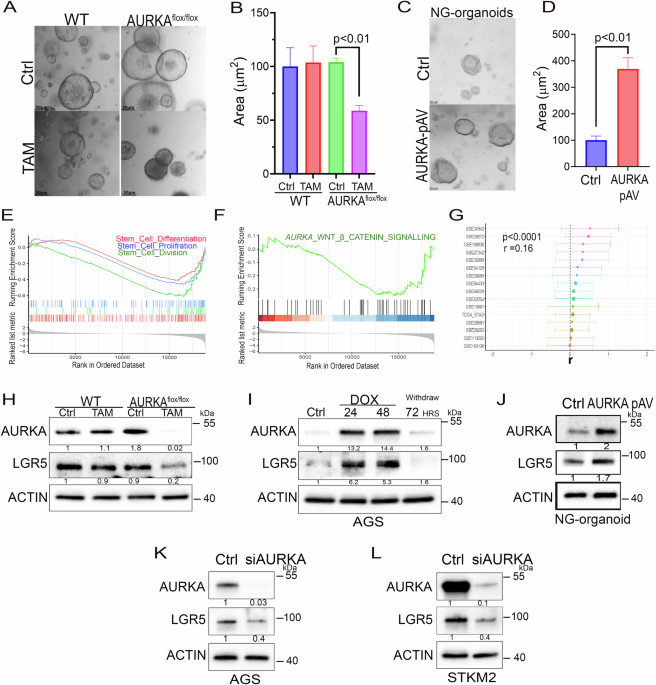
GSEA was utilized to analyze the AURKA-associated signaling pathways. The “limma” package was used to identify the differentially expressed genes (DEGs) between AURKA high-expression samples compared to AURKA low-expression samples in TCGA and GEO datasets (GSE122401, GSE54129, GSE66229, GSE15459, GSE110875, GSE26899, GSE26901). The Normalization Enrichment Score (NES) and false discovery rate (FDR) of each gene set were calculated using the “clusterProfiler” package (v3.12.0) [43]. A gene set was considered significant when the FDR was less than 25%. Reference gene sets, including hallmark gene sets, and GO biological processes gene sets, were downloaded from MSigDB (https://www.gsea-msigdb.org/). All data were analyzed using R software (R 3.6.1; https://www.r-project.org/). To evaluate the overall association between AURKA, LGR5, and STAMBP expression across multiple independent datasets, a meta-analysis of correlation coefficients was conducted using the “metafor” package in R. Each dataset provided an estimate of the Pearson correlation coefficient (r) between the two genes of interest, along with the corresponding sample size (n). Before meta-analysis, the correlation coefficients were transformed using Fisher’s r-to-z transformation to stabilize variance and normalize the distribution: \(z=\frac{1}{2}{\mathrm{ln}}\left(\frac{1+r}{1-r}\right)\), The standard error (SE) for each transformed correlation was calculated as \({{SE}}_{z}=\,\frac{1}{\sqrt{n-3}}\). A random-effects model was fitted using the rma() function in the metafor package, incorporating between-study heterogeneity using the DerSimonian-Laird method. The model estimated the overall effect size (Fisher’s z) and 95% confidence interval, which were subsequently back transformed to correlation coefficients for interpretation. Statistical heterogeneity across studies was assessed using Cochran’s Q statistic and the I² statistic. An I² value > 50% was considered to indicate substantial heterogeneity.
Immunofluorescence
For paraffin-embedded tissue sections, the slides were deparaffinized, followed by antigen retrieval in a pressure cooker for 30 min in 1X-TE buffer (pH 9). Blocking was performed for 1 h at room temperature in 5% BSA (bovine serum albumin) in 1X PBS. Next, the tissue sections were incubated with a combination of three primary antibodies: anti-LGR5 (1:100), anti-STAMBP (1–100), and anti-AURKA (1:100) overnight at 4 °C. The following day, sections were washed and incubated with secondary antibodies, donkey anti-mouse IgG conjugated to Alexa Fluor 488 and donkey anti-rabbit IgG conjugated to Alexa Fluor 568 (Thermofisher), diluted in blocking buffer (1:500) for 1 h at room temperature. For nuclear staining, sections were washed three times and incubated with Hoechst (10 μg/ml) for 10 min. The tissue sections were mounted, and images were captured using the Leica Stellaris 5 confocal microscope.
For immunofluorescence on cells, cells were seeded for 24 h prior to transfection with Silencer™ Select Negative Control siRNA (ThermoFisher Scientific) or Silencer™ Pre-Designed AURKA siRNA for a total of 72 h. AURKA or control siRNA were plated in the 8-well chambers and co-cultured with Helicobacter pylori strains 7.13 and J166 for 6 h at a multiplicity of infection (MOI) of 50:1 when applicable. Cells were washed with PBS and fixed with 4% paraformaldehyde for 20 min at room temperature. After a permeabilization step, cells were blocked using 10% nonimmune goat serum blocking solution (Invitrogen) for 1 h at room temperature and incubated with primary antibodies anti-LGR5 (1:100), anti-STAMBP (1-100) or anti-AURKA (1:100) overnight at 4 °C. After 4× washes with washing buffer (PBS-3% Bovine Serum Albumin-0.1% Triton X100), cells were incubated with donkey anti-rabbit/anti-mouse IgG secondary antibodies conjugated to AF488 and AF568 (Invitrogen) and incubated for 1 h at room temperature. Cells were then washed 4× times with washing buffer and mounted with Vectashield Mounting Medium containing DAPI (4′,6-diamidino-2-phenylindole) (Vector Laboratories). Images were acquired using a Leica Stellaris 5 Confocal Microscope and analyzed using the Leica Application Suite X (LAS X).
Animal care
All animal studies followed our approved protocol (UM-23-110) by the Institutional Animal Care and Use Committee of the University of Miami (Miami, FL).
Mice infection with H. pylori
Mice were infected with H. pylori following the guidelines outlined in the Guide for the Care and Use of Laboratory Animals published by the National Institutes of Health. All procedures were approved by the Institutional Animal Care and Use Committee (IACUC) at the University of Miami. C57BL/6 mice obtained from Charles River Laboratories were divided into two groups (8 per group) for either H. pylori infection or control (PBS). The study was designed to provide adequate sensitivity (n = 32; n = 8/group in a 2×2 factorial design), under standard ANOVA assumptions to detect large, biologically relevant effects consistent with prior studies of AURKA‑deficient gastric epithelium challenged by H. pylori. The design provided 80% power (α = 0.05) to detect main‑effect differences of approximately Cohen’s d ≈ 1.02 ( ≈ 1 SD shift) and an interaction effect of Cohen’s f ≈ 0.53, a magnitude aligned with known genetic × infection divergences in epithelial injury, cytokine induction, and proliferative responses. Additionally, Krt19CreErt/Aurkaflox/flox mice [15] aged 8–10 weeks were divided into four groups (8-9 per group) for treated with or without Tamoxifen (50 mg/kg, IP) for 10 days. In the case of H. pylori infection, mice received orogavage of either PBS (uninfected control) or the mouse-adapted wildtype H. pylori strain PMSS1 (109 CFU/mouse) at the age of 10–12 weeks [44]. Subsequently, the mice were euthanized two weeks post challenge, and gastric tissues were collected for pathology, Western blots, and real-time PCR analyses. All histological changes were evaluated by our gastrointestinal pathologist (OGM). All mice used in the experiment were included in the data analysis; no inclusion or exclusion criteria were applied. Mice were randomly allocated to the two experimental groups using simple randomization, ensuring equal probability of assignment for each animal given their matched age and background. No formal stratified randomization was performed, as baseline covariates were already similar across the cohort.
Mouse generation and experimental design
Compound mutant mice were generated by crossing Krt19CreErt/Aurkaflox/flox mice [15] with Tff1-/- mice [45] to produce Krt19CreErt/Aurkaflox/floxTff1-/-mice. This model allows for tamoxifen-inducible, stomach epithelium-specific AURKA ablation through Cre-mediated recombination in KRT19-expressing cells. Age-matched Krt19CreErt/Aurkaflox/floxTff1-/-mice (12–14 weeks old) were assigned into two experimental groups (n = 8–10/group). (Control n = 9; Tamoxifen n = 8), the two‑group comparison yields 80% power to detect a large effect of Cohen’s d ≈ 1.46, whereas more modest effects (d ≈ 0.6–0.8) would be underpowered and should be interpreted cautiously (e.g., ~34% power at d = 0.8 and ~49% at d = 1.0). This framing reflects ethical and logistical constraints on additional animals and appropriately aligns the analytic expectations with the likelihood of robust phenotypic changes in AURKA CKO models within the Tff1‑deficient background. The tamoxifen-treated group received 50 mg/kg tamoxifen (intraperitoneal injection) daily for 10 consecutive days. Controls received the vehicle alone. Three months post-treatment, mice were euthanized following the IACUC-approved protocol (UM 23-110). Gastric tissues were collected and processed for histopathological analysis (formalin-fixed, paraffin-embedded), molecular studies (snap-frozen in liquid N₂, stored at -80°C), protein analysis (western blotting), and gene expression profiling (quantitative RT-PCR). All mice used in the experiment were included in the data analysis; no inclusion or exclusion criteria were applied. All mice used in the experiment were included in the data analysis; no inclusion or exclusion criteria were applied. Mice were randomly allocated experimental groups using simple randomization, ensuring equal probability of assignment for each animal given their matched age and genetic background. No formal stratified randomization was performed, as baseline covariates were already similar across the cohort.
Patient-derived xenografts (PDX)
De-identified PDX models of human gastric carcinoma were established using a previously validated protocol [46]. Uniform tumor fragments were subcutaneously implanted into both flanks of immunodeficient mice, and tumor growth was closely tracked over time. Once the mean tumor volume reached approximately 150 mm³, mice were randomly divided into four groups (n = 6 per group): vehicle control, docetaxel alone, AMG-900 alone, and combined AMG-900 and docetaxel treatment. Docetaxel was dissolved in phosphate-buffered saline (PBS) and administered intraperitoneally at 10 mg/kg once weekly for a total of four weeks. AMG-900 was prepared in a solution of 2% dimethyl sulfoxide (DMSO), 2% Tween-80, and 96% sterile water, and delivered intraperitoneally at 15 mg/kg three times per week for four weeks. Tumor length and width were recorded three times per week with a digital caliper, and tumor volume was determined using the formula: volume (mm³) = ½ × (width)² × length. Mouse body weights were measured weekly to assess potential systemic toxicity. Following treatment, mice were monitored for survival until tumors reached a volume of 1000 mm³, at which point mice were euthanized according to humane endpoints. Survival data were analyzed using Kaplan–Meier estimation, with statistical differences assessed by the log-rank (Mantel–Cox) test.
Primary gastric epithelial cells and 3D organoid cultures
Short-term cultures of primary gastric epithelial cells were prepared following our standard protocol [47]. 3D organoid tissue cultures were prepared based on the protocol by Barker et al. [31]. Mouse stomachs were surgically excised, opened, and rinsed with HBSS. The gastric antrum was sectioned and incubated in 5 ml of 5 mM EDTA for 2 h at 4 °C with gentle agitation. After incubation, EDTA was replaced with 5 ml of chelation buffer, and the tissue was shaken vigorously for approximately 2 min to release gastric glands. The resulting suspension was passed through a 70 µm nylon mesh cell strainer (Becton Dickson, Franklin Lakes, NJ) to remove muscle tissue, followed by centrifugation at 150 × g for 5 min. The gland pellet was embedded in an extracellular matrix hydrogel (Trevigen, Inc., Gaithersburg, MD) enriched with IntestiCult Organoid Growth Medium (StemCell Technologies, Vancouver). By day 3, glands formed mature organoids, which were subcultured every 12 days. A 0.1 μM tamoxifen solution was introduced into the culture medium for 48 h, after which the medium was replaced with fresh medium. Organoids were recovered from Matrigel using the Cell Recovery Solution reagent (Gibco) and processed into standard formalin-fixed, paraffin-embedded tissue specimens.
Establishment of patient-derived gastric organoids
De-identified normal and gastric cancer tissue samples were obtained through the Biospecimen Shared Resource (BSSR) at the University of Miami (Miami, FL), with study approval granted by the Institutional Research Ethics Committee. Informed consent was secured from all participants by the BSSR prior to inclusion. The research adhered to the ethical standards set forth in the Declaration of Helsinki. Freshly excised tissue samples (3–5 mm) were placed in human organoid growth medium (IntestiCult Organoid Growth Medium, Stemcell Technologies, RRID: SCR_013642; 06010) and maintained at 4 °C until processing, which was performed within 1 h of surgical removal to optimize organoid formation. Tissue samples were processed as previously described [22]. Specifically, tumor fragments were sectioned into 0.1–0.5 mm pieces using sterile scalpels, washed with medium to remove debris, and primarily used for organoid culture. Remaining fragments were either snap-frozen in liquid nitrogen for RNA or protein extraction or fixed in 10% formalin for histologic evaluation. Organoid culture preparation were established according to the protocol described by Mahe et al. [48]. Briefly, minced tumor tissue was incubated in 2 mL of 5 mg/mL collagenase type II (Gibco; Thermo Fisher Scientific, RRID: SCR_008452, catalog no. 17101-015) dissolved in Advanced DMEM/F12 (Gibco; Thermo Fisher Scientific, catalog no. 12634-010) for 45 min at 37 °C. The digest tissue was then treated with TrypLE (Thermo Fisher Scientific, catalog no. 12604021) before being resuspended in DMEM/F12. To eliminate debris and achieve single-cell suspensions, the resuspension was strained through a 40 μm cell strainer. Approximately 5,000 passage-zero cells were cryopreserved in FBS (Thermo Fisher Scientific, catalog no. A3160402) supplemented with 10% DMSO (Sigma-Aldrich, RRID: SCR_008988; catalog no. D5879) and stored in liquid nitrogen as a patient-derived cell stock. Cell viability was assessed using trypan blue staining, and viable cells were suspended in growth factor–reduced Matrigel (Corning Life Sciences; catalog no. 354230) at a concentration of 20,000 cells/50 μL of 90% Matrigel. Each 50 μL droplet was plated into the center of a 24-well plate and allowed to polymerize at 37 °C for 30 min prior to adding prewarmed (37 °C) organoid growth medium (IntestiCult Organoid Growth Medium, Stemcell Technologies, RRID: SCR_013642; #06010). Organoids were harvested and passaged every 10–14 days depending on growth characteristics. For collection, organoids were gently recovered with ice-cold Cell Recovery Solution, enzymatically dissociated with TrypLE, and reseeded at split ratios of 1:2 or 1:3 in 90% Matrigel to continue culture.
Treatment of patient-derived gastric organoids
As previously described [36], human organoid growth medium (IntestiCult™ Organoid Growth Medium, Stemcell Technologies; RRID:SCR_013642; Cat. #06010) was supplemented into cell suspensions obtained from enzymatically digested human tissue samples, in the presence or absence of test compounds, and each condition was plated in triplicate. The organoids were cultured at 37°C in a humidified incubator with 5% CO₂, and the culture medium was replenished every 2–3 days. The organoids were transduced with the recombinant adenoviral vector pAV[Exp]-EGFP-CMV>hAURKA (VB900148-9083acy) at a multiplicity of infection (MOI) of 25 overnight. The culture medium was then replaced after 24 h. AMG-900 was dissolved in dimethyl sulfoxide (DMSO) to achieve a final concentration of 0.1% (v/v). To assess the effects of the drug treatment on organoid proliferation and viability, the diameter and total count were quantified after 4–5 days of treatment. A minimum of 50 organoids per condition were measured to determine the mean diameter using the Perfect Screen Ruler software. Organoids were counted manually using bright-field microscopy under a 20× objective.
Statistical analysis
Comparison of survival (percent) curves was done using Log-rank (Mantel-Cox) test.
Data are expressed as the mean ± SE or ± SEM for each condition. Statistical significance was determined using the GraphPad Prism statistical software, and the nonparametric Mann-Whitney test was used for comparisons not assuming normal distribution. Differences with p values ≤ 0.05 are considered significant.
