


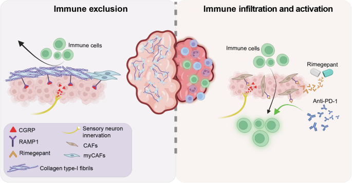
In a recent study published in cell, Zhang et al. demonstrated that calcitonin gene-related peptide (CGRP), secreted by tumor-innervating sensory neurons, alters the structural constituents and the immune landscape of triple-negative breast cancer (TNBC) tumor microenvironment (TME).1 Considering the central role of the nervous system in human ontogeny and homeostasis, its recognition as a key player in tumor development, TME dynamics, and therapeutic response is inevitable.2
For a while, the role of sensory neurons in cancer was indirectly appreciated, such as through the negative influence of stress on cancer patient outcomes. Now, accumulating evidence unveils their impact across multiple facets of tumor biology, underscoring why tumor innervation must be considered for targeted therapeutic intervention.2 Using a large, multi-omic repository of patient datasets, Zhang and colleagues established a correlation between poor clinical outcomes in TNBC patients and a high degree of perineural invasion (PNI), which corresponds to areas of malignant cells spreading along nerve sheaths.1 Spatial transcriptomics of TNBC tissues revealed that, in regions surrounding PNI, malignant cells overexpressed neural growth factor (NGF), suggesting an active process of neuronal recruitment. Spatial datasets and machine learning algorithms also highlighted an abundance of myogenic cancer-associated fibroblasts (myCAFs) surrounding PNIs. Pharmacological activation (low-dose capsaicin) and genetic ablation (Nav1.8-DTA) of sensory neurons in orthotopic mouse TNBC grafts showed that PNIs are critical for sustaining a fibrotic TME phenotype, marked by enhanced collagen deposition and immune cell exclusion. Further investigation revealed that NGF present in TNBC-conditioned medium induced overexpression of the neuropeptide CGRP in sensory neurons. RAMP1, the primary receptor for CGRP, is enriched in TNBC patients’ myCAF subpopulations, and in vivo treatment of murine TNBC with the RAMP1 inhibitor rimegepant reduced collagen type-I fibrosis and restored immune infiltration (Fig. 1). Zhang et al. recognized that such a structural remodeling of the TME may clear a major obstacle to the efficacy of immune checkpoint inhibitors. Thus, combining rimegepant with an anti-PD-1 antibody in vivo drastically reduced TNBC burden while increasing cytotoxic T-cell and macrophage infiltration compared with anti-PD-1 monotherapy (Fig. 1). This approach sheds light on the underappreciated role of neuronal influence on the structural constituents of the TME and on how direct targeting of myCAFs in fibrotic tumors remains ineffective since it does not eliminate the underlying influence of neuropeptides such as CGRP.
Fig. 1
RAMP1 inhibition alleviates sensory neuron-driven immune exclusion in breast tumors. CGRP secreted by tumor-innervating neurons binds to its receptor RAMP1 on cancer-associated fibroblasts (CAFs) to induce myofibroblastic differentiation (myCAFs). In turn, myCAFs upregulate collagen type-I deposition, fashioning a pro-tumorigenic microenvironment that fosters immune cell exclusion. Treatment with the RAMP1 antagonist rimegepant inhibits myCAF differentiation, consequently increasing immune infiltration. RAMP1 blockade synergizes with immune checkpoint inhibitors (e.g., anti-PD-1 antibody) to promote cytotoxic immune activation within the tumor mass. The figure was created with BioRender.com
The findings by Zhang et al. align with recent observations about the neuro-immune axis in the TME. Specifically, an article by Wei and colleagues in Nature shows that lung adenocarcinomas can hijack tumor-brain crosstalk to suppress anticancer immunity.3 Vagal sensory neurons in the lung use neuropeptides to signal the brain for respiratory regulation and to mediate responses to allergens or bacterial infections.3 In lung adenocarcinoma, however, vagal sensory neurons are integral to a bidirectional tumor–brain communication axis that restrains anticancer immunity by increasing circulating noradrenaline levels. In the lung TME, noradrenaline binds the ADRB2 receptor on alveolar macrophages, thereby polarizing them toward an immunosuppressive phenotype. Mouse knockout experiments showed that suppressing monocytic ADRB2 expression and ablating Npy2r+/Trpv1+ sensory neurons severed this tumor-brain connection and restored anti-tumor immunity. Thus, the findings by Wei et al. underscore the importance of tumor-brain crosstalk in shaping immune cell representation in the TME and open the door to inhibiting the sensory-sympathetic axis in future oncology treatments. In fact, both above-mentioned studies firmly support the prospect of combinatorial therapies targeting the source of neuron-mediated immune evasion and enhancing tumor immunogenicity with checkpoint inhibitors, which could be highly effective in future clinical investigations.
Other recent studies showed that monoamine neurotransmitters, such as dopamine and serotonin, and their associated transporters, are druggable regulatory axes to modulate antitumor immunogenicity. For example, the dopamine transporter (DAT) was associated with self-renewing and tumor-initiating functions in colorectal tumors.4 Blocking DAT with vanoxerine (GBR-12909) suppressed stem-like attributes by altering G9a-dependent chromatin configuration and reactivating endogenous transposable elements. This, in turn, increased type-I interferon response and associated infiltration of CD8 + T cells and macrophages into syngeneic mouse colorectal tumors. Moreover, serotonin transporter (SERT) expression by CD8 + T cells was documented as part of an endogenous negative regulatory loop of the antitumor immune response, and associated with poor survival outcomes in several types of non-neural solid tumors. Indeed, SERT blockade with the selective serotonin reuptake inhibitor fluoxetine stimulated autocrine serotonin signaling in CD8 + T cells, synergistically bolstering T-cell receptor signal transduction through the MAPK pathway. In mouse melanoma models, SERT inhibitors enhanced CD8 T cell proliferation, effector function, tumor infiltration, and anti-PD-1-induced immunogenicity, while limiting T cell exhaustion.5
There is growing evidence that the peripheral nervous system is a critical contributor to TME dynamics. Much of this has centered on how neuropeptides contribute to immune evasion through localized interactions with immune cells. The studies by Wei and Zhang took the concept one step further: (1) Demonstrating how tumors can hijack preexisting tissue-brain neural circuits in effecting system-wide alterations resulting in tumor immune evasion, and (2) revealing that neuronal influences on the stromal compartment of the TME can elicit large-scale structural alterations to impart immune exclusion.1,3 As cancer neuroscience expands beyond neuro-originating malignancies to describe the impact of neurotrophic and neurogenic factors on TME alterations, it will be interesting to consider the influence of neurodiversity on cancer progression and therapeutic response. Neurodiversity encompasses conditions marked by fundamental differences in sensorial perception and information processing, such as autism spectrum disorder (ASD), language-based learning differences (LBLD), and attention-deficit/hyperactivity disorder (ADHD), but also psychosis-related states, including schizophrenia. Neurodiversity is robustly associated with inherited genetic factors, including copy number variants and single-nucleotide polymorphism (SNPs). Just as genetic diversity contributes to differential patient outcomes, the neuro-TME interactome in neurodiverse individuals could yield a similar array of consequences. SNPs and larger-scale genomic rearrangements contributing to human neurodiversity and documented to affect the peripheral nervous system could alter a tumor’s capacity to hijack the tissue-brain circuitry or modulate the secretome of tumor-innervating neurons. The availability of genetically engineered murine models of ASD or ADHD, as well as induced pluripotent stem cells derived from neurodivergent human subjects, may help bridge knowledge of the genetic determinants of neurodiversity with the developing field of neuro-oncology once integrated into complex experimental tumorigenesis systems.
While harnessing the neuro-immune crosstalk represents an exciting avenue to develop new precision therapeutic approaches, the vast diversity of neuro- and immune-cell types poses an additional challenge of precisely targeting relevant interactions and functions.2 The potential deleterious impacts from pharmacological neuromodulators on behavior and cognition when used at effective tumor-suppressive dosage also represent a fundamental hurdle to clinical translation.4 Thus, exploring new precision delivery avenues to enhance the bioavailability of neuro-immune modulators in the TME, specifically, could circumvent this challenge.
