


Antioxidant, antimicrobial, and cytotoxic activities of different fractions of Moringa oleifera pericarp
Screening of different fractions of M. oleifera for their antioxidant activity revealed that the ethyl acetate fraction of the pericarp exhibited the highest antioxidant activity with inhibition percentages of 68.7 ± 0.04% compared to the standard ascorbic acid (88.1 ± 0.1%) (Table S1).
When assessed for antimicrobial activity, among all tested fractions, the ethyl acetate extract showed strong antibacterial effects against E. coli, with an inhibition zone of 20.2 ± 0.2 mm and an activity index of 83.3%, and against S. aureus, with a zone of inhibition of 18.17 ± 0.15 mm and an activity index of 78.3% (Table S2). Moderate antifungal activity was also observed against C. albicans, with an inhibition zone of 15.1 ± 0.1 mm and an activity index of 57.7% (Table S2). These findings suggest that the ethyl acetate fraction contains potent bioactive constituents responsible for broad-spectrum antimicrobial activity.
The literature highlights the diverse bioactivities of M. oleifera extracts, including antioxidant, anti-quorum sensing, and DNA damage-preventive properties in aqueous extracts derived from its leaves, fruits, and seeds27. Moreover, acetone extracts of M. oleifera pod husks have shown sustained post-antibiotic effects against both Gram-positive and Gram-negative bacteria, indicating their potential for extended dosing regimens28. In addition, when used in combination with conventional antibiotics, M. oleifera extracts have been reported to target bacterial triggers of autoimmune inflammatory disorders29.
Regarding cytotoxic activity, the ethyl acetate fraction exhibited potent activity among all tested fractions against both liver and colon cancer cell lines, with IC₅₀ values of 2.5 ± 0.08 µg/mL and 4.04 ± 0.13 µg/mL, respectively, as determined by the MTT assay (Tables S10-S13; Fig. 3). These results are more effective compared to the reference drug staurosporine, which showed IC₅₀ values of 8.4 ± 0.41 µg/mL and 7.75 ± 0.35 µg/mL, respectively (Tables S10-S13; Fig. 3). Accordingly, this fraction was selected for further chromatographic separation to isolate its active constituents. Previous studies have reported the anti-proliferative effects of M. oleifera fruit and leaf extracts, which may account for its well-known therapeutic potential as the “miracle tree”30. Specifically, M. oleifera fruit extract has been shown to induce anti-proliferative effects against HepG2 (human hepatocellular carcinoma cells) via ROS-mediated apoptosis and caspase-3 activation11.
Fig. 3
IC50 of cytotoxic activity of different fractions of Moringa oleifera Pericarp extract using MTT assay. All data are presented as mean value ± SD for three independent experiments.
Bio-guided isolation of active constituents from ethyl acetate fraction of Moringa oleifera pericarp
The ethyl acetate fraction of M. oleifera pericarp was subjected to normal-phase silica gel column chromatography, leading to the isolation of three compounds: M1, M2, and M3 (Fig. 4). Compound M1, previously reported from M. oleifera seeds, is identified here for the first time from the pericarp. Its NMR data, Table S3, matched previously published data for 4-(α-L-rhamnopyranosyloxy)-benzaldehyde31, which has also been synthetically prepared32. Compound M2 showed NMR data (Table S4), consistent with 4-(α-L-rhamnopyranosyl) benzyl alcohol, a compound previously isolated from both M. stenopetala33 and M. oleifera seeds34. This study represents the first report of its isolation from M. oleifera pericarp, and notably, in high yield. According to Lewerenz et al. (2021)35, this compound is likely a degradation product of 4-hydroxybenzyl isothiocyanate, produced via myrosinase-catalyzed hydrolysis of sinalbin36. Compound M3 is tentatively identified as 4-(hydroxymethyl) phenol- 1-O-β-D-glucopyranosyl-(1”→3′)-O-α-L-rhamnopyranoside (Table S5). It was previously reported only once from Moringa seeds34; thus, this study marks its second report from nature and the first from the pericarp of M. oleifera. For more details of structure elucidation, see the supporting information (Tables S3-S5, Figures S1-S11). Overall, the isolation of these three glycosylated compounds confirms that the pericarp contains structurally diverse phenolic constituents with potential biological relevance. Among the isolated constituents, M2 was selected as the main scaffold for further semi-synthetic modification based on its abundance and suitable hydroxyl functionality.
Fig. 4
Structures of the isolated compounds from the Pericarp of Moringa oleifera active fractions and semi-synthetic compounds: 4-(α-L-rhamnopyranosyloxy)-benzaldehyde (M1), 4-(α-L-rhamnopyranosyl) benzyl alcohol (M2), 4-(hydroxymethyl) phenol-1-O-β-D-glucopyranosyl-(1”→3′)-O-α-L-rhamnopyranoside (M3), 4-(α-L-rhamnosyloxy) benzyl tetracinnamate derivative (S1M2), 4-(α-L-rhamnosyl) benzyl tetra crotonate derivative (S2M2).
Semi-synthesis of enone derivatives from isolated compound M2
The isolated compound M2 with the skeleton 1, 2-(4-(hydroxymethyl) phenoxy)-6-methyltetrahydro-2 H-pyran-3, 4, 5-triol is considered the main scaffold for our newly semi-synthesized target hybrids S1M2 and S2M2 (Fig. 5). Steglich esterification reaction of cinnamic acid or crotonic acid with alcohol derivative M2 in the presence of dicyclohexylcarbodiimide (DCC) and 4-dimethylaminopyridine (DMAP) afforded our designed target compounds S1M2 and S2M2, respectively, according to the previously reported procedure with some minor modifications37. The structures of the obtained derivatives S1M2 and S2M2 were characterized and confirmed based on the spectroscopic data (1H-NMR, APT, IR and MS) (Tables S6- S9, Figures S12-S20). The spectroscopic data obtained confirmed the incorporation of all free OH functionalities of our starting M2 under the reaction conditions illustrated in Fig. 5 as targeted in our research design rationale.
Fig. 5
Scheme of semi-synthesis of target-designed sugar-based enone derivatives from M2.
Cytotoxic activity of the isolated and semi-synthetic compounds
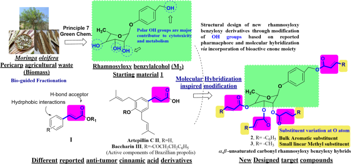
The present study was designed to synthesize two novel potential sugar-based anticancer agents incorporating enone functionality S1M2 and S2M2, using a structure-based drug design approach Fig. 2. These newly synthesized enone hybrids, along with isolated compounds from the pericarp, were evaluated for their in vitro cytotoxicity using the MTT assay against liver (HepG2) and colon (HCT116) cancer cell lines. Among the tested compounds, S2M2 exhibited the most potent cytotoxic effect, with IC₅₀ values of 5.97 ± 0.19 µM and 11.52 ± 0.37 µM against HepG2 and HCT116 cells, respectively, surpassing the reference drug staurosporine. In contrast, the remaining compounds showed moderate cytotoxic activity (Table 1; Tables S14–S17).
Table 1 IC50 values of isolated and semi-synthesized compounds against HepG-2 and HCT116 cancer cell lines, EGFR-TK and CAIX enzyme inhibition. All data are presented as mean value ± SD for three independent experiments.
To evaluate the safety and selectivity of the isolated compound M2 and its semi-synthesized derivatives (S1M2 and S2M2), their cytotoxic effects were assessed against the normal lung fibroblast cell line WI-38 alongside cancer cell lines (Tables S18–S20). The IC₅₀ values against WI-38 were 114.36 ± 1.88, 70.92 ± 3.04 and 68.30 ± 2.31 µM for M2, S1M2, and S2M2, respectively, indicating comparatively lower toxicity toward normal cells (Table S20). Notably, S2M2 exhibited the highest selectivity indices, with values of 11.43 (HepG2) and 5.93 (HCT116), compared to the reference compound, which showed selectivity indices of 2.90 and 3.16, respectively (Table S20, Fig. 6). These findings demonstrate that semi-synthetic modification of M2, particularly in the case of S2M2, enhances selective cytotoxicity toward cancer cells while maintaining a favorable safety profile. S2M2 showed potent activity, especially against HepG2 cells, with reduced toxicity toward WI-38 cells, highlighting its potential as a promising lead compound for drug development.
Fig. 6
Selectivity index (SI) values of M2 and its semi-synthetic derivatives (S1M2 and S2M2) against HepG2 and HCT116 cancer cell lines in comparison with the reference compound staurosporine.
Previous studies have demonstrated the chemopreventive potential of Moringa oleifera Lam pods. Compound M1, previously isolated, was tested for its cytotoxicity against doxorubicin-resistant human breast cancer cell lines (MCF-7/Adr), where it demonstrated promising activity38. These findings align with our results, which highlight the cytotoxic activity of M. oleifera and its isolated compounds.
The anticancer potential was further supported by additional in vitro assays, including EGFR TK and CAIX inhibition, confirming the therapeutic promise of M. oleifera as a source of anticancer agents.
EGFR-TK inhibition assay of the isolated and semi-synthetic compounds
The inhibitory activity of the tested compounds against EGFR tyrosine kinase was evaluated in comparison with erlotinib as the reference inhibitor39. Among the isolated and semi-synthetic compounds, M2, M3, and S2M2 showed the most notable inhibitory effects, with IC50 values of 0.39 ± 0.004, 0.12 ± 0.002, and 0.40 ± 0.008 µM, respectively, while the remaining compounds exhibited weaker activity (Table 1; Table S21). The potent activity of M3 suggests that glycosylated phenolic scaffolds from Moringa oleifera may interact effectively with the EGFR active site and may provide a useful framework for further optimization. In contrast, the comparable activity of M2 and S2M2 suggests that semi-synthetic modification of the parent scaffold does not compromise EGFR inhibitory potential and may, in addition, improve its drug-like properties.
CAIX enzyme inhibition assay of the isolated and semi-synthetic compounds
The inhibitory activity against carbonic anhydrase IX (CAIX) was evaluated in comparison with acetazolamide as the reference inhibitor40. Among all tested compounds, S2M2 exhibited the highest inhibitory activity with an IC50 value of 0.27 ± 0.01 µM, exceeding that of the reference inhibitor acetazolamide (0.49 ± 0.01 µM) (Table 1; Table S22). This result indicates that crotonate derivatization of M2 significantly improved its affinity toward CAIX and supports the value of introducing an α, β-unsaturated carbonyl moiety into the parent scaffold24,25.
The superior CAIX inhibition shown by S2M2 may be attributed to its optimized fit within the enzyme active site and its ability to engage in multiple noncovalent interactions. In contrast, the parent compound M2 displayed lower activity, suggesting that free hydroxyl-rich glycosylated scaffolds are less effective than their semi-synthetic enone derivatives in this assay. The activity of M3 also suggests that naturally occurring glycosylated phenolics from Moringa oleifera can retain meaningful enzyme inhibitory potential. CAIX is a hypoxia-inducible enzyme that contributes to tumor pH regulation, progression, and resistance to therapy, making it a relevant anticancer target. In this context, dual targeting of EGFR and CAIX is particularly attractive because it may interfere with both tumor proliferation and the hypoxic microenvironment that supports malignant growth24,25,26.
Overall, these findings identify S2M2 as the most promising dual EGFR/CAIX inhibitor among the compounds investigated and provide a strong basis for the subsequent docking and molecular dynamics analyses.
Molecular docking study of isolated and semi-synthetic compounds targeting EGFR-TK and CAIX
To further rationalize the observed biological activity, molecular docking was performed to examine the binding modes of the most active compounds within the active sites of EGFR-TK and CAIX. The binding modes of compounds M3, S1M2, and S2M2 against wild-type EGFR tyrosine kinase (EGFRwt TK) revealed high binding affinities, with binding energies of − 7.15, − 7.17, and − 7.71 kcal/mol, respectively. Compound M3 formed four hydrophobic π-interactions with Leu694, Val702, Ala719, and Lys721. Additionally, it established two hydrogen bonds with Asp831 and Met769, with bond distances of approximately 2.59 and 2.95 Å, respectively (Table 2). In contrast, Compound S2M2 exhibited nine hydrophobic π-interactions involving His781, Leu820, Leu768, Met769, Val702, Leu694, and Cys773. It also formed three hydrogen bonds with Cys773 and Lys692, at distances of 2.13, 2.45, and 2.48 Å, respectively. For comparison, Erlotinib, displayed a binding affinity score of − 7.31 kcal/mol. Erlotinib formed eight hydrophobic π-interactions with Leu694, Ala719, Leu820, Lys721, and Val702, and a single hydrogen bond with Met769 at a distance of 1.97 Å (Table 2).
Table 2 Molecular docking scores and type of binding interaction for EGFR-TK enzymes.
The binding mode of compound S2M2 against carbonic anhydrase IX (CAIX) exhibited a binding energy of − 8.55 kcal/mol, indicating a strong interaction (Table 3). Compound S2M2 formed eight hydrophobic π-interactions with Pro201, Leu197, Val130, Leu91, His64, Leu140, Val121, and Val142. These interactions were further stabilized by two hydrogen bonds and one ion–metal interaction with Met1, Thr199, and the Zn²⁺ ion at position 301, with bond distances of 3.00 Å and 2.97 Å, respectively. In comparison, the co-crystallized ligand in the CAIX complex (PDB ID: 6S03) showed a binding affinity of − 7.45 kcal/mol. It established ten hydrophobic π-interactions involving Trp208, His96, Leu91, Val130, Pro201, Val134, and Leu197. Additionally, it formed two hydrogen bonds with Thr198, at distances of 2.09 Å and 1.84 Å.
Table 3 The molecular docking scores and type of binding interaction for Carbonic anhydrase IX inhibition.
Notably, the EGFR binding pattern of S2M2 is in line with the behavior of established EGFR inhibitors such as erlotinib, which exert activity through stable occupation of the kinase domain. Likewise, the CAIX binding profile supports the interpretation that the enone derivative is able to exploit the structural features of the CAIX active site more efficiently than the parent scaffold. Overall, the docking results provide a structural explanation for the superior dual inhibitory activity of S2M2 and strengthen the view that semi-synthetic modification of M2 enhanced its target engagement across both enzymes. These findings also justify the subsequent molecular dynamics analysis of S2M2, which was used to verify the stability of the docked complexes over time.
Molecular dynamics (MD) simulation study of compound S2M2
MD simulation was processed for 100 ns to evaluate the molecular stability of compound S2M2 with the CAIX and EGFR-TK target pockets. The displacement of the ligand within the target pockets was measured by using root mean square deviation (RMSD). Finally, candidate S2M2 interactions were also analyzed and evaluated in detail.
Protein and ligand RMSD and RMSF analysis
In the present study, compound S2M2 was docked against the CAIX and EGFR-TK target proteins and showed a promising binding affinity among other tested compounds. Therefore, compound S2M2 was selected for further molecular dynamics (MD) simulation studies. The structural stability of the protein structures was monitored through the C atoms (blue line) of the protein concerning their initial position. To validate this interaction, the simulation to 100 ns was processed. At first, the complex of CAIX /Compound S2M2 was inserted into the simulation system. MD simulation analyses of compound S2M2 in complex with CAIX reveal a protein structure that remains predominantly stable over a 100 ns trajectory, as indicated by average protein backbone RMSD values fluctuating between 1.5 and 2.2 Å. The ligand RMSD, calculated by fitting to the protein, initially remains low (< 1.5 Å) and stable within 3.2 to 4.8 Å, but showed minor fluctuation after approximately 65–70 ns, suggesting an increase in ligand conformational mobility or a possible shift in binding orientation during the later stage of the simulation as shown in Fig. 7A. Additionally, Root mean square fluctuations (RMSF) assessed on a per-residue basis demonstrates that most CAIX residues display limited mobility (< 2.0 Å RMSF), except for certain terminal and loop regions, which are inherently more flexible. Compound S2M2 showed minor fluctuations and many movements inside the pocket of CAIX at 10–30, and 130–150 amino acids areas, which leads to some conformational changes in protein skeleton with minor effect on the protein target interactions (Fig. 7B).
Fig. 7
The RMSD and RMSF of Compound S2M2 against carbonic anhydrase IX (CAIX) over 100 ns.
On the other hand, simulation of compound S2M2 in complex with EGFR tyrosine kinase exhibited that the protein showed structural stability over the simulation time, and Cα RMSD values that ranged between 1.8 and 2.4 Å. The RMSD profile shows only moderate fluctuation, with a slight increase in the latter part of the simulation, but no indication of protein unfolding or major deviation from the starting structure. The ligand RMSD, measured after fitting on the protein, remains stable and comparable to the protein backbone, generally below 1.5 Å ( within 2.5 to 3.5 Å), indicating that compound S2M2 retains a consistent binding mode for most of the simulation time. A modest increase in ligand RMSD after ~ 75 ns suggests some degree of conformational flexibility or minor pose adjustments within the binding site, without any effect on ligand protein interactions (Fig. 8A).
Fig. 8
The RMSD and RMSF of Compound S2M2 against EGFR-TK over 100 ns.
Root mean square fluctuation (RMSF) analysis reveals that most protein residues have restricted mobility (< 1.5 Å), with elevated flexibility observed in selected loop regions and termini, as expected for EGFR TK (Fig. 8B).
Histogram of protein ligand interactions analysis
Ligand interaction analysis of compound S2M2 with carbonic anhydrase IX (CAIX) reveals a multifaceted and persistent interaction network within the active site. The most frequent and sustained contacts are ionic interactions with Glu106, evident from an interaction fraction exceeding 1.0, indicating that multiple ionic interactions are maintained for nearly the entirety of the simulation. Additionally, compound S2M2 forms persistent hydrogen bonds with His96 and Thr199, as well as hydrophobic contacts with Pro201 and Thr198. Water-mediated bridges further stabilize the complex, notably with Thr199 and His96 (Fig. 9).
Fig. 9
Histogram analysis describes the binding interactions of Compound S2M2 against carbonic anhydrase IX (CAIX) during the simulation time (100 ns), green column represents hydrogen bond, blue column represents polar water linkage and violet column represents hydrophobic π-interactions.
Additionally, MD simulation of S2M2/EGFR tyrosine kinase complex demonstrated sustained ATP-binding site engagement through a multivalent interaction network combining dominant hydrophobic contacts with water-mediated hydrogen bonding. Val702 emerged as the primary sharing residue (91% interaction fraction within the P-loop), supported by another hydrophobic interactions with Leu694 (43%), Phe699 (36%), and Ala719 (35%), establishing a gradient of Van der Waals complementarity across the adenine-binding cleft. Moreover, water molecules functioned as critical mediating agents in the protein-ligand interface, with Cys-773 forming water-bridge contacts (32% occupancy), hydrogen bonding (70% occupancy) and Asp-776 establishing a highly persistent water-mediated hydrogen bond (85% occupancy) in the hinge region. Collectively, these findings establish compound S2M2 as an ATP-competitive inhibitor achieving high-affinity EGFR binding through hydrophobic (Fig. 10).
Fig. 10
Histogram analysis describing the binding interactions of Compound S2M2 against EGFR-TK during the simulation time (100 ns), green column represents hydrogen bond, blue column represents polar water linkage and violet column represents hydrophobic π-interactions.
To analyze the frequency of the interactions, a heat map was used to monitor these interactions by plotting the number of interactions over time (Figs. 11 and 12), where the dark color indicates more interactions. From the heat map of Fig. 11, it was observed that the highest number of conformations of the protein of CAIX formed up to five interactions. The most interacted amino acids of CAIX with Compound S2M2 are His96, Glu106, His119, Val142, Thr198, Thr199, Pro201, and Trp208. Additionally, the heat map of compound S2M2 with EGFR TK showed persistent primary anchoring residues including Val702, Cys773, and Asp776 demonstrate continuous orange coloration, reflecting uninterrupted binding and validating their critical roles in stabilizing the ligand within the ATP pocket Fig. 12.
Fig. 11
Heat map describing the total interactions within Compound S2M2 against carbonic anhydrase IX (CAIX) during the simulation time.
Fig. 12
Heat map describing the total interactions within Compound S2M2 against EGFR-TK during the simulation time.
MM-GBSA calculations
The molecular mechanics, with a generalized Born and surface area solvation (MM–GBSA), were carried out to calculate both the ligand binding strain and free energies for the docked ligand over 100 ns. The ΔG binding energies, Coulombic energies, hydrogen bond energies, Van der Waals forces and lipophilic energies were recorded.
The results observed in Figs. 13 and 14 provide an analysis of Compound S2M2 against CAIX and EGFR-TK. Throughout the simulation, Compound S2M2 maintained a consistent total binding free energy (∆G), with an initial value of − 59.48, and -86.25 kcal/mol at 0 ns, for CAIX and EGFR-TK, respectively, which remained stable at 100 ns. This persistence in binding energy demonstrates the sustained stability of the Compound S2M2 over the course of the simulation.
Fig. 13
MM-GBSA energies for Compound S2M2 against carbonic anhydrase IX (kcal/mol).
Fig. 14
MM-GBSA energies for Compound S2M2 against EGFR-TK (kcal/mol).
Structure–activity relationship analysis
The semisynthetic modification of the parent glycoside M2 was designed to modulate the physicochemical and biological properties associated with its free hydroxyl groups. Polyhydroxylated glycosides are typically characterized by high polarity and extensive hydrogen-bonding capacity, which can limit membrane permeability and drug-like behavior13,14. To address these limitations, we employed a targeted esterification strategy that masks the hydroxyl functionalities and introduces an α,β-unsaturated carbonyl (enone) pharmacophore. This approach was intended to enhance lipophilicity and incorporate a Michael acceptor motif with reported anticancer relevance.
Our structure–activity relationship (SAR) analysis revealed that biological activity depended largely on scaffold modification and the incorporation of the α,β-unsaturated carbonyl group. Compared with the parent alcohol M2, both enone derivatives (S1M2 and S2M2) displayed improved cytotoxic activity, indicating that esterification significantly enhanced the biological profile. Notably, S2M2 was the most active compound, likely because the smaller crotonate enone fits more favorably within the EGFR-TK and CAIX active sites than the bulkier cinnamoyl group of S1M2. This structural advantage is consistent with the superior dual inhibitory activity observed for S2M2, which exhibited IC₅₀ values of 0.40 µM against EGFR-TK and 0.27 µM against CAIX, surpassing the reference inhibitor in the CAIX assay. While M3 also exhibited strong EGFR/CAIX inhibition, it showed only moderate cytotoxicity, suggesting that glycosylated phenolic scaffolds can retain target-specific activity without necessarily inducing high cell death. In addition, the glycosylated nature of these Moringa‑derived compounds may support prodrug behavior and improve aqueous solubility and selectivity, as documented for glycosylated anticancer agents41. These experimental observations are consistent with our docking and molecular dynamics results, which supported stable binding of S2M2 in both targets and confirmed its superior dual inhibitory profile. Collectively, these data validate our design strategy and identify the enone-modified M2 scaffold, particularly S2M2, as the most promising lead for further development.
