


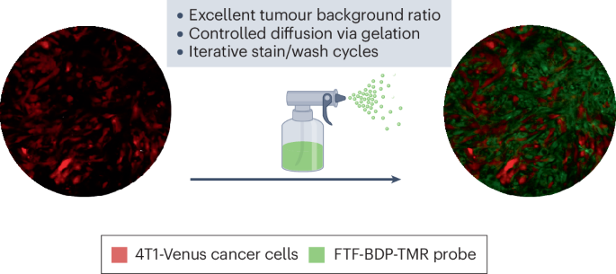
Surgical resection remains the main treatment for many solid tumours, yet outcomes are often limited by uncertain margins during an operation. Cancer cells can extend beyond the visible tumour mass into surrounding tissue and visual inspection is often insufficient to detect these microscopic extensions, necessitating re-excision. Fluorescence-guided surgery is an approach that contrasts healthy tissue against tumorous tissue to improve margin detection. Current methods rely on intravenously administered imaging agents that suffer from a low tumour to background ratio (TBR) at the target tissue due to the low sensitivity of systemic application. In the surgical setting, this can result in a high personal and economic cost for both patients and healthcare workers.
A central design decision underlying this strategy is the molecular target. The study focuses on fibroblast activation protein (FAP), which is expressed by cancer-associated fibroblasts in the tumour microenvironment. Unlike many tumour-cell markers that vary substantially across cancer types and even within a single tumour, FAP expression is relatively consistent and enriched at the interface between the tumour and neighbouring healthy tissue. From a chemical perspective, this targeting strategy exploits a spatially well-expressed and predictable feature of tumour tissue, increasing the likelihood that a topically applied probe will selectively engage its intended binding site and that the maximal signal will arise at the margins rather than deep within the tumour.
