


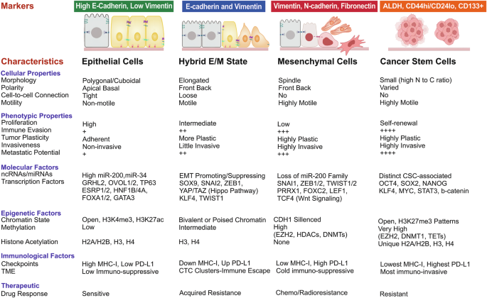
Epithelial-mesenchymal transition (EMT) is a cell-biological program that contributes to tumor invasion and metastasis if it is aberrantly activated. Although EMT is commonly associated with the aggressive properties of carcinoma, its role extends beyond cancer progression. There is evidence supporting the correlation between EMT and autoimmune diseases, as well as fibrosis.1,2 This suggests that the pathophysiology of autoimmune disorders and the emergence of fibrotic illnesses may be influenced by EMT. Evidently, many biological processes, such as embryogenesis, tissue morphogenesis, and adult wound healing, depend critically on EMT. During embryogenesis, EMT is involved in gastrulation, which is the process of forming endoderm, mesoderm, and ectoderm that are vital in giving rise to different tissues and organs.3 In compliance with cancer, however, EMT is often activated, leading to the progression of malignancies and increased aggressiveness of cancer cells, which can permeate nearby tissues and travel to other parts of the body.4 As a result, the epithelial cells no longer have tightly connected characteristics and develop the migratory and invasive properties commonly observed in mesenchymal cells.5 Epithelial cells undergoing EMT also express specific markers associated with the non-epithelial cell state, the mesenchymal state. The markers, which include neural cadherin (N-cadherin), together with vimentin and fibronectin.6 The cell adhesion molecule N-cadherin, which mainly occurs in neural tissues, increases its expression throughout EMT. The intermediate filament protein vimentin provides cellular structural support and normally exists in mesenchymal cells, while the extracellular matrix protein fibronectin helps cells stick to their surrounding environment and shows increased expression during EMT (Fig. 1). The understanding of cellular mechanisms that control junction stability helps researchers understand EMT while creating approaches to stop or reverse this process. The preservation of cellular junctions could prevent cancer cells from invading tissues and spreading throughout the body, which would lead to better outcomes for patients.7 The process of EMT shows an increase in N-cadherin levels along with a reduction of E-cadherin levels. In addition to tumor progression, tumor cells can suppress the immunological response by inducing EMT to upregulate immunosuppression molecules, such as immune checkpoints, leading to exhausted immune cells.8,9 EMT’s well-established function in tumor initiation, progression, and therapeutic resistance is accompanied by its ability to facilitate the cancer cells’ separation from the origin site and invasion of adjacent tissues, yet it should be noted that EMT alone is insufficient for successful colonization, which marks the final phase of the metastatic process.10 The term “colonization” describes a cancer cell’s capacity to form new tumors at various locations throughout the body. In order to proceed, cancer cells must adapt to the new environment, survive, avoid immune surveillance, and create a niche that will enable the creation of new tumors.11 Evidently, the activation of EMT is a prevalent characteristic observed in numerous cancer types since it helps cancer cells acquire aggressive properties and promotes their proliferation, making it an important process in the progression of malignancies.12 It is worth noting that the main EMT-inducer is EMT-inducing transcription factors (EMT-TF), namely Zeb1, Twist1,2, Snail (SNAI1), and Slug (SNAI2). The cellular function of EMT-TF is cell proliferation, programmed cell death, and differentiation, and it has a critical role in binding cells together to form tissues, modulating the activation of genes linked to cell motility, allowing cells to migrate within tissues or to disparate locations, as well as breaking down components in the extracellular matrix.13 What is more, external signaling can activate the EMT-TFs, like WNT, Sonic Hedgehog (Shh), and NOTCH signaling pathways, and mitogen factors, such as epidermal growth factor (EGF), platelet-derived growth factor (PDGF), vascular endothelial growth factor (VEGF), and, more importantly, transforming growth factor-β (TGF-β). In carcinoma cells, which are cancerous cells derived from epithelial tissues, the activation of EMT is not a common intrinsic process. Instead, it implies that external factors or signals from the tumor microenvironment may be required to trigger EMT in these cells. These signals can come from neighboring cells, immune cells, or the extracellular matrix surrounding the tumor.14
Fig. 1
Dynamic changes in cellular, molecular, epigenetic, and immunological properties during epithelial–mesenchymal transition (EMT), metastasis, and cancer stemness. This schematic illustrates the progressive transformation of epithelial cells into mesenchymal and stem-like states during cancer progression. Upon EMT induction, epithelial cells lose apico-basal polarity and intercellular adhesion, acquiring mesenchymal traits that promote motility, invasiveness, and survival. This phenotypic conversion is accompanied by extensive reprogramming at multiple levels, including cellular architecture, transcriptional and epigenetic regulation, metabolic adaptation, immune modulation, and signaling network rewiring. The culmination of these coordinated changes enables metastatic dissemination and the acquisition of cancer stem cell–like properties, conferring enhanced self-renewal capacity, resistance to apoptosis, immune evasion, and tolerance to therapeutic interventions. Figure created using BioRender
EMT and tumor immunology have become major subjects for researchers during the last ten years of scientific exploration. The reason behind this is that EMT affects cancer cell physical features, while simultaneously influencing immune system recognition and response to these cells. Tumor cells change EMT properties to avoid immune surveillance before continuing their growth and spread.15 The scientific community originally believed that EMT functioned as an on/off switch to transform cells completely from an epithelial state to a mesenchymal state.16 A significant amount of research demonstrates that EMT usually remains partial instead of complete, resulting in cells that display both epithelial and mesenchymal features as hybrid epithelial/mesenchymal (E/M) states or quasi-mesenchymal (qM) phenotypes. The qM cells maintain specific epithelial characteristics, which include E-cadherin expression, while simultaneously developing mesenchymal traits of enhanced movement and apoptosis resistance.17,18 Therefore, the understanding of EMT has evolved, and it is now considered a pleiotropic and flexible change.16 In the context of EMT, pleiotropy refers to the multiple effects of a single gene or process, manifesting as diverse outcomes and levels of transition in different cells or tissues, while flexibility refers to the cellular ability to undergo partial EMT or revert back to their original epithelial state.19 The main reason for emphasizing the higher importance of studying EMT in malignancies would be to pave the way for comprehending cancer behavior, as the non-genetic processes, particularly EMT programs, are considered responsible for driving the various stages of the invasion-metastasis cascade.20 In essence, the prevailing belief that genetic factors primarily contribute to cancer development has been challenged by evidence demonstrating that the progression and invasion of nearly all types of cancer involve a phenomenon known as EMT.21 Additionally, the significance of qM in conferring treatment resistance to cancer cells has also been underscored, necessitating further investigation into cancer dynamics and the development of precision therapies.21 The remarkable correlation between immune cells and EMT has been indicated, such as the correlation between EMT and Treg cells in non-small cell lung cancer (NSCLC),22 increased expression of PD-L1 in bladder cancer to enhance immune escape,23 and elevated levels of PD-L1 and Treg resulting in T cell exhaustion in breast cancer.24 Additionally, ovarian cancer has demonstrated an undesirable association between EMT and tumor growth.25 Studies have shown that tumors with a higher degree of mesenchymal characteristics (qM tumors) tend to have immunosuppressive cells like M2-like macrophages and Treg cells in their microenvironment.26 Furthermore, these tumors have a tendency to displace CD8+ T cells towards the periphery of the cancer cells, and exhibit increased resistance to anti-CTLA4 therapy.27 To effectively address malignancies, it is crucial to examine how tumor cells, defense cells, and their interactions contribute to the progression of cancer. With the ongoing advancements in technology and laboratory tools like multi-omics/NGS technologies, CRISPR/Cas, multiple single-cell RNA sequencing, and organoid models, among others, it is anticipated that the discovery of new EMT biomarkers and the development of targeted and personalized therapies will be accelerated.28,29,30,31,32 There is no doubt that signaling transductions play a key role in this regard, and as already mentioned, EMT holds paramount significance in all aspects of tumor pathogenesis, ranging from the initiation of tumor, progression, and therapeutic resistance.33,34,35 Therefore, studying the EMT program and molecular mechanisms opens the doors to developing therapeutic applications, such as immune checkpoint inhibitors (ICIs), CAR-T cells, and chemotherapy. As an illustration, the connection between WNT5A, SNAIL, and vimentin can result in the suppression of E-cadherin.36 Moreover, the canonical ligand of WNT, WNT3A, can repress the expression of E-cadherin in HER-2 in breast cancer sufferers. Also, other signaling pathways, such as TLR4/IL-10, can assist in inducing the EMT program by tumor-associated macrophages (TAMs) in pancreatic cancer.37 Coculture with TAMs promotes EMT in gastric cancer cells through the production of forkhead box Q1 (FOXQ1).
This review aims to provide a comprehensive analysis of the role of EMT in tumor initiation, progression, and therapeutic resistance, with particular emphasis on its emerging interplay with cancer immunity. We further discuss the key molecular drivers that promote EMT in cancer cells, including transcription factors, epigenetic regulators, and signaling pathways, and explore their implications for immune evasion. The identification of novel EMT-associated biomarkers is critical for early cancer detection, patient stratification, and the development of targeted therapeutic strategies. Advancements in transcriptomics and spatial profiling technologies have opened new avenues for dissecting EMT dynamics in the tumor microenvironment, reinforcing the urgent need for translational research in this area.
Research history of EMT in cancer
Greenburg and Hay found that epithelial cells could be transformed into mesenchymal-like cells in a three-dimensional culture of collagen gels; therefore, they termed the transformation as epithelial-mesenchymal transformation. The scattering effect visualization was attributed to Hepatocyte Growth Factor (HGF), indicating that specific growth factors play a role in inducing epithelial-mesenchymal transitions.38 In the 2000s, major transcription factors such as Snail, Twist, and ZEB became regarded as regulators of EMT through promoting mesenchymal features and inhibiting epithelial markers.38 Since the 2010s, EMT has been recognized for its role in cancer metastasis, stemness, and drug resistance, leading to its consideration as a therapeutic target in oncology.33,34,39 In the 2020 s, studies traced the reversibility of EMT (mesenchymal-epithelial transition or MET) and defined some of its consequences regarding metastasis and tumor heterogeneity.38
EMT is a process of trans-differentiation wherein epithelial cells develop mesenchymal characteristics, lose their cell-cell adhesion, as well as apical-basal polarity, and gain migratory and invasive abilities.17,40 This dynamic cellular program plays multiple roles in cancer progression. During tumorigenesis, for example, the function of EMT allows early-stage epithelial tumor cells to dissociate from the primary tumor niche, breach the basement membrane, and enter into the surrounding stroma.41 In the context of metastasis, EMT also promotes intravasation of cancer cells into blood vessels, affects their survival in the circulation, and subsequently aids in extravasation to distant sites, followed by a potential mesenchymal-epithelial transition (MET) for colonization.42 Mesenchymal-like tumor cells have lower proliferative indices, enhanced anti-apoptotic signaling responses, and cancer stem cell-like (CSC) features, thus contributing to the development of therapeutic resistance associated with EMT.43 On the other hand, EMT has an effect on immune evasion by regulating immune checkpoints as well as antigen presentation.44
EMT is a complex and tightly regulated cellular process in which a wide range of molecular regulators operate at different levels (Fig. 2). Transcription factors (EMT-TFs), Snail, Slug, Twist, and ZEB1/2, are at the forefront of this very regulatory architecture. These EMT-transcription factors downregulate epithelial markers such as E-cadherin, while they upregulate mesenchymal ones such as N-cadherin and vimentin. MicroRNAs (miRNAs), especially members of the miR-200 family, miR-34, and miR-205, are known to post-transcriptionally fine-tune the effectiveness of EMT-TFs, i.e., they usually act as brakes on EMT. Long non-coding RNAs, e.g., MALAT1, HOTAIR, and ZEB2-AS1, enhance EMT by modulating chromatin states or serving as miRNA sponges.45 The epigenetic regulations constitute yet another vital layer wherein DNA methyltransferases (DNMT1, for instance), histone deacetylases (HDACs), and methyltransferases (such as EZH2) operate on chromatin to silence genes associated with epithelial identity or to activate programs of mesenchymal genes.46 With alternative splicing, one can see a downregulation of splicing factors specific to epithelial cells (such as the ESRP1 and ESRP2 splicing factors) and upregulation of mesenchymal ones (such as RBFOX2) during the EMT.47 Any hallmark of post-translational mechanisms appears to be involved in the EMT: an example would be GSK3β-mediated phosphorylation responsible for Snail degradation, while deubiquitination by USP10 stabilizes Snail protein.48
Fig. 2
Hallmarks of epithelial–mesenchymal transition (EMT) in cancer. This figure summarizes the major hallmarks of EMT, highlighting the coordinated cellular, transcriptional, epigenetic, metabolic, immune, and phenotypic plasticity changes that collectively suppress epithelial identity and promote malignant progression. EMT is characterized by transcriptional reprogramming driven by EMT-inducing transcription factors, chromatin remodeling, altered metabolic states, immune evasion mechanisms, and enhanced cellular plasticity. Together, these changes enable increased invasion, acquisition of stem-like traits, resistance to therapy, immune suppression, and successful metastatic colonization at distant sites. Figure created using BioRender
Numerous signaling cascades converge on the EMT program, forming an intricate web of pathway interactions and crosstalk. TGF-β is one of the main EMT inducers acting through both the canonical SMAD-dependent signaling and non-canonical pathways (PI3K/AKT, MAPK, and Rho GTPases).49,50 The Wnt/β-catenin pathway promotes EMT by stabilizing β-catenin, which then acts as a transcriptional co-activator for EMT-TFs. The Notch signaling axis induces Snail and Slug expression and potently collaborates with hypoxic and inflammatory signals. Especially by the GLI1 signaling component, Hedgehog (Hh) activates ZEB1 for the activation of the epithelial-mesenchymal transition process in different tumors.51 Additionally, activation of the NF-κB pathway, usually by chronic inflammation, synergizes with the EMT-TFs to provide conditions for tumor survival.52 The Hippo pathway is regulated by its downstream effectors YAP and TAZ that integrate mechanical and oncogenic signals to the activation of EMT and persistence in stemness.53 Besides these, receptor tyrosine kinases (RTKs) such as EGFR may also activate the RAS/MAPK and PI3K/AKT downstream pathways, which are essential for EMT under oncogenic stress.54,55 EMT is activated through metabolic regulation. Precisely, hypoxic conditions, driven by HIF-1α, and metabolic reprogramming towards glycolysis promote EMT and invasion.56,57
EMT plays a central role in various cancers, such as triple-negative breast cancer (TNBC), where often overexpressed EMT-TFs like Snail and Twist mediate plasticity between epithelial and mesenchymal states that increase metastatic potential.58 Moreover, the process of EMT confers resistance to chemotherapeutic agents such as doxorubicin and paclitaxel.59 Furthermore, many hallmarks of this process are associated with the enrichment of CSC-like populations within breast tumors. In the case of NSCLCs, it is assumed that EMT takes part in important resistance toward EGFR tyrosine kinase inhibitors (EGFR-TKIs).60 TGF-β and Wnt signaling induce mesenchymal characteristics to promote the invasion and metastasis of tumors. EMT also occurs alongside reduced immune surveillance to increase immune escape mechanisms.61 Most glioblastomas are described by a mesenchymal subtype, characterized by the activation of an EMT process. Glioma stem cells (GSCs) can be identified by gene signatures associated with EMT and are part of the mechanism underlying therapeutic resistance.62 Ionizing radiation and temozolomide have been found to activate an EMT-like response. At the same time, NF-κB and STAT3 pathways maintain mesenchymal phenotypes and resistance to therapies.63 Table 1, provides a summary of FDA-approved drugs and clinical trials of EMT targeting in cancer.
Table 1 FDA-approved and investigational drugs targeting EMT in different cancers
EMT’s impact: from physiological events to tumor progression
The EMT program extends beyond cellular biological processes and encompasses diverse roles that have implications ranging from physiological events to tumor progression. In this section, we aim to provide a brief summary of the multifaceted functions of EMT. Specifically focusing on its role in tumor pathogenesis, we present an overview of the factors and external signaling pathways that can induce EMT.
EMT in physiological events
The EMT signaling pathway’s contribution to essential developmental processes, including gastrulation, tissue morphogenesis, and wound healing, has been well-documented and confirmed.64,65,66 Furthermore, EMT can be categorized into three distinct types, and studies conducted by Wang et al. and Marconi et al. have highlighted the significant role of type-2 EMT in the wound healing process. These studies demonstrate that type-2 EMT is primarily facilitated by inflammatory cells and fibroblasts.67 Type-1 EMT is primarily involved in embryonic development, while type-3 EMT is associated with the progression of carcinoma.64,68 Oxidative stress mediated by the p38 MAPK (mitogen-activated protein kinase) pathway induces EMT in the amniotic membranes of both humans and mice during pregnancy.69 In other words, EMT was observed in primary human amniotic epithelial cell cultures exposed to oxidative stress, but this process was reversed by the pregnancy maintenance progesterone (P4). TGF–β–activated kinase 1 binding protein 1 (TAB1) and p38 MAPK were found to play a role in the EMT process stimulated by oxidative stress or TGF-β.70 P4, on the other hand, triggered the reverse EMT transition, mesenchymal to epithelial transition (MET), in primary human amnion mesenchymal cells (AMCs) through PGRMC2 and c-MYC.70
One type of cell that lines the surfaces of the body’s tissues is called epithelial cells. These cells are tightly connected through specialized junctions. These junctions include tight junctions, adherens junctions, and desmosomes, which ensure that tissues can withstand mechanical forces, regulate the flow of substances, and facilitate cell communication.71 Tight junctions are situated at the top surface of epithelial cells and seal the space between adjacent cells, preventing substances from passing through the gaps between cells. Moreover, adherens junctions develop through the interaction of cell surface molecules, which are known as epithelial cadherins (E-cadherin). These molecules function as “glue” to bind adjacent epithelial cells together, maintaining the integrity and structure of the epithelial sheet. Desmosomes appear on all lateral aspects of epithelial cells to give extra strength by linking intermediate filaments between cells. Lateral cell-cell junctions play an essential role in upholding the core characteristics and functional properties of epithelial tissues. These junctions control the movement of substances by directing them through particular transport pathways and stop dangerous substances from reaching tissues beneath the surface. The tissue receives structural support from these junctions, which enables it to stay whole and maintain its proper function. However, cell-cell junctions experience disruption throughout the EMT progression. The primary stage of EMT begins with decreased E-cadherin expression, which leads to the destruction of adherens junctions while breaking cell-cell adhesion, thus triggering EMT initiation.72 Epithelial cells achieve mobility through EMT because this process separates them from neighboring cells and grants them invasive abilities. Tissue morphogenesis uses EMT to enable cells to modify their characteristics and move to particular locations, so they help shape and organize developing tissues.73 The development of structures, including the heart, neural tube, and other organs, depends on this process. As an illustration, in wound healing, EMT transforms wound-edge epithelial cells into a mesenchymal phenotype, allowing them to migrate into the wound site and participate in the repair process.67 Following this transformation, these cells develop various tissue-regenerating cell types to restore damaged areas.
EMT in malignancies
Cells experience multiple changes when they activate the EMT process. During this process, the expression levels of epithelial cell markers, including E-cadherin, decrease substantially. The cells that undergo EMT stop producing epithelial markers while beginning to produce mesenchymal markers such as vimentin and fibronectin. Vimentin, together with fibronectin, functions as a structural protein that predominantly exists in mesenchymal cells.74 The cells acquire mesenchymal properties by expressing these markers, which enables them to move more freely and invade tissues.75 The activation of EMT through marker expression changes also triggers the production of specific EMT-inducing transcription factors (EMT-TFs), which include Zeb1, Twist, Snail, and Slug. The regulatory functions of these EMT-TFs control the gene expression patterns that define the mesenchymal nature of carcinoma cells. These transcription factors control gene expression by initiating or preventing the activation of specific genes that drive the transformation from epithelial to mesenchymal states.76 Understanding how EMT-TFs control chromatin structure along with gene regulation could help scientists develop specific therapies for EMT-related diseases like cancer metastasis (Fig. 3). Table 2 provides a comprehensive summary of the clinical trials that have focused on targeting the EMT in cancer.
Fig. 3
Schematic representation of epithelial–mesenchymal transition (EMT) and its consequences in cancer progression. The figure depicts the molecular and phenotypic alterations associated with EMT in cancer cells. During EMT, epithelial cells downregulate junctional and adhesion markers, including E-cadherin, Claudin-1, and Occludin, while upregulating mesenchymal markers such as N-cadherin, Fibronectin, and Vimentin. These changes result in enhanced migratory and invasive capacities. EMT also contributes to the conversion of stromal components, including the activation of cancer-associated fibroblasts (CAFs), promotes multidrug resistance through efflux transporter expression, and facilitates the emergence of cancer stem cells (CSCs) characterized by markers such as CD44, CD133, EpCAM, and ALDH1. Collectively, EMT sustains tumor invasion, metastatic dissemination, stemness, and resistance to conventional therapies
Table 2 A clinical trial list of therapies targeting EMT
Beyond its role in metastasis, EMT is increasingly recognized as a key driver of therapeutic resistance. Mesenchymal-like tumor cells exhibit features such as enhanced drug efflux, apoptosis resistance, immune evasion, and acquisition of stem-like phenotypes, traits that severely limit the effectiveness of current therapies.77 Targeting EMT and its regulatory circuits, therefore, represents a promising strategy to overcome resistance and improve patient outcomes. Table 3 summarizes the molecular mechanisms of EMT-induced resistance and current or emerging therapeutic strategies aimed at reversing them. A deeper mechanistic analysis of these strategies, including EMT transcription factors, epigenetic regulators, signaling pathways, and tumor microenvironment components, is provided in subsequent sections.
Table 3 EMT-induced therapeutic resistance and strategies to overcome it
Hybrid E/M states and MET
The EMT is not a binary switch, but rather a dynamic continuum of cellular plasticity. Cells often exist in hybrid epithelial/mesenchymal (E/M) states, co-expressing both epithelial and mesenchymal markers, which enables enhanced migratory capacity, stemness, and immune evasion. In TNBC, Bisphenol S (BPS), an environmental pollutant, activates the SHH-Gli1 axis, promoting CSC-like traits and reinforcing hybrid phenotypes.78 Agents like BPS, BPA, and PFNA similarly induce co-expression of KRT8 and KRT14, hallmarks of luminal and basal identity, respectively. These hybrid states are enriched in circulating tumor cells and basal breast cancers, where they correlate with immune evasion, particularly through CD38-mediated PD-L1 expression.79 Inhibition of CD38 has been shown to enhance antitumor immunity and sensitize tumors to immune checkpoint blockade. In NSCLC, hybrid tumors exhibit impaired NK cell infiltration via chemokine suppression (CXCL1, CXCL8), contributing to poor prognosis.80
At the other end of the plasticity spectrum lies mesenchymal–epithelial transition (MET), also referred to as reverse mesenchymal transition (RMT). MET is a reversion to epithelial phenotypes that facilitates metastatic colonization by restoring cell-cell adhesion and epithelial polarity.81 Although the regulation of EMT is well-characterized, the mechanisms driving MET remain less understood. Transcriptional and post-transcriptional regulators known to promote MET include OVOL1, OVOL2, and members of the miR-200 family, which repress mesenchymal gene expression and induce epithelial gene programs. A study by Fan et al. demonstrated that OVOL1 promotes degradation of the TGF-β type I receptor, thereby reducing EMT signaling and limiting invasiveness in breast cancer cells.82 Morris et al. investigated the interplay between the actin-binding protein fascin (FSCN1) and EMT-TFs in pancreatic ductal adenocarcinoma (PDAC).83 Their human tissue-based study revealed early upregulation of EMT-TFs such as Slug/SNAI2 in low-grade PanIN lesions, followed by FSCN1 elevation in advanced stages. These findings indicate a hierarchical EMT cascade where EMT-TFs initiate the transition, possibly priming cells for actin cytoskeletal remodeling via FSCN1, thereby driving invasiveness and disease progression. The study also supports FSCN1 as a prognostic marker, potentially reflecting EMT progression. In mesenchymal TNBC, suppression of the transcriptional repressor ZHX2 restores E-cadherin expression and promotes a hybrid MET phenotype with reduced invasiveness.84 This reversible shift offers a therapeutic opportunity to constrain plasticity. Consistently, differential regulation of CDH1, CDH5, and ZEB1 defines plasticity in various cancers.85 The SNAIL-miR-200-ZEB axis functions as a molecular rheostat, modulating cell state transitions along the epithelial–hybrid–mesenchymal spectrum.86
EMT-TFs
Epithelial–mesenchymal transition (EMT) is orchestrated by a core set of transcription factors that reprogram epithelial cells toward a motile, invasive phenotype. Key EMT transcription factors, including Snail, Slug, Twist, and ZEB proteins integrate diverse tumor microenvironmental signals to promote metastasis and therapeutic resistance in cancer (Fig. 4).
Fig. 4
Tumor microenvironment–derived cellular signals relayed through immune cell populations to drive EMT, metastasis, cancer stemness, and therapeutic resistance. This schematic illustrates how signals originating from the tumor microenvironment are transmitted through diverse immune cell types to activate transcriptional programs that induce, maintain, and stabilize EMT. Cytokines, chemokines, growth factors, and inflammatory mediators released by immune cells converge on key transcription factors and signaling pathways within tumor cells, promoting EMT initiation, invasive behavior, metastatic competence, cancer stemness, and resistance to anticancer therapies. The figure emphasizes the dynamic crosstalk between tumor cells and immune components as a central driver of malignant progression. Figure created using BioRender
Zinc finger E-box binding homeobox (ZEB)
Zinc finger E-box binding homeobox (ZEB) is a protein encoded by the ZEB1 gene, which is located on chromosome 10p11.2 in humans. ZEB1 proteins play a crucial role in regulating various important cellular functions such as the cell cycle, cellular aging, and programmed cell death. Low levels of protein arginine methyltransferase 1 (PRMT1) result in inhibited cell growth during the G1 phase of the cell cycle, leading to tetraploidy. PRMT1 was found to activate the transcription of ZEB1, which in turn promoted an EMT program. Additionally, the activation of ZEB1 by PRMT1 also prevented cellular senescence. In other words, ZEB1 supports oncogenes in activating apoptosis and cellular senescence, which plays a significant role in cancer progression and invasion.
What is more, ZEB1 plays a critical role in chromatin regulation, which controls gene expression through chromatin structure modifications. Through direct DNA interaction, ZEB1 suppresses the expression of epithelial genes, including E-cadherin, which helps preserve epithelial characteristics. In addition to its direct DNA binding activity, ZEB1 interacts with Sirt1 by forming a complex that functions as a histone deacetylase enzyme. The removal of acetyl groups from histone proteins by HDACs produces tighter and more transcriptionally repressive chromatin structures. Through its interaction with Sirt1, ZEB1 recruits the histone deacetylase enzyme to the E-cadherin promoter region, which leads to its expression silencing in prostate, pancreatic, and colorectal cancer cells.87 Furthermore, the protein ZEB1 functions by recruiting HDAC1 and DNMT1 enzymes that modify chromatin structure at the E-cadherin promoter. The histone-deacetylating activity of HDAC1 and the DNA methylation function of DNMT1 result in gene repression. Research findings demonstrate that ZEB1 forms complexes with the NuRD protein complex, which performs chromatin structure modifications and controls gene expression processes. The researchers found that members of the chromodomain helicase family are crucial for the survival of metastatic NSCLC.88 This suggests that ZEB1 expression is linked to a negative clinical prognosis in various epithelial cancers. Further expanding on the functional hierarchy of EMT-TFs, Lien et al. demonstrated that ZEB1 is an early and indispensable regulator of spontaneous EMT in primary breast carcinomas.89 Knockdown of ZEB1 not only reversed EMT but also downregulated other EMT-TFs like ZEB2, suggesting a potential ZEB1-ZEB2 axis with cooperative functionality. Interestingly, studies also highlighted the context-dependency of EMT-TFs, noting that TWIST1 and SNAI1/2 did not consistently correlate with EMT markers in their dataset.90 These results provide key insights into EMT heterogeneity and the intrinsic vs. microenvironmental drivers of EMT-TF expression.90
Cancers are very aggressive with a dismal prognosis despite the standard of care, including surgical resection, radiation therapy, chemotherapy (traditional therapies), and immunotherapy.91 Combining in-vitro created dendritic cells (DC) with cytokine-induced killer cells (CIK), primarily NKTs, DC-CIK is a widely applicable, easily produced immunotherapy that is not MHC-restricted.92,93 In a study by Siebzehnrubl et al., it was demonstrated that ZEB1 is expressed in invasive glioblastoma cells.94 Precisely, ZEB1 seems to dominate these processes in up to 50% of patients with glioblastoma. Although β-catenin is known to induce the expression of ZEB1, in this study, however, the authors did not detect nuclear accumulation of β-catenin in our samples. EMT was shown to upregulate tissue factor expression in glioma cells through the miR-200a/ZEB1 axis.95 However, one study showed that the absence of ZEB1 makes cancer stem cells less responsive to the standard treatments typically used to kill cancer cells. The study findings suggest that when cancer stem cells lose ZEB1, they become resistant to chemoradiation used to treat cancer.96 miR-451 inhibits the PI3K/Akt/Snail signaling pathway to prevent EMT and metastasis; this miRNA is implicated in the invasion and metastasis processes in glioblastoma. ERK5 represses E-cadherin, and miR-200b-3p inhibits the ERK5 pathway, which lowers EMT and glioma cell growth. By inhibiting the Wnt receptor FZD7, miR-504 can impede the Wnt pathway, which encourages EMT and invasion. miRNAs can also regulate the expression of several matrix metalloproteinases, including TIM3.97
Extending its influence to melanoma, genome-wide analyses have identified ZEB1 as a lineage-defining transcriptional regulator that governs phenotypic switching.98 ZEB1 represses the SOX10-MITF axis, which drives melanocytic differentiation, while simultaneously activating AP-1-dependent transcriptional programs linked to invasiveness and stemness. Single-cell and spatial transcriptomic analyses further confirm ZEB1’s enrichment in mesenchymal and neural-crest-like subpopulations within human melanomas, underscoring its central role in intra-tumoral heterogeneity and EMT-associated dedifferentiation. This multifaceted role highlights ZEB1’s ability to establish cancer-type-specific transcriptional circuits, orchestrating not only cell identity transitions but also resistance to therapy. In hepatocellular carcinoma (HCC), ZEB1 likewise acts as a central regulatory node in metastasis and cancer progression.99 Precisely, post-transcriptionally regulated by the frequently downregulated miR-200 family, ZEB1 protein levels are stabilized via USP22-mediated deubiquitination.100 Functionally, ZEB1 reinforces liver cancer stem cell (CSC) phenotypes by upregulating key stemness-related genes (CD13, CD24, and EpCAM) and promotes angiogenesis through direct transcriptional activation of vascular endothelial growth factor A (VEGFA). Moreover, recent insights have implicated ZEB1 in metabolic reprogramming via transcriptional induction of PHGDH, a rate-limiting enzyme in the serine synthesis pathway (SSP), thereby linking EMT, CSC maintenance, and oncogenic metabolism.101 These findings position ZEB1 as a critical effector of metabolic plasticity and resistance mechanisms, particularly given that targeting PHGDH may sensitize tumors to CAR-T cell therapy, as shown in glioblastoma models. In contrast to its largely pro-tumorigenic functions in melanoma and HCC, ZEB1 exhibits context-dependent, even divergent, roles in colorectal cancer (CRC).102 In KRASG12D-driven CRC, ZEB1 correlates with aggressive EMT-like features and poor prognosis, whereas in BRAFV600E-mutant tumors, ZEB1 expression associates with reduced EMT signatures, diminished metastatic potential, and more favorable clinical outcomes.103 Mechanistically, ZEB1 knockdown in KRAS-mutant CRC cells promotes apoptosis, while paradoxically enhancing clonogenicity in BRAF-mutant counterparts. This mutation-dependent bifunctionality underscores the need for molecular stratification when considering ZEB1-targeted interventions. In addition, in AML, Cuevas et al. found that while bioinformatic analyses initially suggested low ZEB1 expression in some AML subtypes, functional studies revealed that simultaneous deletion of Zeb1 and Zeb2 significantly improved survival in MLL-AF9 AML mouse models.104 The findings indicate that ZEB1/2, although potentially underrepresented in bulk RNA data, are critical drivers of leukemia progression in blast populations. Collectively, these insights converge on the view that ZEB1 is not merely an EMT inducer but a contextual modulator of cancer evolution, shaping lineage identity, stromal dynamics, immune evasion, and metabolic adaptation. Future therapeutic strategies should integrate tumor genotypes (e.g., KRAS vs. BRAF), expression profiles of ZEB1-enriched cancer-associated fibroblasts (CAFs), and downstream metabolic or immune checkpoints to design more precise and effective ZEB1-targeted therapies.
Emerging evidence underscores the pivotal role of non-coding RNAs, including circular RNAs (circRNAs) and microRNAs (miRNAs), in regulating ZEB1 expression and consequently orchestrating EMT, ferroptosis, and metastatic progression in diverse cancer types. In bladder cancer (BCa), the circular RNA circNIPBL (hsa_circ_0001472) has been identified as a novel oncogenic driver.105 Clinical data show its significant upregulation correlates with poor patient prognosis, and functional assays confirmed its role in promoting metastatic potential both in-vitro and in-vivo. Mechanistically, circNIPBL acts as a competing endogenous RNA (ceRNA), directly sponging miR-16-2-3p, thereby derepressing Wnt5a expression. This activation of Wnt/β-catenin signaling ultimately leads to ZEB1 upregulation, which facilitates the mesenchymal transition and dissemination of BCa cells.106 These findings reveal a novel circNIPBL/miR-16-2-3p/Wnt5a/ZEB1 axis, establishing a mechanistic framework in which circRNAs mediate EMT through finely tuned post-transcriptional control. In a distinct context, ZEB1 has also been implicated in regulating ferroptosis via microRNA-mediated mechanisms in osteosarcoma.106,107 Specifically, miR-144-3p is markedly downregulated in osteosarcoma tissues and cell lines, inversely correlating with ZEB1 overexpression. Ectopic overexpression of miR-144-3p significantly impairs osteosarcoma cell viability and metastatic capabilities by inducing ferroptosis. Mechanistic investigations revealed that miR-144-3p directly targets the 3′UTR of ZEB1 mRNA, repressing its translation. The suppression of ZEB1 disrupts redox homeostasis and iron metabolism pathways, thereby sensitizing OS cells to ferroptosis.
TWIST
Twist is a protein that has a significant impact on embryogenesis, specifically in the control of gene expression. It is a member of the protein family known as basic helix-loop-helix (bHLH) transcription factors.108 The bHLH proteins have a characteristic structure consisting of two functional domains: the basic region and the helix-loop-helix region. The basic region sequence-specific DNA binding, while the helix-loop-helix region facilitates protein-protein interactions.109 It binds to specific DNA sequences and engages in protein interactions to control the expression of genes. In embryogenesis, Twist is involved in determining cell fate and tissue differentiation. Although throughout development, certain tissues originating from the mesoderm and ectoderm usually express twist proteins, they are expressed at a significantly elevated level in various types of malignancies in humans, including carcinomas, sarcomas, gliomas, neuroblastomas, and melanomas.110 High expression levels of Twist have been strongly associated with the advancement and progression of these tumors, particularly their ability to metastasize or spread to other parts of the body. It is thought that there is a correlation between Twist expression and tumor metastasis during the EMT program, which is associated with increased tumor cell invasion and metastasis.111 Similarly, Twist can induce EMT by downregulating the expression of a protein called E-cadherin. When Twist levels are elevated, it leads to a decrease in E-cadherin levels, resulting in weakened cell-cell adhesion and increased cell mobility. Norozi’s review focuses on the abnormal expression of Twist in hematopoietic malignancies, including leukemias, lymphomas, and myelodysplastic syndrome. This aberrant expression is associated with a negative prognosis and resistance to treatment in these diseases.112
According to one study, EMT transcriptional regulator proteins such as TWIST (encoded by the SNAI2 gene) and SLUG are almost exclusively expressed in pericytes of glioma vascular proliferations, but not in endothelial or glioma cells, nor in “normal” pericytes covering vessels in the nearby healthy brain. Pericytes play a crucial role in maintaining the integrity of the blood-brain barrier (BBB) and regulating the vascular structure within the brain. The results of a study confirmed that in glioblastoma, the expression of PDGFR-β and smooth muscle actin (αSMA) closely correlates with SLUG expression.113 Increased levels of stemness factors and improved resistance to radiation and temozolomide were linked to the genetic overexpression of SLUG. Additionally, a group in the REMBRANDT dataset that had downregulated SLUG showed a significant survival improvement (P < 0.001). The scientists discovered that SLUG may have a role in the emergence of treatment resistance and glioma recurrence, in addition to regulating glioma invasion at the infiltrating tumor edges.114 As Nordfors et al. approved in a study, Twist and Zeb1 are significant regulators, and Twist is upregulated in glioblastomas in particular.115 Therefore, in glioma patients, the considerable role of Twist regarding poor prognosis should not be overlooked.
In NSCLC, recent findings by Kumar et al. revealed that TWIST1 is directly stabilized by the MET receptor tyrosine kinase, positioning it downstream of the HGF/MET signaling cascade, commonly hyperactivated in tyrosine kinase inhibitor (TKI)-resistant tumors.116 TWIST1 overexpression was notably observed in patient-derived xenograft (PDX) models at the time of resistance to MET TKIs. Mechanistically, TWIST1 represses the cell cycle inhibitor p27, thereby enhancing proliferative capacity and driving resistance to MET-targeted therapy. Importantly, genetic or pharmacologic inhibition of TWIST1 reversed resistance both in vitro and in vivo, marking it a promising target for overcoming acquired resistance in MET-amplified NSCLC and potentially other oncogene-driven tumors exhibiting MET bypass activation. Lee et al. provided evidence that pelitinib, an irreversible EGFR TKI, suppresses HCC invasion and migration through inhibition of Twist1. By dampening Twist1-mediated EMT, pelitinib exerts its antimetastatic effects, supporting its repositioning as an anti-EMT agent in HCC. In HCC, TWIST1 functions as a downstream effector of NEIL3-driven EMT activation via the BRAF/MEK/ERK/TWIST cascade. Lai et al. demonstrated that NEIL3 not only induces transcription of EMT-related genes but also interacts directly with TWIST1 to promote HCC cell migration, invasion, and stem-like properties.117 This dual regulation, post-translational stabilization in NSCLC and transcriptional activation via upstream oncogenic pathways in HCC, highlights TWIST1’s context-specific plasticity in sustaining malignancy. In esophageal squamous cell carcinoma (ESCC), TWIST1 has been implicated in the acquisition of CSC traits and evasion of apoptosis. Shegal et al. identified carcinogenic regulatory signatures using network motif–driven pathway analysis to computationally prioritize candidate regulatory proteins.118 Khales et al. showed that TWIST1 binds directly to E-box motifs in the promoters of stemness-associated genes (CD44, SALL4, NANOG, SOX2), establishing a CSC-like transcriptional program. In parallel, TWIST1 enhances cell survival by upregulating Bcl-2 and repressing Bax and further contributes to chemoresistance by increasing the expression of ABC transporters (ABCG2, ABCC4). Collectively, these findings position TWIST1 as a central mediator of stemness, survival signaling, and drug efflux in ESCC. In prostate cancer (PCa), TWIST1, co-expressed with SNAI1, correlates with higher Gleason scores, poor overall survival (OS), and reduced progression-free survival (PFS).119 Said et al. reported that elevated TWIST1 expression predicts resistance to androgen deprivation therapy (ADT) and next-generation anti-androgens (abiraterone, enzalutamide), yet notably not to taxane-based chemotherapies.120 This differential pattern suggests that TWIST1’s contribution to therapeutic resistance is treatment-specific and may depend on tumor context and the mechanism of drug action. In stomach adenocarcinoma (STAD), Chen et al. identified a novel Rab31/STAT3/MUC1 axis upstream of TWIST1. Rab31 overexpression promotes STAT3 phosphorylation and represses MUC1, leading to TWIST1 activation, EMT induction, and cisplatin resistance.121 Silencing TWIST1 reversed these effects, restoring cisplatin sensitivity, suppressing metastasis, and limiting tumor growth in-vivo. Notably, Rab31 overexpression failed to rescue the phenotype in TWIST1-deficient models, emphasizing TWIST1’s role as a non-redundant bottleneck in this resistance pathway.
Despite its broad oncogenic activities, TWIST1’s prognostic and predictive utility is context-dependent. Its impact on drug resistance varies not only by tumor type but also by treatment modality, underscoring the importance of tumor-specific molecular profiling. Future studies should explore TWIST1-targeted inhibitors, dissect signaling dependencies across cancer types, and investigate TWIST1’s potential crosstalk with immune-modulatory circuits to advance personalized therapeutic strategies.
SNAIL and SLUG
The Snail family of transcription factors, including SNAI1 (Snail), SNAI2 (Slug), and SNAI3 (Smuc), is characterized by the presence of a highly conserved C2H2 zinc-finger C-terminal region and a more diverse amino-terminal region.122 The C2H2 zinc fingers enable these transcription factors to recognize and bind specific DNA sequences called E-box elements. Additionally, all Snail family members have a highly conserved N-terminal SNAG repressor domain. Research has shown that the Snail family primarily functions as transcriptional repressors in various developmental and epithelial-mesenchymal transition-related pathways.123 They regulate gene expression by binding to specific DNA sequences and inhibiting the transcription of target genes. This repression activity allows the Snail family to play crucial roles in controlling cell fate determination, tissue development, and tumor progression.
Glutamine depletion activates the EMT transcriptional program via the transcription factor SLUG. This program involves the specific gene-induced, that are correlated with the development of mesenchymal characteristics, such as motility, invasion, and resistance to therapy by cancer cells.124 Glutamine serves as a central metabolic substrate for cancer cells, contributing to nucleotide, amino acid, and lipid biosynthesis. SLUG has been validated as a key inducer of the EMT program in various tumors, including head and neck squamous carcinoma, triple-negative breast cancer, and colorectal carcinoma.87,88,89 Table 4 presents a concise summary of the mechanisms by which EMT-TFs contribute to cancer pathogenesis. It outlines the various molecular mechanisms and signaling pathways through which EMT-TFs influence tumor progression and metastasis. This summary provides a valuable resource for understanding the complex role of EMT-TFs in cancer development, helping to advance our knowledge in this field.
Table 4 Overview of EMT-TFs implicated in cancer pathogenesis
Signaling network and mitogenic growth factors involved in EMT
The pathways involved in activating or inhibiting EMT are complex, and each path can be easily overshadowed by external signals and agents, such as alcohol and UV light, or hypoxia. As discussed above, EMT is crucial for embryonic development as well as adult wound healing. However, as long as EMT is activated abnormally, the harmful effects of EMT on the development of cancer will be evident. EMT-TFs are factors inducing EMT during cancer initiation, metastasis, and therapeutic resistance.13 Hence, it is required to study the pattern of signaling transduction and molecular mechanisms involved in EMT. Here, we will discuss specifically the role of extracellular signals and autocrine factors that activate EMT.
Transforming growth factor-β (TGF-β)
Although multiple pathways regulate EMT-TFs, TGF-β remains one of the most well-established and dominant inducers across various tumor types. Table 5 provides an overview of clinical trials that investigate the use of TGF-β inhibitors in the treatment of cancer. It summarizes the different trials conducted to explore the efficacy and safety of TGF-β inhibitors as potential therapeutic interventions for various types of cancer. Under the role of TGF-β, non-activated macrophages undergo differentiation to become a phenotype similar to TAMs.125,126 TAMs play a pivotal role in orchestrating the tumor microenvironment by promoting EMT through the secretion of TGF-β, IL-6, and TNF-α. Among these, TGF-β is a major EMT-inducing cytokine that facilitates immune evasion and metastatic dissemination.127 In addition to the differentiation of macrophages, TGF-β has a key role in expressing PD-L1 (through the PI3K/Akt pathway) and inhibiting the complement system’s response by expressing CD59.128,129 TGF-β-induced EMT increases the vulnerability of cells to be targeted and killed by natural killer (NK) cells. In this case, TGF-β induces EMT in specific cells, leading to increased expression of CADM1, a protein that is significant in the attachment of cells to each other or the extracellular matrix and communication.130 Increased CADM1 expression activates the NK ligand, which is a molecule on the surface of cells that can be recognized by NK cells. This highlights the complex interplay between different immune cell types and their ability to recognize and eliminate abnormal cells in the body.
Table 5 Clinical trials investigating the use of TGF-β inhibitors in the treatment of cancer
The TGFβs ligands bind to TGFβ receptor type 1 (TGFβR1) and TGFβR2, which initiate a series of phosphorylation events.131 TGFβ ligand binding induces phosphorylation of receptor-regulated SMAD proteins, specifically SMAD2 and SMAD3, which are critical mediators of intracellular signal transduction from the cell membrane to the nucleus. There are several types of SMAD proteins, including SMAD1, SMAD2, SMAD3, SMAD4, SMAD5, and SMAD6.132 Once phosphorylated, SMAD2 and SMAD3 form a trimeric complex with SMAD4. This complex translocates to the nucleus, where it regulates the transcription of target genes involved in cell proliferation, differentiation, and EMT. In the nucleus, these trimeric SMAD complexes function as transcription factors by binding to specific DNA sequences and regulating the expression of target genes.133 The genes regulated by these trimeric SMAD complexes are involved in a wide range of cellular processes, including enhanced motility and invasiveness, cell proliferation, angiogenesis, apoptosis, and cellular senescence, features often associated with tumor progression and EMT. In an in-vitro study by Ramachandran and colleagues, the correlation between SMAD2-SMAD3 and SMAD1-SMAD5 is crucial in modulating EMT in carcinoma cell types.134 Crosstalk between TGFβ and β-catenin signaling pathways facilitates coordinated transcriptional regulation of target genes, particularly those implicated in EMT, stemness, and oncogenic transformation. SMAD complexes, formed upon activation of TGFβ receptors, can interact with β-catenin to co-regulate the expression of target genes involved in cell adhesion and metastasis.135 This cooperative interaction enhances the specificity and efficiency of transcriptional regulation, leading to either the activation or repression of genes critical for epithelial–mesenchymal transition and metastatic progression. The convergence of TGFβ and β-catenin signaling underscores the complexity and interconnectedness of pathways that govern cellular behavior and tissue development.
In gastric cancer, lysyl oxidase (LOX) secreted by CAFs activates the TGF-β/IGF1 signaling axis, thereby promoting EMT, enhancing glycolytic flux, and upregulating PD-L1 expression.136 The resulting lactate accumulation leads to histone H3 lactylation at the PD-L1 promoter, effectively linking metabolic reprogramming to both immune evasion and EMT. These findings provide a mechanistic rationale for targeting CAF-mediated pathways in immunotherapeutic strategies.
Zhao et al. demonstrated that miR-675 directly targets TGF-β1, downregulating EMT signaling and leading to reduced invasiveness and impaired metastatic potential.137 These results position miR-675 as a promising therapeutic candidate for disrupting the TGF-β/EMT axis in ovarian malignancies. Post-transcriptionally, miR-122 serves as a key inhibitor of both WNT/β-catenin and PI3K/AKT pathways in NSCLC, thereby suppressing EMT and metastatic dissemination.138 Beyond lung cancer, miR-122 also impedes colon cancer progression by targeting the oncoprotein metadherin (MTDH). However, its tumor-suppressive effects are antagonized in HCC by lncRNAs such as LINC01094.139 Acting as a ceRNA, LINC01094 sequesters miR-122-5p, derepresses TGF-β1, and amplifies TGF-β/SMAD signaling, promoting EMT and metastasis. A similar ceRNA-based regulatory loop is observed in breast cancer, where the lncRNA X-inactive specific transcript (XIST) facilitates tumor progression through the miR-455-3p/HOXC4 axis.140 XIST knockdown suppresses TGF-β/SMAD-driven EMT and induces cell cycle arrest, underscoring its role in oncogenic signaling and tumor cell plasticity.
TGFβ can regulate the expression of microRNAs (miRNAs) and long non-coding RNAs (lncRNAs).141 miRNAs are small RNA molecules that can bind to messenger RNAs (mRNAs) and inhibit their translation into proteins. The miR-200 family is a group of miRNAs that can inhibit the synthesis of the ZEB1 protein.142 ZEB1, a transcription factor, is significant in suppressing miR-200 through the repression of miR-200 transcription, resulting in a double negative feedback loop. miR-675 inhibits primary ovarian tumor growth and metastasis by directly suppressing TGFβ1 and downregulating EMT signaling. The resulting phenotype includes reduced invasion and impaired metastatic capacity, suggesting miR-675 as a promising therapeutic candidate targeting the TGFβ-EMT axis in ovarian cancer.137 The presence of TGFβ further decreases the availability of miR-200, ultimately causing an elevation in ZEB1 protein levels. This increase in ZEB1 promotes EMT by repressing the expression of genes involved in maintaining epithelial characteristics. Additionally, the correlation between TGFβ and certain lncRNAs that also contribute to promoting EMT in carcinogenesis was observed.143 These lncRNAs regulate key aspects of EMT, including cell migration, invasion, and extracellular matrix (ECM) remodeling. On the other hand, in another study, it was found that SMAD3, a downstream signaling molecule of TGFβ, interacts with SETDB1.144 SETDB1 is a histone-lysine methyltransferase, can affect gene expression.145 In this scenario, SMAD3 recruits SETDB1 to epigenetically silence SNAIL expression, thereby acting as a brake on TGFβ-induced EMT during tumor development. Notably, this critical role of SETDB1 in modulating TGFβ in breast cancer has been demonstrated.144 The finding suggests that SETDB1 acts as a counteractive force against the initiation of EMT by TGFβ, acting to restrain the activation of EMT in cancer cells.
Epidermal growth factor (EGF)
Epidermal growth factor (EGF) is a key regulator of cell proliferation, differentiation, and survival. EGF promotes EMT by activating the MEK–ERK and PI3K/Akt signaling pathways.146 This pathway is involved in transmitting signals from the cell surface to the nucleus, resulting in changes in gene expression and cellular dynamics. EGF-induced activation of the MEK–ERK pathway leads to the downregulation of E-cadherin, a key adhesion molecule responsible for maintaining epithelial cell–cell junctions and tissue integrity. In addition to the MEK-ERK pathway, a study conducted by Wang et al. showed EGF also activates the Janus kinase 2 (JAK2)-signal transducer and activator of transcription 3 (STAT3) pathway, contributing to the regulation of key oncogenic processes such as proliferation, survival, and migration in head and neck squamous cell carcinoma (HNSCC).147 Activation of JAK2-STAT3 signaling by EGF has been observed in promoting EMT in several cancer types, including ovarian carcinoma and squamous cell carcinoma.148 In colorectal cancer, EGF signaling has been shown to stabilize ZEB1 protein via inhibition of proteasomal degradation rather than transcriptional induction. This stabilization facilitates EMT in dense cultures with intact cell-cell contacts, providing insight into microenvironmentally dependent regulation of metastasis-related transcription factors.149
In breast cancer cells, it has been demonstrated that EGF induces the nuclear colocalization of SNAIL and phosphorylated SMAD2 and/or SMAD3. Additionally, inhibiting the expression of SMAD2 and SMAD3 was found to suppress the EGF-induced expression of SNAIL, N-cadherin, and vimentin. In ovarian carcinomas, EGF-induced EMT has been observed to coincide with an elevation in IL-6 levels.148 IL-6, known for its potent ability to induce EMT, suggests the presence of a positive feedback loop in the autocrine form. Additionally, TGFβ can synergize with EGF to further promote EMT in this context.150 FDX1, a gene associated with cuproptosis, is downregulated in colorectal cancer and negatively correlated with EMT markers such as TWIST1 and fibronectin 1 (FN1). Overexpression of FDX1 suppressed EMT, migration, and invasion in colorectal cancer cells.151 Rescue experiments using EGF treatment reversed this phenotype, confirming that FDX1 inhibits EMT progression and is involved in cancer suppression through modulation of epithelial and stromal marker expression.152 Mechanistically, FDX1 bound to the FMR1 protein and upregulated its expression, subsequently restraining Bcl-2 and N-cadherin expression and enhancing ALCAM, Cleaved Caspase-3, and E-cadherin expression.153
Fibroblast growth factor (FGF) and Hepatocyte growth factor (HGF)
Studies suggest that FGF and HGF exert a substantial influence in inducing the EMT program, and overexpression of FGF2 has been observed in various carcinoma types and is associated with a poor prognosis and reduced OS.154 While the specific mechanisms are still not fully understood and require further investigation, recent studies have shed light on their involvement in this process.155,156 Maehara et al. indicate that in esophageal squamous cell carcinoma, FGF2-mediated ERK signaling through the FGF receptor (FGFR) plays a crucial role in the formation of cancer stem-like cells.157 Consequently, inhibiting either FGFR or ERK pharmacologically induces MET in laboratory settings and leads to delayed tumor growth in animal models. Hepatocyte growth factor (HGF), which acts as the ligand for the MET tyrosine kinase receptor, has been demonstrated to be a potent inducer of EMT by upregulating the expression of SNAIL. In addition to its role in EMT induction, HGF also boosts the mobile and aggressive characteristics of carcinoma cells, thereby promoting tumor metastasis.
WNT signaling pathway
The WNT signaling pathway operates as a sophisticated collection of molecular connections that control fundamental biological activities, including developmental stages, tissue regulation and pathological conditions.158 The signaling pathway includes three fundamental pathways which comprise the canonical WNT pathway together with the non-canonical WNT/PCP pathway and the non-canonical WNT/Ca2+ pathway. A WNT ligand triggers the canonical WNT pathway through its binding to a Frizzled family receptor.159 β-catenin is a key component of the canonical WNT signaling pathway, which plays a crucial role in cellular fate specification and tissue development. Wound healing initiates with β-catenin signaling activation which drives epithelial cells to migrate from the wound edges in order to restore the wound surface through re-epithelialization. The migration of cells during wound healing depends on the flexible actin cytoskeleton organization which creates lamellipodia and filopodia membrane protrusions to direct cell movement toward the wound area. After migrating cells cover the wound surface β-catenin signaling activates their proliferation and the formation of new tissue.160
The WNT signaling pathway plays a pivotal role in the malignant progression of various carcinomas,161 primarily by regulating EMT and cancer stem cell (CSC) properties. By promoting EMT, WNT signaling enhances the invasive capacity of cancer cells, facilitating local tissue infiltration and distant metastasis.162 On the other hand, CSCs are a small subpopulation of cancer cells that resemble stem cells and are assumed to be in charge of tumor development, expansion, and therapeutic resistance. Within the tumor, these cells demonstrate the capability to undergo self-renewal and differentiate into multiple cell lineages.163,164 It has been demonstrated that the WNT pathway contributes to the maintenance and control of CSCs in various carcinomas. In other words, the WNT pathway can aid in the development and aggressiveness of cancer by encouraging CSC traits, including self-renewal and resistance to treatment.165 It forms complexes with β-catenin at the cell membrane, effectively sequestering β-catenin and preventing its accumulation in the nucleus.166 As mentioned earlier, β-catenin is a key component of the canonical WNT pathway, and its accumulation in the nucleus leads to the activation of genes, which have roles in cell differentiation and survival. Therefore, E-cadherin acts as a suppressor of the WNT signaling pathway by limiting the availability of β-catenin for nuclear signaling.167 To summarize, in cancer, the decrease or suppression of E-cadherin is frequently seen, resulting in higher levels of unbound β-catenin in the cell’s cytoplasm. This permits β-catenin to move into the nucleus and trigger specific genes that encourage the proliferation and survival of cells.168 The dysregulation of the WNT pathway due to the loss of E-cadherin contributes to the malignant progression of carcinoma by enhancing the activation of target genes involved in EMT and CSC maintenance.
In HCC, microRNAs such as miR-5188 and miR-197 play pivotal roles in modulating Wnt/β-catenin signaling and EMT. Overexpression of miR-5188 promotes nuclear accumulation of β-catenin by targeting and downregulating FOXO1, a key suppressor of Wnt signaling. This upregulation drives interstitial gene expression and supports tumor proliferation and invasion. In parallel, miR-197 accelerates EMT in HCC by targeting several negative regulators of Wnt signaling, including Axin-2, NKD1, and DKK2, enhancing TCF/LEF activity and facilitating β-catenin nuclear translocation. The therapeutic potential of Wnt/β-catenin pathway inhibition has been exemplified by several agents. Asiaticoside has demonstrated efficacy in NSCLC by impairing Wnt signaling and blocking EMT.169 Within this regulatory network, the FAM83 gene family has gained attention for its oncogenic properties. FAM83A, targeted by miR-1, modulates Wnt signaling by regulating the stability and degradation of β-catenin. This regulation is further influenced by its antisense lncRNA FAM83A-AS1. In TNBC, METTL3-driven m6A modification of FAM83D enhances tumor progression via Wnt/β-catenin signaling, underscoring the epi-transcriptomic regulation of EMT.170 In colorectal and medullary thyroid carcinoma, dihydroartemisinin (DHA) has shown promise by inhibiting EMT through distinct pathways.171 In CRC, DHA acts through the GSK3β/TCF7/MMP9 axis, whereas in thyroid carcinoma, it targets IL-6-induced Hippo signaling to suppress Wnt/β-catenin activity.171,172 Further evidence from TNBC indicates that FBXW7 suppresses carcinogenesis by degrading CHD4, thereby disrupting Wnt signaling. In CRC, hirsutine (HT) impedes EMT by inhibiting β-catenin translocation and downstream targets such as c-Myc and Cyclin D1.173 Additional studies implicate lncRNAs like RP11-417E7.1 and exosomal miR-92a-3p from CAFs in Wnt-driven EMT and metastasis, suggesting a multifaceted regulatory network.174 CircFADS1, through interaction with GSK3β, enhances EMT and lenvatinib resistance in HCC, further emphasizing the clinical relevance of noncoding RNAs in modulating this pathway.175
The non-canonical planar cell polarity (PCP) pathway is one of the non-canonical pathways regulated by Frizzled receptors. It is involved in the regulation of cellular orientation and spatial organization, cell migration, and reorganization of the cytoskeleton.176,177
NOTCH signaling pathway
The role of the NOTCH pathway in regulating cell fate control, differentiation, and proliferation has been approved.178 The NOTCH receptor is found in four different types (NOTCH1–NOTCH4) that can bind to the delta-like ligand 1-4 (DLL1-4), and Jagged-1-2 (JAG1-2). When these ligands bind to the NOTCH receptor, the process of breaking down proteins into smaller fragments or individual amino acids through the action of proteases, enzymes that specifically target peptide bonds, occurs to generate the active, intracellular fragment of NOTCH (NOTCH-ICD). Regarding EMT, there is a correlation between NOTCH-ICD and EMT activation since it is expressed in embryonic regions where EMT occurs. As a result, it is expected to consider the enrollment of the NOTCH pathway in both normal and tumor cells, such as breast, pancreatic ductal carcinoma, lung adenocarcinoma, hepatocellular cancer, ovarian cancer, colorectal cancer, and head and neck squamous carcinoma.179
CD44 is involved in various cellular processes, including cell adhesion, migration, and signaling, and the overexpression of CD44 has been associated with increased tumor invasiveness, metastasis, and resistance to therapy.180 Additionally, CD44 can activate intracellular signaling pathways, such as PI3K/Akt and MAPK/ERK, which play a key role in cell survival and proliferation.181 Natsuizaka and colleagues discovered the positive correlation between overexpression of NOTCH-ICD and CD44high cells by inducing EMT in patients with squamous cell carcinoma.182 It is believed that NOTCH can activate EMT by repressing the expression of E-cadherin and inducing mesenchymal markers, such as vimentin, fibronectin, β1 and β2 integrins.
The inhibition of NOTCH signaling pathways are NUMB, siRNA-mediated inhibition, a disintegrin and metalloproteinase domain-containing protein 10 (ADAM10) or ADAM17 (also called tumor necrosis factor-alpha convertase, TACE), Sarcoendoplasmic reticulum Ca2 + -ATPase (SERCA), and γ-Secretase inhibitors (GSIs) by inhibiting the interplay between NOTCH receptors and their ligands, preventing downstream signaling and inhibiting tumor growth.183 From the outside looking in, the inhibition of NOTCH signaling pathways represents a promising therapeutic strategy for malignancies; however, additional investigation is required to gain a comprehensive understanding of the processes involved in NOTCH signaling in these types of malignancies and to develop effective and safe targeted therapies.
Notably, the interactions of signaling pathways with outside inputs to acquire the mesenchymal characteristic are neglected. For example, a study approved the efficacy of TGF-β in EMT activation through the NOTCH pathway.184 Another study approved the more robust efficacy of TGF-β and EGF in inducing EMT.150 In NSCLCs, NOTCH3 has been found to transcriptionally activate the expression of ZEB1. Moreover, inhibiting NOTCH3 using siRNA effectively prevents TGFβ-induced EMT.182 In addition to TGF-β and EGF, two other growth factors, namely FGF and HGF play in inducing EMT through several pathways in various types of malignancies.10 Hence, it is urgent to consider the synergies pathways when it comes to understanding the signaling pathways involved in tumor initiation, proliferation, and metastasis.
Parallel research has unraveled the role of NOTCH signaling in chemoresistance and EMT. Notably, inhibition of the γ-secretase complex, a key regulator of NOTCH activation, has emerged as a therapeutic target.185 Structural insights into γ-secretase inhibition support the development of anticancer compounds targeting this pathway.186 Disruption of HES1, a downstream target of Notch signaling, in TAMs has been shown to enhance CD8⁺ T cell infiltration and reduce tumor growth.187 Imatinib, traditionally used as a tyrosine kinase inhibitor, suppresses EMT and Notch activity by inhibiting p300 acetyltransferase, reducing H3K18Ac and H3K27Ac, and downregulating HES1 and related proteins such as AKT and p21.188
Sonic Hedgehog (Shh) signaling pathway
The sonic hedgehog (Shh) signaling pathway is a complex molecular pathway that is involved in various biological processes during embryonic development. It is named after the protein called Sonic Hedgehog, which performs the functions of a molecule of signaling molecule in this system.189 Stem cell maintenance refers to the ability of stem cells to self-renew and differentiate into different cell types. The Shh signaling pathway helps regulate this process by providing signals that promote stem cell self-renewal or direct their differentiation into specific cell types. During embryonic development, the Shh signaling pathway plays a critical role in regulating cell proliferation, differentiation, and tissue patterning.190 It provides positional information to cells, guiding them to develop into different tissues and organs in the correct locations and proportions. This pathway is particularly important in the development of the central nervous system, limbs, and various other organs. Moreover, it is essential for maintaining stem cell populations and orchestrating proper tissue morphogenesis. Disruptions in Shh signaling can result in congenital malformations and are implicated in a range of developmental disorders and diseases.
The expression of Shh and its downstream effector Gli1 has been shown to promote EMT. This association between Shh–Gli1 signaling and EMT has been well established in pancreatic cancer cell lines.191 The SHH–GLI signaling pathway has been shown to induce SNAIL expression and repress E-cadherin in breast carcinoma and bladder cancer, thereby promoting EMT.192 However, inhibition of Shh signaling has been shown to suppress EMT-related processes, thereby potentially limiting the progression and invasiveness of muscle-invasive bladder cancer. In esophageal squamous cell carcinoma, dysregulated Shh signaling is associated with lymphatic metastasis, indicating its role in sustaining metastatic potential in this malignancy.193 These findings suggest that aberrant activation of the Shh signaling pathway is associated with increased tumor aggressiveness and metastatic potential. In gastric cancer, Shh signaling has been shown to induce EMT via activation of the PI3K/Akt pathway and upregulation of matrix metalloproteinase 9 (MMP9), thereby facilitating tumor invasion and metastasis.189 The phosphoinositide 3-kinase (PI3K)/Akt pathway regulates a wide range of cellular processes, including cell growth, survival, and migration.194 In the context of EMT, the PI3K/Akt signaling pathway plays a pivotal role in orchestrating the molecular reprogramming that underlies this process. Activation of PI3K/Akt triggers multiple downstream effectors that promote EMT, including the upregulation of mesenchymal markers, repression of epithelial markers, and enhancement of migratory and invasive capabilities.194 A key downstream target of Akt activation is the transcription factor Snail, a master regulator of EMT. Akt phosphorylates Snail, resulting in its stabilization and nuclear translocation, where it represses E-cadherin expression, a hallmark epithelial marker. In addition, the PI3K/Akt pathway modulates the activity of other EMT-inducing transcription factors, including Twist and ZEB1/2. Akt-mediated phosphorylation enhances their stability and transcriptional activity, leading to the downregulation of epithelial genes and upregulation of mesenchymal markers, thereby facilitating EMT and promoting invasive tumor phenotypes.195 A study found that activation of the Shh-Gli1 signaling pathway could induce EMT in ovarian cancer cell lines, resulting in increased invasion and migration abilities of these cells.196 The Hedgehog/Gli2 axis plays an essential role in EMT and malignant transformation, particularly in OSCC.197 Gli2 activation promotes EMT and Wnt/β-catenin signaling, enhancing tumor proliferation and metastasis. Gli2, therefore, presents a viable therapeutic target. Further mechanistic studies have identified diverse modulators of Hedgehog signaling, including Smad4, MYBL2, PARD3, HERC4, USP5, and EHMT2, all contributing to EMT and aggressive tumor phenotypes across cancers such as pancreatic, renal, liver, ovarian, and osteosarcoma. In OSCC, allotrypine and m6A modifications have also been implicated in Hedgehog-driven EMT.197 YY1, a zinc finger transcription factor, contributes to EMT, migration, and stemness in NSCLC through direct transcriptional activation of Shh.198 Elevated expression of YY1 and Shh correlates with poor survival and aggressive tumor behavior. Functional assays confirm that YY1 facilitates EMT and tumor growth by enhancing Shh expression, establishing the YY1-Shh axis as a candidate for therapeutic intervention. Figure 5 provides a schematic review of the signaling network and regulation of the EMT program.
Fig. 5
Comprehensive overview of signaling networks regulating epithelial–mesenchymal transition (EMT). This schematic depicts the complex signaling landscape governing EMT regulation. Major signaling pathways, including TGF-β, Wnt/β-catenin, Notch, Hedgehog, PI3K/AKT, MAPK/ERK, and NF-κB, are shown converging on EMT-inducing transcription factors (EMT-TFs) such as Snail, Twist, and ZEB. These EMT-TFs orchestrate transcriptional repression of epithelial genes and activation of mesenchymal gene programs, driving phenotypic plasticity. Regulatory components, including microRNAs and post-translational modifiers, fine-tune EMT signaling outputs. Red arrows indicate immunosuppressive signaling, while light blue arrows denote immunostimulatory pathways. SUFU suppressor of fused homolog, GSK3β glycogen synthase kinase-3 beta, GLI glioma-associated oncogene, PI3K phosphoinositide 3-kinase, ERK extracellular signal-regulated kinase, NF-κB nuclear factor-κB, miR microRNA, TCF-LEF T-cell factor/lymphoid enhancer-binding factor, CSN constitutive photomorphogenic 9 (COP9)
Tumor microenvironment
CAFs
In carcinomas, the tumor microenvironment consists of stromal cells that secrete cytokines and chemokines, which act on nearby carcinoma cells in a paracrine fashion, meaning they signal to cells in close proximity. The combination of these paracrine signals can induce a process of EMT in carcinoma cells. During carcinoma progression, these stromal fibroblasts, types of connective tissue cells found in various organs, play a crucial role in promoting the formation of myofibroblasts, which are specialized cells involved in tissue repair and remodeling.199 One of the key molecules secreted by stromal fibroblasts is TGF-β. In this context, TGF-β acts in an autocrine fashion, meaning it signals back to the same cells that secreted it. Autocrine TGF-β signaling induces the expression of TGF-β itself in stromal fibroblasts, establishing a positive feedback loop that amplifies and sustains TGF-β production within the tumor stroma. Additionally, TGF-β induces the expression of stromal-derived factor 1 (SDF1), a chemokine that plays a key role in cell migration and recruitment. SDF1 is critical for the mobilization and activation of various cell types involved in tissue repair, regeneration, and remodeling of the tumor microenvironment.200 During carcinoma progression, the formation of myofibroblasts involves the activation and differentiation of stromal fibroblasts into myofibroblast-like cells. Autocrine signaling loops involving TGF-β and SDF1 play a pivotal role in driving this phenotypic transition. The sustained secretion of TGF-β by stromal fibroblasts promotes their activation and differentiation into myofibroblasts. Simultaneously, SDF1-driven signaling further enhances this process by attracting other cells to the site and facilitating their activation into myofibroblasts. Mezawa and team approved the existence of myofibroblasts in CAFs, which play an important role in releasing TGF-β and SDF-1.201 Having secreted TGF-β and SDF-1, the chances of proliferation and invasion are high in human breast cancers.201
CAFs play a crucial role in remodeling the ECM and significantly contribute to tumor progression and metastasis. By secreting pro-tumorigenic factors that promote angiogenesis, invasion, immune evasion, and tumor growth, CAFs emerge as both key facilitators of malignancy and promising targets for therapeutic intervention.202 A deeper understanding of the molecular crosstalk between CAFs and carcinoma cells is essential for designing effective strategies to disrupt these interactions and improve cancer treatment outcomes.10 In immunocompetent CRC models, fibroblast-specific deletion of ZEB1 revealed a dual role for this transcription factor in the tumor microenvironment. Loss of ZEB1 in CAFs impaired ECM integrity, thereby suppressing tumor invasion and metastasis. Paradoxically, ZEB1 deletion also enhanced inflammation-driven tumorigenesis by promoting NF-κB-mediated cytokine production, which led to increased lymphocyte infiltration and upregulation of immune checkpoint molecules. This immunologically active microenvironment ultimately rendered tumors more responsive to immune checkpoint inhibitor therapy. These findings emphasize the plasticity of ZEB1 function in CAFs, revealing its potential as both a prognostic biomarker and therapeutic target for enhancing immunotherapy efficacy. Meantime, Wang et al. approved the effects of CAFs in inducing EMT through the production of SDF-1 in lung adenocarcinoma by upregulating CXCR4, β-catenin, and PPARδ.203 Indeed, the co-culture of myofibroblast and breast carcinoma leads to the induction of EMT by expressing and inhibiting the expression of the EMT-TF vimentin and E-cadherin, respectively. One of the key factors involved in EMT is vimentin, which is a marker of mesenchymal cells, and myofibroblasts can induce the expression of vimentin in breast carcinoma cells, enhancing the acquisition of mesenchymal characteristics, thereby promoting the transition of these cells to a mesenchymal phenotype. On the other hand, myofibroblasts can also inhibit the expression of E-cadherin, which is a marker of epithelial cells. The evidence suggests that a decrease in the expression of E-cadherin is associated with an increase in invasiveness and a loss of cell-cell adhesion. Specifically, there is currently no evidence demonstrating that normal fibroblasts, those not activated or transformed by carcinoma-derived signals, can induce EMT. In contrast, the ability of CAFs, including myofibroblasts, to promote EMT is believed to arise from their activation and dynamic interactions with carcinoma cells within the tumor microenvironment. CAF-derived signals have been shown to alter gene methylation patterns in cancer cells, thereby directly influencing gene expression and contributing to tumor growth and metastatic progression.204 According to the summary by Fiori et al., CAFs play a significant role in promoting resistance to therapy.205 Under therapeutic pressure, CAFs secrete cytokines that activate multiple signaling cascades in tumor cells, ultimately promoting therapy resistance and disease relapse. Specifically, in ESCC, IL-6 secreted by CAFs has been identified as a key mediator of treatment resistance. IL-6 activates the STAT3/NF-κB signaling pathway in ESCC cells, leading to upregulation of the chemokine receptor CXCR7, which is associated with enhanced survival and resistance to therapy.206 Both in-vitro studies and subcutaneous xenograft models have demonstrated that elevated CXCR7 expression induces a chemoresistant phenotype in ESCC cells following cisplatin treatment. Furthermore, the study revealed that patients with ESCC who developed chemoresistance exhibited significantly higher levels of CXCR7 expression in their tumor tissues compared to individuals who remained responsive to chemotherapy. This illustrates the broader role of IL-6 in cancer progression, including its ability to induce EMT. Table 6 summarizes key mechanistic studies supporting IL-6-induced EMT across various cancer types.
Table 6 Mechanistic studies linking IL-6 signaling to EMT in various cancers
Recent work has identified CFD+ inflammatory CAFs (iCAFs) as drivers of CRC metastasis via secretion of SFRP1, activating the FGFR2-HIF1 axis to maintain stemness.207 While SFRP1 plays a suppressive role in HNSCC by restraining proliferation and migration, its dichotomous functions across cancer types underscore the complexity of EMT regulation. Another CAF-secreted molecule, TIAM1, promotes OSCC progression through Zeb2 activation,208 while CTHRC1+ CAFs facilitate EMT in CRC by upregulating WNT5A and inducing mesothelin (MSLN).209 The latter, a potent EMT and cancer stemness inducer in pancreatic cancer, is targeted by Amatuximab to restore gemcitabine sensitivity, illustrating therapeutic potential. CAF-derived exosomal cargoes also profoundly affect tumor biology. Circ_0076535 from ESCC CAFs promotes EMT and tumor progression, whereas SNHG3 carried by CAF-extracellular vesicles drives CRC proliferation by modulating the miR-34b-5p/HuR/HOXC6 axis.210 Moreover, THBS2+ CAFs facilitate oxaliplatin resistance in CRC via COL8A1-mediated activation of PI3K/AKT signaling.211 CD248-expressing CAFs in NSCLC enhance EMT indirectly by polarizing macrophages toward the M2 phenotype. In pancreatic cancer, CAF-derived exosomes promote M2 macrophage polarization and suppress M1 differentiation through PTGS2-mediated NOD1 signaling, highlighting a potent immunosuppressive dimension of CAF-tumor interaction.212
Tumor-associated macrophages (TAMs) and neutrophils
Tumor-associated macrophages (TAMs) and circulating tumor cell (CTC)–associated neutrophils represent two key immune accomplices that reinforce epithelial–mesenchymal transition (EMT) and facilitate metastatic dissemination. Within the tumor microenvironment, M2-polarized TAMs secrete a repertoire of cytokines, including TGF-β, IL-6, and TNF-α, that activate the SMAD, STAT3, and NF-κB signaling pathways in carcinoma cells.213 This cascade induces canonical EMT transcription factors such as Snail, Slug, and ZEB1, while repressing epithelial markers like E-cadherin, thereby promoting cytoskeletal remodeling, motility, and acquisition of stem-like properties. Once tumor cells intravasate, they often circulate as CTC–neutrophil clusters, where neutrophils provide both structural and paracrine support.214 Through the release of IL-8, neutrophil elastase, and neutrophil extracellular traps (NETs), neutrophils sustain ERK/AKT-dependent EMT signaling, shield CTCs from immune surveillance, and facilitate vascular adhesion and extravasation at secondary sites (Fig. 6). Together, TAMs act as initiators and sustainers of EMT within the primary tumor niche, while CTC–neutrophil interactions stabilize these aggressive phenotypes during hematogenous transit, collectively driving metastatic colonization and therapy resistance.
Fig. 6
Molecular mechanisms underlying the role of tumor-associated macrophages (TAMs) and circulating tumor cell (CTC)–neutrophil clusters in EMT and metastasis. This illustration outlines how TAMs and CTC–neutrophil clusters initiate and sustain EMT during cancer progression. TAM-derived cytokines, growth factors, and inflammatory mediators activate EMT-related signaling pathways in tumor cells, promoting invasion, stemness, immune evasion, and drug resistance. Concurrently, CTC–neutrophil clusters enhance tumor cell survival in circulation, facilitate metastatic seeding, and reinforce EMT programs. Together, these interactions represent critical hallmarks of cancer invasion and metastasis and highlight potential therapeutic targets to disrupt EMT-driven disease progression
One recent study demonstrated that TAM-derived exosomes enhance EMT in HNSCC cells by upregulating the long noncoding RNA MIR4435-2HG, identifying it as a potential therapeutic target in HNSCC.215 This exosome-mediated transfer of oncogenic cargo from TAMs to tumor cells underscores the importance of intercellular communication in EMT induction. Therapeutic interventions aimed at reprogramming TAMs away from the M2-like immunosuppressive phenotype are gaining traction. Shu et al. developed a non-invasive therapeutic strategy utilizing ultrasound-mediated nanobubbles (UMNBs) loaded with STAT6 siRNA to specifically target TAMs in lung squamous cell carcinoma (LUSC).216 This approach effectively inhibited M2-like TAM polarization and suppressed the TGF-β1-driven EMT axis, thereby halting tumor progression.216 In a similar vein, Hu et al. described an IL-6–STAT3–C/EBPβ positive feedback loop between TAMs and lung adenocarcinoma (LUAD) cells. TAM-secreted IL-6 activated STAT3 signaling in both TAMs and LUAD cells, promoting C/EBPβ expression. This loop facilitated EMT in LUAD cells through repression of E-cadherin, and induction of N-cadherin and vimentin, ultimately enhancing migratory and invasive behavior.217 A study in CRC demonstrated a reciprocal interaction between tumor-derived exosomes and TAMs. CRC cells released exosomes containing miR-106b-5p, which, upon uptake by M2-TAMs, were re-delivered to CRC cells, activating EMT and promoting metastasis.218 Precisely, miR-106 b-5p downregulated PDCD4 expression in TAMs, activating the PI3Kγ/AKT/mTOR axis, which not only enhanced EMT and intravasation of tumor cells but also fostered the generation of CTCs, promoting liver and lung metastasis.218 The involvement of epigenetic regulators in TAM-EMT crosstalk was explored by Jiang et al., who demonstrated that histone deacetylase 6 (HDAC6) facilitates LUAD progression through dual roles: inducing EMT and promoting M2-like TAM polarization.219 HDAC6 activated the PI3K/AKT/mTOR signaling axis, thereby upregulating EMT-TFs. This cascade increased cell motility, cytoskeletal remodeling, and invasion. Simultaneously, HDAC6-driven mTOR signaling fostered macrophage polarization toward an M2 phenotype, which further reinforced tumor growth and immune evasion. Notably, HDAC6 inhibition reversed M2 polarization, favoring M1-like phenotypes and reactivating anti-tumor immunity.219
MicroRNA-based modulation of TAM phenotype and EMT has also been documented in NSCLC. MiR-135a-5p was shown to directly inhibit STAT6 expression, a driver of M2-TAM polarization. This suppression led to decreased IL-4 production and inhibited EMT, indicating that miR-135a-5p can modulate TAM polarization and EMT progression in NSCLC, making it a viable biomarker for predicting tumor behavior.220 Collectively, these studies highlight the multifaceted roles of TAMs in driving EMT, immunosuppression, and metastatic dissemination via exosomal communication, signaling feedback loops (e.g., IL-6–STAT3–C/EBPβ), and epigenetic regulation. Therapeutically, strategies targeting TAM reprogramming, such as RNA-loaded nanobubbles or microRNA mimics or most recently developed nanobots, offer promising avenues to disrupt TAM-induced EMT and enhance immunotherapeutic efficacy.221,222
With respect to immune cells, namely T lymphocytes and macrophages, several studies indicate the activation of the EMT program after co-culturing with adjacent carcinoma cells in-vitro. More specifically, CD8 + T cells can induce the EMT program by decreasing E-cadherin expression through the expression of vimentin and ZEB1 in pancreatic carcinoma cells.223 In addition to pancreatic adenocarcinoma, in patients with breast carcinoma, the transition of epithelial to mesenchymal was induced by T cells.224 In one study, the activation of the EMT program by M2 macrophages by binding TGF-β and its ligand was approved.225 CD68 + TF macrophages refer to a specific type of immune cell called macrophages that express the CD68 marker and possess tumor-fighting (TF) properties. These macrophages have been found to be significant in inhibiting two important processes in cancer progression: EMT and CSC formation. It has been demonstrated that CD68 + TF macrophages obstruct EMT, preventing cancer cells from acquiring invasive characteristics and reducing their ability to metastasize.226 Additionally, these macrophages can inhibit CSC formation, limiting the pool of therapy-resistant cancer cells.226 Moreover, CD68 + TF macrophages can also induce an adaptive immune response. The presence of CD68 + TF macrophages in tumors has been associated with improved OS in cancer patients. These macrophages not only directly impede cancer progression but also improve the efficacy of the immune cells’ response against the cancerous growths. Furthermore, CD68 + TF macrophages have shown the ability to reverse the negative impact of tumor buds on survival outcomes. Tumor buds are small clusters or single cancer cells detached from the primary tumor, that is associated with increased invasiveness and poor prognosis. CD68 + TF macrophages can counteract the detrimental effects of tumor buds, potentially improving patient survival.226 Meanwhile, other reports indicate the capability of tumor differentiation and proliferation through EMT by macrophage-derived cytokines, such as IL-10 and IL-6.227 One study suggests that gastric cancer-associated mesenchymal stem cells (GC-MSCs) contribute to the division of macrophages into a tumor-promoting phenotype, which then promotes the metastasis of gastric cancer by inducing EMT.228 This polarization is facilitated by the secretion of certain cytokines, specifically IL-6 and IL-8, by GC-MSCs. Therefore, this study highlights the importance of the interaction between GC-MSCs, macrophages, and gastric cancer cells in driving tumor progression and metastasis.
MDSCs
Myeloid-derived suppressor cells (MDSCs) are a type of immature myeloid cell that can suppress the immune system and facilitate tumor immune evasion. They play a role in regulating the immune response and can inhibit the activation and function of immune cells, such as T cells and natural killer cells.10 Once in the tumor microenvironment, MDSCs secrete various soluble factors that can promote tumor progression and contribute to the induction of EMT. These factors include IL-6, IL-23, HGF, and VEGF. These factors have been associated with poor responses to chemotherapy in different types of tumors, including colorectal cancer.229 MDSCs are recruited to primary tumors through secreted chemoattractants, most importantly CXCL5, from myeloid cells.230 CXCL5 is abnormally expressed in different types of tumors, including gastric cancer, prostate cancer, endometrial cancer, squamous cell cancer, hepatocellular carcinoma, and pancreatic cancer.231 In these tumors, increased expression of CXCL5 has been associated with more advanced tumor stages, local invasion, neutrophil infiltration, and metastatic potential. This hypothesis is that CXCL5 may have a role in promoting tumor growth and invasion. The exact mechanisms by which CXCL5 contributes to cancer progression are not fully understood. However, it is thought that CXCL5 may enhance tumor cell survival and proliferation, promote angiogenesis, and recruit immune cells that can promote tumor growth and invasion. Targeting CXCL5 or its receptors has emerged as a target therapy option for cancer treatment.232 In preclinical studies, blocking CXCL5 or its receptor has been shown to inhibit tumor growth and metastasis in animal models.233 Additionally, targeting CXCL5 could potentially enhance the effectiveness of other cancer treatments by modulating the tumor microenvironment.
According to one study, CXCL12 treatment increased the expression of Twist, N-cadherin, phosphorylated ERK (p-ERK), phosphorylated AKT (p-AKT), and MMP9 in U87 glioblastoma cells, while concurrently reducing E-cadherin levels. These findings suggest that the CXCL12/CXCR4 axis promotes EMT in human glioblastoma by upregulating Twist through activation of the ERK and PI3K/AKT signaling pathway.
Deciphering interaction patterns in the EMT program across malignancies
The network of signals that regulate EMT is intricate. Understanding the intricate mechanisms involved in EMT can enhance the effectiveness of various treatment approaches, including ICIs, chimeric antigen receptor T-cell therapy (CAR-T), chemotherapy (CT), and targeted therapy.234 The breakdown of the basement membrane allows cells to invade and move into surrounding tissues. This process is triggered by the expression of matrix metalloproteinases (MMPs), which are enzymes that break down proteins in the extracellular matrix. In essence, the activation of MMPs by Snail and ZEB2 facilitates the degradation of the basement membrane, enabling the invasive behavior of cells into the surrounding tissues.235 In addition, much evidence has shown the correlation between the expression of Snail and ZEB2 with the repression of the E-cadherin-encoding gene, CDH1, and inducing the expression of vimentin and N-cadherin. More importantly, growing evidence shows new genes associated with EMT-TFs in cancer, such as a study conducted by Yalim-Camci and team that there is a link between ZEB2 and transcription factor (ETS1) during the EMT program, whose mechanisms remain unclear.236 Meantime, one study showed the involvement of TGFβ in expressing the Zeb EMT-TF to promote cell migration through Smad3 which is mediated by ETS1.133 Interestingly, a similar mechanism was observed in breast cancer via the interaction between Twist and TGFβ/Smad3 signaling pathway by downregulating the E-cadherin and activating the EMT.
Targeting the signaling pathway of TGF-β holds promise as a potential therapeutic strategy for these disorders. Mounting evidence suggests that increased TGF-β signaling is a shared feature in various diseases and conditions that involve the process of EMT. The signaling analysis of vascular endothelial (VE)-cadherin indicated the role of SMAD2 in promoting tumor progression and metastasis via TGF-β in patients with breast carcinoma.237 It is worth mentioning that TGFβ’s function is not regulated by the Smad family. According to recent research, the Shh signaling pathway and epigenetic dysregulation contribute to the survival and proliferation of cancer stem cells in lung, glioblastoma, and breast malignancies.238,239,240 These cells are difficult to treat due to their resistance to drugs and ability to self-renew. The vague mechanism of TGFβ and Shh signaling pathway highlights the necessity of further study to comprehend tumors’ pathogenesis, but it is not yet understood how Shh signaling induced by TGF-β1 relates to the development of EMT and aggressive tumors in bladder cancer.241 What is more, the Shh signaling pathway is not restricted by TGFβ, as evidenced by studies showing the correlation of Gli1 expression with Shh in inducing EMT in pancreatic carcinoma and esophageal squamous carcinoma.191,193 Yoo et al conducted a study on 173 patients with gastric cancer and found that 66% of men and 47% of women had positive Shh expression in metastatic gastric cancer. They also discovered that Shh can activate the EMT program and MMP-9 through the PI3K/Akt pathway.189 Interestingly, the cross-talk between PI3K/Akt and Shh signaling pathway in inducing EMT in ovarian cancer through overexpression of Gli1was approved.196 In other words, the Shh/Gli 1 signal can regulate ovarian cancer cells’ progression and migration ability by inducing the EMT program through the PI3K/Akt pathway.
The interaction between TGFβ, Wnt, and Shh signaling pathways in oncogenic processes, including EMT, also deserves attention.242 Another analysis revealed that TGFβ, Shh, and Wnt components are critical nodes in feedback loops that maintain the EMT steady state.243 In hepatocellular carcinoma, Steinway and the team supported the potential of TGFβ in activating Wnt and Shh signaling pathways to promote tumor progression and metastasis. The other way to induce EMT is a non-canonical pathway in which TGFβ activates PI3k/Akt and MAPK.
Besides autocrine TGFβ, EGF, and HGF signaling, other factors like fibroblast growth factors (FGFs), and insulin-like growth factors 1 and 2 (IGF1/2) also play a role in promoting EMT through autocrine production. Recent studies have revealed that various components of the platelet-derived growth factor receptor (PDGF-R) signaling pathway, including STAT1 and STAT3b, are upregulated during TGFβ-induced EMT. This leads to the formation of an autocrine loop involving PDGF and its receptor (PDGF-R), which is essential for the complete manifestation of the EMT phenotype. The autocrine loop not only facilitates EMT but also provides significant advantages, such as increased protection against apoptosis and the development of metastases in-vivo. Notably, these effects are self-regulated by the cells themselves, contributing to the overall dynamics of EMT progression.
The levels and stability of β-catenin are influenced by various factors. If E-cadherin is degraded or its transcription is repressed, it leads to an increase in β-catenin levels and stability. Additionally, the activity of glycogen synthase kinase 3β (GSK-3β), which normally targets β-catenin for degradation, can be suppressed by activated PI3K, a signaling enzyme downstream of receptor tyrosine kinases (RTKs) or Ras. This suppression of GSK-3β activity results in the stabilization and accumulation of β-catenin. The function of β-catenin is mediated through its interaction with T cell factor/lymphocyte enhancer factor (TCF/LEF) transcriptional regulators. When β-catenin levels are enhanced, it can translocate to the nucleus and bind to TCF/LEF to activate target genes involved in various cellular processes. Moreover, β-catenin/LEF signaling can cooperate with transforming growth factor beta receptor (TGFβ-R)/Smad signaling pathways through multiple mechanisms.244 For instance, TGFβ signaling induces the expression of Snail, which represses E-cadherin, thereby promoting the release of β-catenin from the E-cadherin complex and facilitating its accumulation and translocation into the nucleus. Furthermore, the interaction between Smad and TCF/LEF enables synergistic regulation of target gene expression, thereby coordinating the cellular responses mediated by both signaling pathways.245 The regulation of β-catenin levels, stability, and its functional interaction with TCF/LEF transcriptional regulators involves a complex interplay between various signaling pathways and transcription factors. These mechanisms have important implications in development, tissue homeostasis, and pathogenesis, and further study is needed in malignancies. Studies have provided evidence indicating that the proliferation of leiomyoma cells can be suppressed by inhibiting the canonical Wnt pathway using specific inhibitors that target β-catenin.246 Another research study has provided additional support for the involvement of β-catenin/TCF in patients with HCC metastasis, suggesting that targeting this pathway could hold therapeutic potential for the treatment of HCC.247 The TCF/LEF transcription factors can interact with SNAI2 (Slug) through the Ras/Raf/Mek/Erk signaling pathway. This pathway plays a role in regulating the expression and activity of SNAI2 in various cellular contexts. Similarly, TWIST1 can interact with TCF/LEF through the Ras/PI3K/Akt/NfκB pathway. Activation of this pathway can lead to the modulation of TWIST expression and its downstream effects on cellular processes. Additionally, NOTCH signaling can interact with TCF/LEF through the ligand Jagged. Activation of NOTCH signaling through Jagged binding can influence TCF/LEF-mediated transcriptional activity and downstream signaling events.243 These interactions highlight the cross-talk between different signaling pathways and transcription factors involved in regulating EMT and highlight the complexity of the regulatory network governing this process. The research conducted by Zhao and colleagues established that Formin-like protein 3 (FMNL3) supports breast cancer metastasis through its control of EMT processes. FMNL3 forms a specific interaction with Twist1 during EMT, preventing Rad23B-mediated ubiquitination and subsequent degradation, thereby stabilizing Twist1. The maintained stability of Twist1 allowed it to suppress CDH1 expression, which advanced EMT tendencies and increased metastatic capabilities.
By attaching Wnt ligands to Frizzled receptors, one can increase the amount of β-catenin in the cytoplasm, which then triggers nuclear translocation-related genes and EMT.248 Receptors such as LRP5/6 and FZD on the cell surface can interact with Wnt ligands to generate the Disheveled protein, which is a component of a complex that includes GSK-3β, Axin2, and APC. Subsequently, amassed β-catenin enters the nucleus and activates TCF and LEF family members, thereby promoting more gene target modification.249 Glioma cell migration and EMT are facilitated by increased binding of β-catenin to cadherin, α-catenin to cadherin, or β-catenin. This process is regulated by catenin phosphorylation of certain tyrosine residues triggered by growth factor signaling. Early GBM has elevated EGFR expression, and phosphorylation of β-catenin at serine 641 in glioma cells is caused by EGF ligand/EGFR signaling through extracellular signal-regulated kinases 1/2 (ERK1/2) and casein kinase 2 (CK2). This phosphorylation has been connected to the malignancy of glioblastoma.250 A deficit of nutrients encourages the autophagic machinery to break down β-catenin.251 Interestingly, autophagy activation by β-catenin subcellular re-localization suppresses Wnt/β-catenin signaling in glioblastoma cells. Induction of autophagy is responsible for the development of newly produced N-cadherin-mediated cell–cell junctions and strengthens the interaction between β-catenin and N-cadherin in glioma cells.252 Most notably, Wnt/β-catenin plays a critical part in GBM patients’ chemoresistance.253
Wnt signaling can be categorized into canonical and non-canonical pathways, primarily based on the activation of β-catenin. Canonical Wnt signaling is initiated when Wnt ligands bind to Frizzled (FZD) receptors on the cell surface. Aberrant activation of Wnt signaling, particularly through increased levels of FZD receptors, has been observed in various types of cancer, such as breast cancer, colorectal cancer, and HCC.254 This suggests that the dysregulated activation of Wnt signaling via FZD receptors is implicated in promoting cancer growth. Indeed, a study has indicated that FZD7 plays a critical role in the activation of the canonical Wnt/β-catenin signaling pathway in gastric cancer.255 FZD7’s involvement in aberrantly activating Wnt signaling leads to the stabilization and subsequent nuclear translocation of β-catenin. This activation of the canonical pathway is implicated in various cellular processes and has significant implications in cancer development and progression. The dysregulation of Wnt/β-catenin signaling mediated by FZD7 underscores its potential as a therapeutic target in diseases characterized by aberrant Wnt pathway activation. By inhibiting or modulating FZD7-mediated Wnt signaling, it may be possible to develop novel strategies for treating these cancers. In order to investigate the hypothesis that FZD7 promotes EMT and CSC) activity through the canonical Wnt/β-catenin pathway, the levels of β-catenin and the expression of canonical Wnt pathway target genes, such as c-Myc and Cyclin D1, were examined.255 Through immunofluorescence staining, it was observed that the accumulation of β-catenin in both the nucleus and cytoplasm was significantly reduced in FZD7-silenced cells compared to control cells. These findings suggest that FZD7 plays a crucial role in the activation of the canonical Wnt/β-catenin pathway, leading to the stabilization and nuclear translocation of β-catenin. The attenuation of β-catenin accumulation in FZD7-silenced cells supports the hypothesis that FZD7 is involved in promoting EMT and CSC activity through the canonical Wnt/β-catenin pathway. These findings offer significant fresh perspectives on the molecular pathways behind FZD7’s involvement in the development of cancer, and they could assist in the creation of targeted treatments meant to block this route. Indeed, FZD7 has been identified as a novel prognostic marker and has been implicated in promoting tumor metastasis in ESCC through the WNT and EMT signaling pathways. To be more precise, this study finding is significant and provide important insights into the role of FZD7 in the malignant progression of ESCC. The observation that FZD7, under WNT3A stimulation, induces the nuclear translocation of β-catenin and activates downstream targets of the WNT/β-catenin signaling pathway highlights its crucial role in this pathway’s activation. Indeed, there have been reports suggesting that FZD1 may contribute to drug resistance in breast cancer and ovarian cancer cells.256,257 Moreover, elevated expression levels of Frizzled-4, the WNT/β-catenin receptor, facilitate Snail expression and the development of a mesenchymal phenotype in glioblastoma.258
The integrin-linked kinase (ILK) is a protein that plays a role in cell adhesion and is activated by both cell adhesion and various growth factor receptors through PI3K signaling. It has been identified as an important factor in inducing the repression of E-cadherin and promoting EMT.259 ILK can be induced by TGFβ signaling in a manner dependent on Smad proteins, and this induction of ILK contributes to EMT in renal epithelial cells, which can lead to interstitial fibrosis. ILK phosphorylates AKT and GSK-3β, resulting in the accumulation of nuclear β-catenin. Additionally, ILK is involved in the transcriptional regulation of Snail, a key EMT-inducing transcription factor, possibly through ILK-mediated repression of metastasis-associated protein (MTA3).260 Therefore, ILK appears to have a significant impact on key pathways involved in EMT and the progression of tumors.
Estrogen receptor (ER) plays a role in regulating the expression of Snail, through a mechanism called transcriptional repression, which is mediated by MTA3.261 MTA3 is responsible for inhibiting the transcription of Snail in the presence of ER. On the other hand, STAT3, another transcription factor, controls Snail through a different mechanism. STAT3 activates the transcription of LIV1, a protein that triggers the nuclear localization of Snail. This means that when STAT3 is activated, it promotes the expression of LIV1, which in turn leads to the movement of Snail into the nucleus, where it can exert its transcriptional regulatory functions.262 Therefore, ER and STAT3 represent two distinct pathways that regulate Snail expression. ER represses Snail through the action of MTA3, while STAT3 activates Snail by promoting the expression of LIV1, resulting in its nuclear localization. These regulatory mechanisms contribute to the control of Snail expression and its involvement in cellular processes such as EMT. Additionally, recent findings have revealed that STAT3 exerts a positive regulatory influence on the expression of snail genes. This regulation occurs through the activation of the breast-cancer-associated zinc transporter known as LIV-1.262
In parallel, PI3K/AKT/mTOR (PAM) signaling has been closely linked with EMT-TF activation across several cancer models. For example, PIK3R1 mutations were found to regulate EMT and stemness in renal cell carcinoma via AKT/GSK3β/β-catenin signaling, highlighting cross-talk between metabolic signaling and transcriptional EMT regulation.263 Additionally, MSI2 was shown to enhance pancreatic cancer metastasis through EGF-induced activation of the ZEB1–ERK/MAPK and PI3K/AKT/mTOR pathways, with Nemo-like kinase (NLK) acting downstream to reinforce EMT. These studies highlight the convergence of EMT-TFs and canonical oncogenic signaling cascades in sustaining invasive tumor phenotypes. Therapeutic suppression of these signaling axes also offers promise. For instance, Wu et al. demonstrated that USP1 inhibition via SJB2-043 reduced EMT markers and restored E-cadherin and ZO-1 in NSCLC cells, through concurrent inhibition of PI3K/AKT, MAPK, and Wnt signaling.264 Similarly, TCF7L1-induced upregulation of HSPB6 was found to suppress EMT and PI3K/AKT/mTOR activity in bladder cancer cells, further supporting a transcriptional regulation mechanism that modulates EMT via pathway crosstalk.265 Melatonin inhibits EMT-TFs, and modulates auxiliary EMT-inducing factors such as FOXC2, SOX9, SIX1, and YAP1. Combination therapies targeting EMT through dual inhibition, such as 5-FU with magnolol, demonstrated synergistic anti-metastatic effects in cervical cancer via modulation of the PAM and EMT pathways.266 OTUD4 has been established as a deubiquitinase that stabilizes Snail1, thereby enhancing EMT and metastasis in melanoma and TNBC.267 In GBM, OTUD4 promotes proliferation via CDK1 stabilization and MAPK pathway activation, making it a potential therapeutic target.268
USP13 regulates metastasis and immune evasion through two distinct mechanisms: by stabilizing Twist1 in breast cancer, forming a negative feedback loop,269 and by deubiquitinating WISP1 in ESCC, which activates the Wnt/CTNNB1 pathway and promotes immune evasion. Recent research by Moghbeli et. al. outlines the central role of the PI3K/AKT signaling pathway in modulating EMT in lung tumors. Various upstream effectors and regulatory miRNAs interact with this axis to influence EMT and metastasis. For instance, miR-448 suppresses the PI3K/AKT pathway and EMT by targeting EPHA7 in NSCLC cells. Conversely, brain-derived neurotrophic factor (BDNF) promotes EMT by activating the PI3K/AKT axis through its receptor TRKB and p75NTR. Moreover, miRNA-147 is downregulated in NSCLC and correlates with poor prognosis.270 It suppresses mesenchymal markers like Vimentin and CDH2 while inducing epithelial CDH1. Similarly, miR-625 counteracts resistin-induced EMT by targeting the PI3K/AKT/Snail axis, while miR-126 also attenuates EMT by blocking this signaling route.
Oncogenic regulators such as FAM83A and PAX6 also enhance PI3K/AKT signaling to induce EMT. FAM83A activates EGFR-mediated PI3K/AKT/Snail signaling to drive invasion.271 PAX6 promotes ZEB2 expression and downregulates CDH1, enhancing migratory capacity. Chromatin remodelers and transcriptional regulators add further complexity. HMGA1, targeted by miR-4458, influences EMT through AKT phosphorylation. ING5, an inhibitor of growth family protein, suppresses EMT via EGFR/PI3K/AKT inhibition. SIRT1 and the immune modulator B7H3 engage in reciprocal regulation, promoting EMT and immune evasion via the same axis.272 Additionally, GSK-3β, a downstream target of PI3K/AKT, is inhibited upon AKT or ERK phosphorylation, leading to stabilization of EMT inducers Snail and Slug. Transcription factor ELF3, frequently upregulated in NSCLC, promotes EMT via CDH2, vimentin, Slug, and Snail upregulation and suppresses CDH1 through the PI3K/AKT/GSK-3β pathway.273 Wang et al. identified PIK3CA as an oncogenic driver in bladder cancer regulated by transcription factor CUX1.274 PIK3CA upregulation correlates with poor prognosis and promotes proliferation, invasion, and metastasis by activating EMT. Mechanistically, CUX1 enhances PIK3CA expression, leading to increased Snail, β-catenin, and vimentin, and decreased E-cadherin levels.274 CUX1 overexpression rescues the EMT phenotype even when PIK3CA is silenced, highlighting a functional regulatory axis. RKIP suppresses EMT by downregulating EMT-TFs and maintaining epithelial markers. Bustamante et al. showed that RKIP impairs MAPK-mediated SNAIL expression and stabilizes GSK-3β activity, which leads to β-catenin and Snail1 degradation. RKIP also interferes with NOTCH1–HIF1α signaling, reducing TWIST expression during hypoxia. Furthermore, RKIP inversely regulates PD-L1, a key immune checkpoint molecule. RKIP suppresses MAPK, JAK/STAT, NF-κB, and SOX2/YY1 pathways, indirectly reducing PD-L1 expression and enhancing immune surveillance.275 These findings suggest that RKIP inducers could synergize with ICIs in cancer immunotherapy.
Proprotein Convertase Subtilisin/Kexin Type 9 (PCSK9) is upregulated in colon cancer and promotes EMT and metastasis via Snail1 induction and PI3K/AKT activation.276 Its knockdown reduces tumor cell invasion, inhibits M2 macrophage polarization, and promotes M1-like immune responses by decreasing lactate and macrophage inhibitory factor (MIF), highlighting its role in immunosuppressive TME modulation. PCSK9 knockout inhibited the expression of N-cadherin and Vimentin, indicating that PCSK9 induces EMT in anaplastic thyroid cancer (ATC).277 Collectively, these studies emphasize that EMT-TFs like Twist1, Slug, and ZEB1/2 are not isolated regulators but integrate with proteostasis mechanisms (e.g., Rad23B, USP1), transcriptional repressors, and key oncogenic signaling pathways (e.g., PI3K/AKT/mTOR, Wnt/β-catenin, ERK/MAPK). This complex regulatory network reinforces EMT plasticity, tumor aggressiveness, and resistance phenotypes. Future therapies aimed at dismantling these signaling intersections could achieve superior efficacy by targeting EMT-TFs in both transcriptional and post-translational contexts.
The Janus kinase (JAK)/signal transducer and activator of transcription (STAT) pathway is a pivotal signaling pathway involved in various biological processes that contribute to the formation of human cancers.278 This pathway regulates important functions such as cell proliferation, inflammation, and cell survival. Four distinct domains. Src homology 2 (SH2), pseudokinase, kinase domains, and four-point-one, ezrin, radixin, and moesin (FERM), are present in the JAK family of proteins. The FERM and SH2 domains of JAK proteins are responsible for associating with cytokine receptors and regulating the catalytic activity of the pathway.279 This association allows JAK proteins to transmit signals from cytokines to the downstream STAT proteins. Once activated, the STAT proteins translocate to the nucleus and regulate the expression of target genes involved in tumor development. In the context of tumor metastasis, the activation of IL-6/JAK2/STAT3 signaling pathway has been shown to enhance metastasis by inducing EMT. In prostate cancer, the IL-6/JAK2/STAT3 pathway plays a role in stimulating an autocrine loop where IL-6 produced by tumor cells activates JAK2/STAT3 signaling. The autocrine loop triggers the activation of the STAT3/NANOG/Slug axis, which in turn intensifies the process of EMT. Furthermore, the activation of the insulin-like growth factor receptor (IGF-IR) by IL-6 and IGF contributes to the promotion of the EMT program in prostate and pancreatic cancer.280 In summary, the JAK/STAT pathway is a vital component in tumor development, exerting influence over diverse biological processes associated with tumor progression. These processes include proliferation, inflammation, metastasis, and EMT. Zheng et al. study focuses on investigating the IL-6 and IGF-1R in the process of EMT in NSCLC.281 To induce EMT, they utilized IL-6 in NSCLC cells that are sensitive to EGFR-TKI (epidermal growth factor receptor tyrosine kinase inhibitor) and observed the activation of both STAT3 and IGF-1R. Interestingly, when IGF-1R was inhibited, the activation of STAT3 and JAK1 was also blocked, implying that IGF-1R might signal through the JAK/STAT3 pathway. Furthermore, blocking STAT3 resulted in inhibition of IGF-1R and AKT activation, suggesting that the blockade of STAT3 could potentially provide an inhibitory feedback mechanism to inhibit IGF-1R.281 Indeed, the induction of a mesenchymal phenotype by insulin-like growth factor-1 (IGF-1) contributes to the progression of multiple myeloma (MM) through the regulation of the PI3K/Akt pathway. Similarly, in those suffering from pancreatic cancer, a similar mechanism was observed, where stimulation of IGF-I resulted in the phosphorylation of insulin-like growth factor-I receptor (IGF-IR) and activation of the PI-3K/Akt signaling cascade. However, it was found that IGF-I does not effectively induce EMT in pancreatic cancer cell lines. Furthermore, the stimulation of the IGF-I/IGF-IR signaling pathway did not lead to changes in cell differentiation associated with EMT. This suggests that the role of IGF-I in EMT induction may vary across different cancer types.282 The EMT program is activated via the EGFR/ERK1/2 signaling pathway in head and neck squamous cell carcinoma. In colon carcinoma, IL6, a potent EMT inducer, is predominantly found in CD11b+Ly6GhiLy6C- polymorphonuclear myeloid-derived suppressor cells (PMN-MDSCs), which actively promote cancer cell dissemination by inducing EMT through the TGFβ, EGF, and HGF signaling pathways.230,283
EMT and its implications in therapeutic resistance
Overcoming therapy resistance is crucial for improving patient outcomes, especially considering the association between resistance and increased metastasis, which is a major cause of cancer-related deaths. Therapy resistance can manifest in various forms, including innate and acquired drug resistance, as well as resistance to treatments such as chemotherapy, radiotherapy, immune therapies, and targeted therapies.283 These types of resistance pose significant challenges in the effective management of cancer. To address therapy resistance and enhance treatment efficacy, the development of new targeted-therapy strategies is of utmost importance. Targeted therapies are designed to specifically inhibit or modulate molecular pathways that are critical for tumor growth and progression. By addressing the specific mechanisms underlying therapeutic resistance, these strategies offer the potential to overcome treatment failure and improve clinical outcomes. Additionally, combining therapeutic modalities such as chemotherapy, radiotherapy, immunotherapy, and targeted therapy may provide greater clinical benefit by addressing distinct aspects of tumor biology and the surrounding microenvironment. Such an integrated approach enhances the potential to overcome therapeutic resistance and reduce the risk of metastasis. One mechanism by which EMT contributes to drug resistance is increased drug efflux, wherein cancer cells acquire an enhanced capacity to expel chemotherapeutic agents, thereby lowering intracellular drug concentrations and diminishing treatment efficacy.10 This is often mediated by upregulation of drug efflux transporters, such as P-glycoprotein (P-gp), encoded by the MDR1 gene.284 The study conducted by Kamioka et al. investigated the role of ezrin–radixin–moesin (ERM) proteins in regulating P-gp activation during Snail-induced EMT in HCC827 lung cancer cells.284 They demonstrated the role of P-gp in resistance to paclitaxel through Snail-induced EMT. Interestingly, the enhancement of P-gp activity occurred without a corresponding increase in its expression level, suggesting post-translational regulation or functional modulation during EMT. ERM proteins are important in connecting the actin cytoskeleton to the plasma membrane as well as cell adhesion, motility, and signaling.284 In addition, several studies have elucidated a correlation between Frizzled-1 (FZD1) expression and treatment resistance in specific cancer types. Zhang and team showed the role of FZD1 in modulating the Wnt/β-catenin signaling pathway in breast cancer, coinciding with multidrug resistance (MDR)-1/P-gp expression.256 The MDR1 gene, a direct target of the Wnt/β-catenin signaling pathway, is implicated in the regulation of tumor growth and (MDR). Silencing FZD1 has been shown to restore sensitivity to chemotherapeutic agents. Additionally, the observed reduction in both cytoplasmic and nuclear β-catenin levels following FZD1 silencing suggests that FZD1 may contribute to MDR by regulating the Wnt/β-catenin signaling pathway. Similarly, in ovarian cancer, elevated FZD1 expression has been linked to resistance to platinum-based chemotherapy, a cornerstone of standard treatment for this malignancy.257 Further research is needed to fully understand the mechanisms by which FZD1 regulates the Wnt/β-catenin pathway and its implications in MDR.
Targeting EMT-TFs to treat metastasis and overcome associated drug resistance is one of the significant therapeutic strategies.285 In brief, EMT can induce therapeutic resistance through increased drug efflux, dysregulated cell proliferation, evasion of apoptosis, activation of survival signaling pathways, and immune evasion. Wang et. al. highlighted the critical role of TGFβ in promoting resistance to anti-tumor therapies.286 In premalignant cells, TGFβ can initially act as a tumor suppressor by inducing the expression of cyclin-dependent kinase (CDK) inhibitors, specifically p15, p21, and p57. These CDK inhibitors are essential for regulating cell cycle progression and preventing uncontrolled cellular proliferation. By inducing the expression of these CDK inhibitors, TGF-β helps to inhibit cell cycle progression and maintain cell growth control in premalignant cells. Cancer cells exhibiting a slow-cycling phenotype demonstrate reduced metabolic activity and reduced sensitivity to therapeutic agents, resulting in enhanced resistance to treatment.
Elevated levels of Snail are closely associated with reduced therapeutic responsiveness to paclitaxel (Taxol) in breast cancer cells, indicating its role in mediating chemoresistance.287 Twist, another transcription factor involved in EMT, can maintain the stemness of cancer cells and contribute to the development of drug resistance.288 Various studies found the significant role of the PI3K/Akt signaling pathway in mediating resistance to malignancy therapies. However, the precise connection between deregulated activation of Akt, altered cancer metabolism, and therapy resistance requires further investigation to elucidate the underlying mechanisms. The deregulated activation of Akt has been associated with various cellular processes, including enhanced cell survival, increased proliferation, and resistance to apoptosis. Additionally, metabolic reprogramming in cancer cells, such as the Warburg effect, can alter energy production and nutrient utilization, thereby influencing cellular responsiveness to therapy.289,290 Another TF-targeted strategy to overcome drug resistance and improve the clinical outcomes involves targeting Twist. In addition, targeting Snail1 may enhance the sensitivity of these cancers to cisplatin therapy, offering a promising strategy to improve treatment outcomes in affected patients. A study demonstrated that FOXC2, a transcription factor, can enhance the cisplatin resistance of NSCLC cells by activating the AKT/GSK3β/Snail/EMT signaling pathway.291 By inhibiting this pathway, it may be possible to enhance the effectiveness of cisplatin treatment in NSCLC patients.291 Downregulation of Snail1 has been shown to sensitize lung adenocarcinoma, head and neck squamous, and ovarian cancers to cisplatin.292,293
CCL2 has been implicated in promoting resistance to EGFR-TKIs in NSCLC by activating the AKT signaling pathway, which drives EMT and reinforces mesenchymal traits associated with therapeutic resistance. Silencing CCL2 reversed these effects, supporting its role as a key mediator of EMT-related drug resistance.294 A parallel study on NSCLC organoids revealed that both ZEB1 upregulation and E-cadherin loss contribute to EGFR-TKI resistance. The use of bevacizumab and transfection with miR-200c improved resistance profiles and reversed EMT traits, supporting therapeutic targeting of EMT in resistant NSCLC phenotypes.295 In addition, PD-L1 overexpression is a key player in primary EGFR-TKI resistance in EGFR-mutant LUAD. Mechanistic investigations revealed that PD-L1 induces autophagy through MAPK pathway activation, promoting tumor progression and gefitinib resistance. Importantly, PD-L1 expression was correlated with poor prognosis, while combination therapy with chemotherapy and EGFR-TKIs mitigated resistance, suggesting a potential therapeutic avenue.296
During the epithelial-mesenchymal transition, the acquisition of a mesenchymal phenotype is linked to the suppression of epithelial genes, including E-cadherin and the miR-200 family of microRNAs, in which ZEB1 and ZEB2 are implicated. Importantly, ZEB1 has been strongly associated with a therapy resistance phenotype in various cancers.297,298,299,300 In breast cancer cells, the development of chemoresistance is associated with the overexpression of ZEB1, which induces and sustains constitutive activation of the ATM kinase, leading to the recruitment of transcriptional coactivators p300/PCAF to the ATM promoter. On one hand, the presence of a positive feedback loop indicates that the excessive activation of ATM contributes to the stabilization of ZEB1, which subsequently inhibits the poly-ubiquitination of endogenous CHK1 by directly interacting with the deubiquitinating enzyme USP7.301 This mechanism may contribute to the attenuation of CHK1 phosphorylation and potentially promote chemoresistance. On the other hand, Song et al. suggest that by inhibiting ZEB1, there is an increase in CHK1 phosphorylation, leading to cell cycle arrest in the interphase and enhancing the sensitivity of p53-mutated pancreatic cancer cells to therapy with an ATR inhibitor.302 As research in this area progresses, a more comprehensive understanding of the interplay between ZEB1, ATM, CHK1, and other associated factors will help unravel the precise mechanisms involved in therapy response and resistance. Docetaxel is the primary treatment for advanced castration-resistant prostate cancer. Hanrahan et al. conducted a study that confirmed the involvement of ZEB1 as a key driver of both EMT and resistance to docetaxel in prostate cancer cells that have developed resistance to this drug.303 Ongoing research into the intricate relationship between EMT, therapy resistance, and the tumor microenvironment is crucial to advance our comprehension of this phenomenon. By delving deeper into these mechanisms, we can lay the groundwork for personalized treatment approaches and combination therapies that effectively target both EMT and therapy-resistant cells. Ultimately, these endeavors have the capacity to greatly improve patient outcomes in the realm of cancer therapy.
Recent investigations into papillary thyroid carcinoma and gastric cancer have highlighted the involvement of EMT through the FAK/ERK/PI3K signaling axis. In thyroid cancer, miR-142-3p exerts tumor-suppressive effects by targeting FN1, which inactivates focal adhesion kinase (FAK) and its downstream ERK/PI3K pathways, thereby attenuating EMT and limiting tumor progression.304,305 Similarly, in gastric cancer, the fibrinogen α chain (FGA) suppresses metastasis and induces autophagic cell death by inhibiting ITGA5, leading to downregulation of the FAK/ERK cascade. These findings support the centrality of FAK-mediated signaling in EMT regulation across multiple cancer types. Further expanding on this pathway, PROTAC-based FAK degradation has emerged as a viable strategy to impair AKT and ERK activation, thus inhibiting EMT and reversing multidrug resistance.306 In thyroid carcinoma, KLK7 has been shown to activate MAPK/ERK signaling, thereby inducing EMT and promoting tumor invasiveness.307
PTEN has been identified as a key negative regulator of EMT in thyroid cancer, exerting its effects through suppression of the Wnt/β-catenin signaling pathway. Functional assays demonstrated that PTEN inhibits both the EMT and the invasive and migratory capabilities of thyroid cancer cells by blocking Wnt/β-catenin pathway activation.308 Beyond its canonical function, PTEN is also modulated by a regulatory network involving its pseudogene PTENP1 and miR-21. A dynamic Boolean network model revealed that the PTENP1/miR-21/PTEN axis orchestrates crucial cell fate decisions in the DNA damage response, including EMT, apoptosis, and drug resistance across several cancers, including breast and liver cancers.309 This modeling underscores the importance of lncRNA-mediated modulation of PTEN in controlling EMT and therapeutic outcomes.309 In HCC, CD63-high tumor-associated macrophages secrete exosomes enriched in miR-6876-5p, which directly target PTEN, thereby activating the PI3K/Akt signaling cascade. This axis promotes EMT, cancer stemness, and tumor progression both in vitro and in vivo, highlighting a novel exosome-mediated mechanism by which macrophages foster malignancy via PTEN suppression.310 Similarly, PTEN is subject to proteasomal degradation mediated by the E3 ligase FBXO32 in lung adenocarcinoma. FBXO32 promotes EMT and cancer progression by facilitating PTEN degradation, thereby activating the PI3K/AKT/mTOR axis. This oncogenic function is context-dependent, as FBXO32 has contrasting roles in different cancer types.311
RHOJ, a small GTPase, was identified as a mediator of chemoresistance in EMT tumor cells. Debaugnies et al. revealed that RHOJ enhances DNA damage response and replicative stress resistance by regulating nuclear actin dynamics, thus enabling EMT tumor cells to evade chemotherapy-induced death.312 Khair and Karagöz demonstrated that miR-21-5p enhances EMT by downregulating RhoB in lung adenocarcinoma.313 Its knockdown inhibits migration, invasion, and spheroid formation by restoring RhoB expression, pointing to miR-21-5p as a key regulator of lung cancer EMT progression.
Animal models
Animal models remain indispensable tools for elucidating the biological role of EMT and EMT-TFs in tumor initiation, progression, and metastasis. These models allow for the mechanistic dissection of EMT events in a physiologically relevant context and serve as platforms for evaluating the efficacy and safety of therapeutic interventions targeting EMT and its signaling networks.314 Although essential for preclinical drug testing, it is important to acknowledge the limitations of animal models and interpret findings in light of human data to ensure translational relevance. Integrating data from animal models, in-vitro assays, and clinical observations provides a more comprehensive understanding of EMT and its relevance in malignancy. Several types of animal models have been employed in EMT research. Genetically engineered mouse models (GEMMs), such as the Twist1-inducible squamous cell carcinoma model, offer insight into EMT induction and its reversibility during metastasis. In this model, Twist1 activation is sufficient to induce EMT and enable carcinoma cells to enter the circulation, while deactivation of Twist1 facilitates MET at distant sites, allowing colonization and metastatic outgrowth.315,316,317,318 Knockout models, such as the Fancd2 KO mouse, provide opportunities to investigate early tumorigenesis and EMT events in epithelial ovarian cancer (EOC). This model supports the hypothesis that EOC may arise from the premature loss of germ cells in primordial follicles. This study sought to provide genetic evidence supporting the use of the Fancd2 KO animal model as a valuable tool for studying early epithelial ovarian cancer. Zebrafish (ZF) xenograft models are gaining traction due to their high-resolution live imaging capabilities and rapid in-vivo assessment of EMT plasticity. Chen et al. demonstrated that targeting EMT-TFs such as Snail1 and Zeb1 suppressed metastatic outgrowth in ZF models of prostate cancer, highlighting the model’s value for functional and microenvironmental studies. These findings suggest that the zebrafish microenvironment plays a role in regulating the plasticity of CSCs and EMT in human prostate cancer cells, thereby promoting the initiation of metastasis. Syngeneic mouse models, such as 4T1 in BALB/c mice, and orthotopic transplantation models allow the study of tumor–immune interactions in EMT. For example, CD11b⁺Gr1⁺ MDSCs in murine models of metastasis were shown to secrete IL-6 and IL-6Ra, activating JAK/STAT3/Snail pathways to promote EMT in tumor cells.319
Furthermore, in models of squamous cell carcinoma, the EMT-TF Twist1 was shown to drive intravasation and dissemination when activated, while its repression was necessary for MET and secondary tumor formation.318 These observations reinforce the dynamic plasticity of EMT/MET and the critical role of temporal regulation. In cervical cancer, xenograft models have been employed to study the role of apurinic/apyrimidinic endonuclease 1 (APE1), a multifunctional protein overexpressed in metastatic tumors. APE1 enhances ZEB1 recruitment to the E-cadherin promoter, suppressing E-cadherin expression and thereby promoting EMT. This implicates the redox activity of APE1 as a novel therapeutic target.320 Phytochemicals and dietary botanicals, such as ellagic acid and others, exhibit antioxidant and anticancer activities by inhibiting the ROS-induced JNK/p38 MAPK signaling pathway.35
Emerging technologies and future directions in EMT research
Deciphering epithelial–mesenchymal transition (EMT) plasticity requires an integrative framework capable of capturing both rare cellular intermediates and their adaptive trajectories under therapeutic stress. A multimodal strategy combining single-cell transcriptomics, functional organoid systems, and in vivo genetically engineered mouse models (GEMMs) provides unprecedented resolution of EMT dynamics (Fig. 7). Through this convergence, researchers can identify stable hybrid EMT states, that sustain metastasis, foster immunosuppressive microenvironments, and drive therapeutic resistance.
Fig. 7
Integrative single-cell and organoid-based approaches to map EMT plasticity, therapeutic response, and cancer prognosis. This figure illustrates the integration of single-cell technologies and patient-derived organoid models to dissect EMT heterogeneity and plasticity. Single-cell transcriptomic, epigenomic, and proteomic analyses enable high-resolution mapping of EMT states, while organoid platforms recapitulate tumor architecture and microenvironmental interactions. Together, these approaches facilitate the identification of EMT-associated biomarkers, prediction of therapeutic responses, and improved prognostic stratification in cancer. Figure created using BioRender
Single-cell RNA sequencing, exome sequencing and other platforms delineate the transcriptional continuum from epithelial to mesenchymal phenotypes, defining high-resolution EMT and immune interaction signatures across heterogeneous tumor ecosystems relevant for clinical applications.31,321,322,323 Complementary hybrid EMT–specific organoid assays enable high-throughput testing of drug vulnerabilities, linking molecular state to functional response. When integrated with lineage-traced GEMMs, these datasets validate the prognostic significance of hybrid EMT subsets and confirm that therapeutic responses correlate with real-time modulation of EMT plasticity.90
Studying the complexity of cancer EMT requires experimental models that can retain the dynamic, reversible, and context-dependent nature of EMT (Fig. 8). Traditional models, such as in vitro trans-well assays or subcutaneous xenografts, provide limited insight into the spatiotemporal plasticity of EMT or its interaction with the TME. Moreover, EMT is not a binary change but rather a continuum with different hybrid E/M forms, often missed by static endpoint assays. To overcome these limitations, recent advances in genetic engineering, real-time imaging, organoid technology, and single-cell multi-omics have dramatically reshaped EMT research in vivo and ex vivo. Recent genetically engineered mouse models (GEMMs) have shown how metabolic signaling intertwines with EMT regulation. For instance, in KRAS-driven pancreatic cancer GEMMs, conditional deletion of the mitochondrial calcium uniporter (MCU) reversed EMT and reduced metastasis.324 Similarly, in a prostate cancer GEMM, spontaneous liver metastases provided a platform to study EMT-driven metastatic progression.325 These findings underscore the value of GEMMs in dissecting intracellular metabolic and signaling pathways that sustain mesenchymal phenotypes. Advanced lineage tracing and fluorescent reporter systems also offer powerful tools to map EMT states in their native microenvironments. For example, macsGESTALT allows high-resolution lineage tracing of EMT dynamics in vivo.325 The Tri-PyMT model further enables lineage tracking through Cre-mediated fluorescent switching, confirming that most lung metastases arise from epithelial, rather than mesenchymal, tumor cell populations.
Fig. 8
Experimental models employed to study epithelial–mesenchymal transition (EMT). This figure summarizes commonly used experimental systems to investigate EMT, including two-dimensional (2D) models such as wound-healing, transwell migration, and co-culture assays, as well as three-dimensional (3D) platforms such as spheroids, organoids, scaffold-based matrices, and microfluidic systems. Each model offers unique advantages and limitations in recapitulating tumor architecture, cell–cell interactions, mechanical cues, and therapeutic responses, thereby providing complementary insights into EMT induction, progression, and drug resistance
In recent years, CRISPR/Cas9 technology has enabled precise gene editing to interrogate EMT pathways. Huo et al. targeted pre-miR-21 using CRISPR/Cas9, leading to EMT inhibition and reduced ovarian cancer cell proliferation and invasion. CRISPR-based lineage tracing and barcoding approaches (e.g., EvoCaP model) have traced hybrid EMT states in metastatic prostate cancer.326 Furthermore, Nadalin et al. combined CRISPR barcoding with single-cell multi-omic profiling in triple-negative breast cancer to reveal that EMT plasticity and chromatin accessibility at EMT loci predicted drug resistance and metastatic capacity.327
Tumor-derived organoids have emerged as physiologically relevant 3D models to study EMT (Fig. 9). Organoids retain the genetic and histological characteristics of the primary tumor, and co-cultures with CAFs, immune, or endothelial cells further replicate the TME. CTCDOs (circulating tumor cell-derived organoids) from CRC patients showed hybrid EMT states and unique drug responses.328 Organoid morphology was also linked to EMT status and tumor grade, with TGFβ1 driving full EMT and upregulating S100A4 as a potential biomarker.329 In liver and colon cancer, RBP7 and other EMT-associated proteins are emerging as diagnostic markers in organoid studies.330
Fig. 9
Experimental and clinical platforms for studying epithelial–mesenchymal transition (EMT). This infographic highlights key experimental, translational, and clinical platforms used to dissect EMT in cancer research. It integrates in-vitro assays, in vivo models, single-cell technologies, patient-derived samples, liquid biopsy approaches, and computational analyses to study EMT dynamics, therapeutic resistance, and clinical outcomes. Collectively, these platforms enable comprehensive investigation of EMT across molecular, cellular, and clinical dimensions, facilitating biomarker discovery and therapeutic development. Figure created using BioRender
Patient-derived xenograft (PDX) models also offer key insights into EMT in vivo. PDX models retain EMT-related characteristics, including TF expression and hybrid phenotypes, especially when implanted orthotopically. They allow for longitudinal tracking of EMT plasticity under therapeutic pressure and are especially valuable in cancers like SCLC or triple-negative breast cancer, where resection samples are scarce. Studies have shown that PDXs with mesenchymal phenotypes exhibit resistance to EGFR-TKIs and immunotherapy.331 Modern PDXs now incorporate transcriptomic and spatial profiling technologies, revealing how tumor location and TME modulate EMT induction.332 Nonetheless, challenges remain, such as the lack of immune context due to immunodeficient mouse hosts and scalability limitations.
From an immunotherapeutic perspective, cytokine-induced killer (CIK) cells represent a promising strategy to counteract epithelial–mesenchymal transition (EMT)–driven tumor progression and immune escape. EMT endows cancer cells with stem-like properties, enhanced invasiveness, resistance to apoptosis, and reduced susceptibility to conventional therapies, while simultaneously reshaping the tumor immune microenvironment toward immunosuppression.333 Notably, EMT-associated tumors frequently exhibit downregulation of antigen presentation machinery and upregulation of immune checkpoint molecules, limiting effective antitumor immunity. CIK cells, characterized by their MHC-unrestricted cytotoxicity, rapid expansion ex vivo, and dual T cell–NK cell phenotype (CD3⁺CD56⁺), can bypass several EMT-mediated immune evasion mechanisms.334 Emerging evidence suggests that CIK cells effectively target EMT-high and cancer stem–like populations through perforin–granzyme–mediated killing and NKG2D-dependent recognition of stress ligands, which are often upregulated during EMT.335 Therefore, integrating CIK cell–based immunotherapy with EMT-targeting strategies holds potential to overcome therapeutic resistance, eradicate metastatic and stem-like tumor cells, and improve clinical outcomes in aggressive cancers.336
Future directions and remaining challenges in EMT research
Single-cell sequencing holds great potential in advancing our understanding of EMT in the future.337 By analyzing individual cells during EMT at a single-cell resolution, this technique can provide insights into the heterogeneity of cell populations, identify transitional states, and uncover key regulatory factors involved in EMT progression.325,338 To gain a comprehensive understanding of the EMT program mechanism in cancer, it is imperative to urgently analyze multiple single-cell RNA sequencing (scRNA-seq) datasets, examining the interplay between various aspects of cancer progression such as EMT, CSC traits, and cell-cell signaling. With single-cell sequencing, researchers can profile the gene expression patterns of thousands of cells simultaneously, allowing them to identify specific molecular changes associated with EMT. This approach can reveal dynamic gene expression signatures and potential driver genes that play crucial roles in promoting or reversing EMT. The intermediate cell states (ICSs) that arise during the process of EMT are increasingly recognized as crucial contributors to cancer invasion and metastasis, displaying a combination of epithelial and mesenchymal characteristics along with CSC properties like proliferation and resistance to drugs. Furthermore, single-cell sequencing can help uncover rare subpopulations of cells that undergo EMT, providing a deeper understanding of the cellular plasticity and heterogeneity associated with this process. To gather in vivo proof of EMT in metastasis, a model called EMT lineage tracing (Tri-PyMT) was developed, where tumor cells experiencing EMT would permanently change their fluorescent marker from RFP+ to GFP+ due to the expression of mesenchymal-specific Cre. Surprisingly, Lourenco et al.‘s findings revealed that lung metastases primarily originated from the epithelial portion of breast tumors.339 By characterizing these rare EMT cells, researchers can gain insights into their functional properties, metastatic potential, and potential therapeutic vulnerabilities. In addition to gene expression analysis, single-cell sequencing can be combined with epigenetic profiling techniques to investigate the role of chromatin modifications and DNA methylation in regulating EMT-related gene expression.340 Taken together, single-cell sequencing has the potential to revolutionize our understanding of EMT by unraveling the complexities of cellular transitions, identifying novel molecular targets, and paving the way for more precise and personalized therapeutic strategies to combat EMT-related diseases. Importantly, targeting the tumor microenvironment (CAF activation, cytokine gradients, hypoxia) is emerging as a parallel approach to indirectly modulate EMT.341 Newer immunotherapies aimed at mesenchymal-like tumor cells or TME-mediated EMT promotion may restore treatment sensitivity in EMT-high tumors.342 Ultimately, clinical success will require multi-dimensional targeting, embracing EMT’s complexity rather than attempting to suppress it linearly.343 Thus, the major limitations of EMT-targeted research are: spatiotemporal heterogeneity influenced by TME signals, organ-specific variability in EMT-TF function, lack of validated EMT-specific biomarkers for patient stratification, ambiguity in clonal vs. phenotypic origins of EMT states, poor reproducibility across preclinical models, and insufficient understanding of mesenchymal cell persistence and expansion from cancer stem-like pools.344 These challenges necessitate an integrative framework that combines single-cell multi-omics, mathematical modeling, and live-cell tracking to unravel EMT plasticity at a systems level.
The urgent need to identify new biomarkers for predicting the EMT state can be addressed through the utilization of the organoid model, which provides a promising platform for such investigations. The organoid model refers to a three-dimensional cell culture system that attempts to replicate the complexity and architecture of organs in-vitro.345 It involves growing cells in a way that they self-organize into structures resembling small organs or tissues. Organoids are derived from patient samples or stem cells and can mimic the cellular diversity, spatial organization, and functional characteristics of real organs.345,346 By providing a more physiologically relevant environment, organoids offer a valuable tool to study various biological processes, including EMT and its regulation, in a more realistic and controlled manner compared to traditional two-dimensional cell cultures.346 CTCs-derived organoids (CTCDOs) were extensively characterized using various techniques such as proteome profiling, immunohistochemistry, immunofluorescence, flow cytometry, tumor-forming capacity, and drug screening assays. The expression of intra- and extracellular markers observed in CTCDOs was validated by analyzing CTCs isolated from the peripheral blood of CRC patients.328 Remarkably, CTCDOs exhibited several traits associated with CRC CTCs, including a hybrid EMT state and heightened expression of proteins associated with stemness. Furthermore, CTCDOs demonstrated a unique drug sensitivity pattern that could potentially aid in the identification of strategies to counteract metastasis. As already mentioned, exploring new biomarkers to predict the EMT state is an urgent matter, and the organoid model offers a promising avenue for such investigations. By employing an unbiased quantitative proteomics approach, Low et al. examined the relationship between the morphology of murine organoids, the EMT continuum, and tumor grade.329 In a murine model of PDAC, they discovered that organoids with solid morphology underwent partial EMT and formed high-grade tumors upon transplantation. Furthermore, by examining the influence of signals from cytokines in the stromal microenvironment, they discovered that TGFβ1 can induce morphological changes in organoids, resulting in complete EMT and modifications in the expression of S100 family proteins. These findings suggest that S100A4 may serve as a valuable biomarker for predicting the EMT state, disease progression, and survival outcomes. In another study, Elmasry et al. analyzed RBP7 protein, a member of the cellular retinol-binding protein (CRBP) family, as well as mRNA expression in independent tissue collections of colon cancers. This study suggests that RBP7 may serve as a valuable biomarker for the detection or monitoring of colon cancer. Further research is warranted to validate the potential clinical utility of RBP7 as a biomarker in colon cancer patients.
The disruption of EMT and the exploration of new biomarker strategies extend beyond the approaches mentioned earlier. Advanced imaging techniques, such as live-cell imaging and multiphoton microscopy, can provide valuable insights into the dynamic changes associated with EMT.347,348 Epigenetic modifications, such as DNA methylation and histone modifications, exert a profound influence on the regulation of EMT-related gene expression by altering chromatin structure and accessibility, thereby impacting the transcriptional activity and plasticity of cells during the process of EMT.349 Additionally, systems biology approaches, combined with computational modeling, offer a comprehensive understanding of the complex regulatory networks underlying EMT.350 Integrating high-throughput omics data, such as genomics, transcriptomics, and proteomics, with computational modeling techniques can identify key molecular players and signaling pathways involved in EMT.351 This systems-level understanding can guide the development of targeted interventions. By considering these diverse strategies, including advanced imaging techniques, epigenetics, and systems biology with computational modeling, researchers can gain a deeper understanding of EMT and identify novel biomarkers and therapeutic targets that may have significant implications for cancer treatment and personalized medicine.
Studying the complexity of cancer EMT requires experimental models that can retain the dynamic, reversible, and context-dependent nature of EMT. Traditional models, such as in-vitro trans-well assays or subcutaneous xenografts, provide limited insight into the study of the spatiotemporal plasticity of EMT or its interaction with the TME.42 Moreover, EMT is not a binary change but rather a continuum with different hybrid epithelial/mesenchymal forms, the occurrence of which is mostly missed by static endpoint assays.352 To overcome these limitations, recent advances in genetic engineering, real-time imaging, organoid technology, and single-cell multi-omics have dramatically changed the study of EMT both in-vivo and ex-vivo.353,354 The section herein shall cover emerging model systems and tools that aid the forefront in EMT research and enrich the mechanistic and translational understanding of its role in cancer progression and therapy resistance. Recent genetically engineered mouse models (GEMM) studies show metabolic signaling intertwined with EMT regulation.355,356 For instance, in KRAS-driven pancreatic cancer GEMMs, conditional deletion of the mitochondrial calcium uniporter (MCU) induced epithelial reprogramming and an EMT reversal, with lowering of metastatic potential.324 This underlines how powerful GEMMs can be in the functional dissection of intracellular metabolic pathways that sustain mesenchymal phenotypes and tumor progression. Another study underlines how important EMT is for prostate cancer metastasis and demonstrates GEMMs to model and study this process. The development of spontaneous metastases to the liver in this model gives a unique avenue to investigate the mechanisms of EMT-type metastatic progression and to evaluate possible therapeutic interventions against EMT.119
Moreover, advanced lineage tracing and fluorescent reporter systems now offer powerful tools to dissect EMT transitions in their native microenvironment, enabling researchers to map EMT states during tumor progression, metastasis, and therapy resistance with unprecedented precision. For example, the utility of advanced lineage tracing tools like macsGESTALT in unraveling the complex dynamics of EMT during cancer progression.325
In recent years, the revolutionary gene-editing tool known as the clustered regularly interspaced palindromic repeats (CRISPR) and associated protein (Cas) system has emerged, allowing for precise manipulation of specific genes associated with the EMT. This advancement has greatly facilitated the study of the functions and regulatory mechanisms of these genes, as well as the identification and validation of critical genes involved in pathway signaling.357 Undoubtedly, CRISPR/Cas9 genome editing holds immense potential in cancer treatment.358 Numerous ongoing experiments have demonstrated its efficacy in sensitizing tumor cells to various treatment options and inhibiting the EMT program, which plays a crucial role in cancer pathogenesis.359,360 The ability of CRISPR/Cas9 to precisely manipulate specific genes involved in cancer progression opens up new possibilities for targeted therapies and personalized medicine approaches. However, further research and clinical trials are necessary to fully harness the potential of CRISPR/Cas9 in cancer treatment. In one study, Huo et. al. developed four lentiviral CRISPR/Cas9 vectors designed to target different regions of the precursor miR-21 sequence. Their findings demonstrated that the disruption of pre-miR-21 sequences led to the inhibition of EMT in both cell lines. This was evidenced by the upregulation of the epithelial cell marker E-cadherin and the downregulation of mesenchymal marker genes, namely vimentin and Snai2. Consequently, this disruption resulted in reduced cell proliferation, migration, and invasion in ovarian cancer cells. These results provide valuable insights into the potential therapeutic applications of CRISPR/Cas9-mediated gene editing in inhibiting EMT and suppressing ovarian cancer progression.
The identification of hybrid EMT states as key drivers of metastasis highlights the need for therapeutic strategies targeting these intermediate phenotypes. While the study primarily focuses on clonal migration patterns, the methodologies employed, particularly the use of CRISPR-based barcoding and the EvoCaP model, offer valuable tools for investigating the dynamics of EMT in metastatic progression.326 By integrating EMT-specific markers or reporters into this system, future research could elucidate the role of EMT in clonal dissemination and metastatic colonization. A recent landmark study by Nadalin et. al. employed a multi-omics lineage tracing framework integrating CRISPR barcoding, single-cell transcriptomics, and chromatin accessibility profiling to investigate clonal evolution in triple-negative breast cancer.327 The authors discovered that tumor-initiating clones exhibited hybrid E/M states and retained open chromatin at EMT-related loci, predicting drug tolerance and metastatic potential. These findings underscore the utility of lineage tracing in delineating EMT plasticity and identify hybrid EMT states as critical mediators of tumor progression and therapeutic resistance.
In cancer research, tumor-derived organoids retain the genetic, epigenetic, and histological features of the original tumors, making them invaluable for studying complex processes like EMT. 3D co-culture models involve growing tumor organoids alongside other cell types such as CAFs, immune cells, or endothelial cells.361 Traditional 2D culture systems fail to replicate the complexity of the TME, underscoring the need for advanced 3D models.362 This setup better recapitulates the TME, which plays a crucial role in regulating EMT.363 The integration of organoid and co-culture technologies has significant implications for drug discovery and personalized therapy. These models can be used to screen for compounds that inhibit EMT or reverse mesenchymal phenotypes, which are often associated with metastasis and therapy resistance. Moreover, patient-derived organoids (PDOs) can be utilized to test individual responses to anti-EMT therapies, paving the way for precision medicine approaches.364 By assessing how a patient’s tumor organoids respond to specific treatments, clinicians can tailor therapies to target EMT-related pathways effectively. Despite their advantages, organoid and co-culture models face challenges such as standardization, scalability, and the need for more complex TME components.365 Future advancements may include the incorporation of vascular structures, neural elements, and mechanical forces to further mimic in-vivo conditions. Additionally, integrating high-throughput screening and omics technologies can enhance the utility of these models in EMT research. By integrating patient-specific tumor and stromal components, co-culture organoid systems pave the way for personalized anti-cancer therapies. These models can be used to screen for effective stroma-targeted treatments, enhancing the precision of cancer therapy development.366
Patient-derived xenograft (PDX) models represent one of the most physiologically relevant in vivo systems for accurately studying EMT in cancer. Unlike conventional cancer cell lines, PDX models retain the original tumor’s genetic, epigenetic, and histological features, including EMT-related characteristics.367,368 This includes preserved expression of canonical EMT transcription factors such as SNAIL, ZEB1, and TWIST1, along with the maintenance of mixed epithelial/mesenchymal phenotypes and intermediate hybrid EMT states.369 This fidelity is especially pronounced in orthotopic PDX models, where tumors are implanted into the anatomical site of origin (e.g., mammary fat pad for breast cancer).370 These models preserve native tumor-stroma interactions and EMT-inducing cues such as TGF-β signaling, hypoxia, and CAF-mediated cytokine release, factors typically lost in subcutaneous models or two-dimensional cultures.367,371 Moreover, PDX models are ideal for studying EMT plasticity under therapeutic pressure. For example, PDX models are critical tools for studying SCLC heterogeneity and plasticity.372 These models are especially valuable because SCLC rarely undergoes surgical resection, making direct patient tissue limited. PDX models enable longitudinal observation of tumor evolution, including transitions from epithelial to mesenchymal states during metastasis and, conversely, MET during colonization at distant sites. These dynamic transitions are difficult to recapitulate in static in-vitro systems.373,374 PDXs are also invaluable for investigating EMT-mediated drug resistance.368 To be more precise, gene-expression profiling of single cells from PDX models of metastatic breast cancer has demonstrated that, before the formation of macroscopic metastases, disseminated tumor cells display stem-like transcriptional signatures, including the activation of the EMT programme; however, those in macroscopic colonies lose signs of EMT-program activation and instead exhibit luminal-like, epithelial differentiation signatures.375 Numerous studies have shown that mesenchymal-like PDX tumors exhibit decreased sensitivity to EGFR-TKIs, chemotherapy, and immunotherapies, particularly in triple-negative breast and lung cancer models.331 This has prompted the use of PDXs in preclinical testing of combination regimens involving EMT pathway inhibitors and checkpoint blockade agents. In addition, modern transcriptomic and epigenomic profiling techniques such as RNA sequencing, ATAC-seq, and spatial transcriptomics can be applied directly to PDX tumor tissue.376 This enables precise mapping of EMT gene expression programs, chromatin accessibility states, and spatial localization of EMT subpopulations. For example, a recent study demonstrated that orthotopic PDX models showed greater transcriptomic fidelity to patient tumors compared to subcutaneous implants and were more responsive to EMT-targeted interventions.332 Importantly, the site of implantation was shown to influence the activation of EMT programs, underscoring the value of orthotopic placement in preserving the tumor microenvironment’s EMT-inducing context. Despite these strengths, PDX models also present limitations. Their reliance on immunodeficient mice limits the study of EMT-immunomodulatory interactions. Additionally, the technical difficulty, high cost, and relatively slow establishment time of orthotopic PDX models may constrain their routine use in large-scale screening studies. Nonetheless, their ability to recapitulate EMT in a patient-specific, in vivo context makes them an indispensable tool for bridging the gap between mechanistic EMT research and translational therapeutic development.377
By combining spatial transcriptomics with bulk RNA sequencing data, the researchers can identify distinct gene expression patterns associated with EMT at specific tumor regions.378 This integrative approach highlights the importance of spatial context in understanding the molecular mechanisms driving EMT. Notably, genes associated with EMT, immune cell migration, and ECM remodeling are highly expressed at the tumor boundary, reinforcing the importance of this region in facilitating tumor invasion and metastasis. Furthermore, the previous study showed that CAFs release growth factors such as TGF-β, VEGF, and FGF, which enhance EMT and tumor proliferation.
Conclusion
During the process of epithelial-mesenchymal transition (EMT), epithelial cells undergo a dynamic transformation into quasi-mesenchymal cells, exhibiting various traits such as stem cell-like properties, enhanced motility, invasiveness, resistance to multiple treatment modalities, and immunosuppressive and immunoevasive features.379,380 Recent studies suggest that EMT generates a spectrum of phenotypic states that share characteristics with both mesenchymal and epithelial cells.381 EMT plays a significant role in therapeutic resistance as it renders cells resistant to diverse treatment modalities, including immunotherapies, targeted therapies, and chemotherapy. This resistance can be attributed to the acquisition of stem cell-like properties during EMT, enabling a small population of cells with enhanced self-renewal capabilities to survive. Additionally, EMT enhances the migratory and invasive capabilities of cancer cells, enabling them to evade the effects of therapies primarily designed for localized tumor control. The changes induced by EMT in the tumor microenvironment can disrupt immune response and recognition, allowing cancer cells to evade immune surveillance and escape immunological-based treatments.44 Gaining a comprehensive understanding of the mechanisms that govern the stability of cell-cell junctions is crucial for unraveling the complexities of EMT and devising approaches to hinder or reverse this process.382 By unraveling these regulatory mechanisms, we can potentially identify strategies to impede or reverse EMT, leading to advancements in the prevention and treatment of various diseases associated with EMT.383 Tumor cells that undergo EMT have the ability to reprogram the immune microenvironment within the tumor. They achieve this by producing signals, such as cytokines and chemokines, that attract or promote the expansion of immunosuppressive cells.380 Conversely, immune cells present in the tumor microenvironment can induce EMT in tumor cells. The reciprocal regulation between EMT and the immunosuppressive activity against tumors needs to be thoroughly investigated.161 This interplay can have detrimental effects, as it may worsen tumor invasion and metastasis. The complexity and adaptability of the EMT process contribute to the difficulties encountered in targeting EMT. EMT can occur in different tumor sites and at different stages of cancer progression, as tumors evolve from initiation to metastasis. The heterogeneity of EMT arises from the fact that various EMT-inducing transcription factors (EMT-TFs) may be involved in driving the EMT program at different stages of tumor growth.
The urgency for further research and partnerships in understanding the role of EMT in cancer treatment cannot be overstated. To develop effective strategies for combating cancer, it is crucial to comprehensively investigate the mechanisms underlying EMT and its interactions with the tumor microenvironment.384 Collaborative efforts among researchers, clinicians, and experts across disciplines are essential to unravel the intricacies of EMT and its implications for treatment. By deepening our knowledge and fostering collaborations, we can pave the way for innovative therapies that target EMT, ultimately improving patient outcomes and reducing the devastating impact of cancer. While the concept of transient EMT-MET switches in metastasis has been proposed in clinical reports, experimental evidence supporting this phenomenon is relatively scarce. The understanding of EMT and its role in metastasis is still an active area of research, and further experimental studies are needed to validate the occurrence of these transient switches.385 By conducting rigorous experiments and investigations (both in silico and in vitro)386,387,388; and integration of artificial intelligence, machine learning and others, scientists can provide concrete evidence to support the existence and dynamics of cancer progression and EMT-MET transitions during metastasis.389,390,391,392,393,394 Additionally, understanding the differential expression of non-coding RNA, regulatory elements, their regulation and their interactome analysis also provides vital information about their diagnostic, prognostic and predictive applications relevant to their characteristics of EMT, metastasis, stemness and therapeutic resistance.33,395,396,397,398,399,400,401 Altogether, this will contribute to a more comprehensive understanding of the metastatic process and potentially open new avenues for therapeutic interventions targeting cancer progression and EMT.
The development of EMT-targeted therapies holds great promise for improving cancer treatment outcomes. By specifically targeting the EMT process, we have the potential to overcome therapeutic resistance, suppress tumor invasion and metastasis, and enhance the efficacy of existing treatment modalities. EMT still plays a crucial role in promoting tumor progression by providing tumor cells with these metastasis-related features. As a result, the aim of this article is to highlight the necessity of more studies to gain a comprehensive role of EMT in malignancies. Understanding the intricate mechanisms and heterogeneity of EMT is crucial for the design of targeted therapies that can disrupt EMT signaling pathways and reverse the stem cell-like properties acquired during EMT. Further research, collaborations, and clinical trials are needed to validate the efficacy and safety of EMT-targeted therapies. Hence, further research and clinical trials are needed to identify novel targets, understand the mechanisms of therapy resistance, and develop effective targeted therapy strategies. By doing so, we can hope to overcome resistance and provide effective treatments for patients with various types of cancer.
