


Ethical approval and consent
This study was performed in accordance with the Declaration of Helsinki. All study procedures were approved by a central or local institutional review board (Supplementary Table 19). All participants provided written informed consent.
Study design and participants
PATHFINDER 2 (NCT05155605) was a prospective, interventional study of the Galleri (GRAIL, Inc.) MCED test, enrolling adults aged 50 years or older from 32 clinical sites in the United States and Canada (Supplementary Table 19).
Inclusion criteria
-
Aged 50 years or older, inclusive, at the time of signing the informed consent form.
-
Capable of giving signed and legally effective informed consent, including compliance with the requirements and restrictions listed in the informed consent form and the protocol. Consent provided by a legally authorized representative was not permitted.
Exclusion criteria
-
Undergoing or referred for diagnostic evaluation due to clinical suspicion of cancer (for example, referred to a medical or surgical oncologist or scheduled for biopsy on the basis of a suspicious imaging abnormality).
-
Personal history of invasive solid tumor or hematologic malignancy diagnosed within the 3 years prior to expected enrollment date or diagnosed more than 3 years prior to expected enrollment date and never treated. Individuals with a diagnosis of nonmetastatic basal cell carcinoma or squamous cell carcinoma of the skin were not excluded.
-
Prior or concurrent concomitant therapy: definitive treatment for invasive solid tumor or hematologic malignancy within the 3 years prior to expected enrollment date. Adjuvant hormone therapy for cancer (for example, for breast or prostate cancer) was not an exclusion criterion.
-
Unable to comply with the protocol procedures.
-
Not currently a registered patient at a participating center.
-
Previous or current participation in another GRAIL-sponsored study, defined as having signed consent and provided a blood sample.
-
Previous or current employee or contractor of GRAIL.
-
Current pregnancy (by self-report of pregnancy status).
Participants were recruited at clinical sites via messages through the electronic health record, at health fairs and through promotional materials in hospitals and clinics. The study protocol and statistical analysis plan are provided in the Supplementary Information. Important protocol deviations are summarized in Supplementary Table 20.
Procedures
At enrollment, participants provided a blood sample, from which plasma was isolated, cfDNA extracted and the MCED test performed as previously described4,5,7. MCED tests were ordered by and results returned to study investigators. The MCED test result was either negative (no cancer signal detected) or positive (cancer signal detected). Positive results included one or two CSO predictions (CSO1 and CSO2), with 21 possible predefined labels (Supplementary Table 21).
For participants with positive MCED test results, clinicians were strongly encouraged to conduct initial workups based on predicted CSO(s) and follow CSO-based targeted diagnostic evaluation recommendations described in the study protocol (Supplementary Table 21), investigating CSO1 first and then, if cancer was not found, CSO2 (if reported). If diagnostic resolution of cancer was not achieved from targeted evaluations, a diagnostic PET/CT was performed (if a PET/CT was not previously conducted during targeted evaluation; Extended Data Fig. 5). The date of diagnostic resolution for participants diagnosed with cancer was based on specimen collection date, date of imaging test or date of clinical investigation that confirmed the cancer diagnosis. A finding of no cancer was established if neither targeted diagnostic evaluation nor diagnostic PET/CT (and/or subsequent workup of abnormal findings) led to a cancer diagnosis. The date of diagnostic resolution for participants who were not diagnosed with cancer was based on the date of the last test or procedure (including diagnostic PET/CT, if performed) that was used to evaluate the positive MCED test result.
Participants were instructed not to interpret a negative MCED test result as absence of cancer and to continue to adhere to all guideline-recommended cancer screenings.
Data collection
Data were collected by clinical site staff. Clinical data were collected from the blood collection tube, test requisition form, test result report, participants’ medical records and self-reported information and participant questionnaires. Clinical data were entered into and stored in the electronic case report forms within Medrio, a validated 21CFR Part 11-compliant electronic data capture system. Data collection was monitored by a contract research organization, PPD/Thermo Fisher Scientific. Categorization by demographic groups, including age, biological sex, ethnicity, race, education, smoking status, alcohol use and body mass index (BMI), was predefined in the statistical analysis plan. Race categories of American Indian or Alaska Native only (this category also included North American Indigenous to accommodate Canadian nomenclature from a clinical site in Canada), Asian only, Black or African American only, Native Hawaiian or other Pacific Islander only, White only or more than one race reported were aligned with US Food and Drug Administration (FDA) guidance. Race and ethnicity identities were determined by the participants.
Clinical outcomes
Cancer was defined as a diagnosis of an invasive solid tumor, excluding nonmetastatic basal cell carcinoma and squamous cell carcinoma of the skin or hematologic malignancy (lymphoma, lymphoid leukemia, plasma cell neoplasm/multiple myeloma, myeloid neoplasm and additional malignant hematologic conditions with behavior code 3 based on ICD-O-3). ICD-O-3 was used as a reference system for what is considered to be a hematologic malignancy. Documentation of the ICD-O-3 behavior code was not required to establish a diagnosis of cancer. Cancer diagnosis was supported by pathologic confirmation of an invasive solid tumor or hematologic malignancy, by imaging confirmation in the absence of pathology, by assigned clinical stage of I−IV by the treating clinician or by other clinical confirmation of disease per treating clinician (for example, biochemical evidence of recurrence of prior cancer).
For stageable cancer types, clinical stage was determined by clinicians through clinical information, pathology and/or imaging for new primary cancers. Recurrences were documented as local, regional (lymph nodes) or distant (metastatic). A cancer type and clinical CSO (that is, as determined by clinical diagnosis) were assigned to each diagnosed cancer. Cancer types not included in the defined CSO list, typically less common cancers, were reported as ‘Other’ and were considered as ‘cancer types without clinical CSO’. Cancers of unknown primary site were not assigned a clinical CSO. Diagnosed cancers were also classified based on ICD-O-3 site and histology codes.
Cancer status assessment on all participants was performed at 1 year and is expected to be performed at 2 years and 3 years (±30 days) after enrollment. Assessment of 12-month cancer status was considered complete (that is, no loss to follow-up) when either (1) a participant’s cancer diagnosis date was within the first 12 months of follow-up or (2) no cancer diagnosis was documented within that time period based on electronic medical records or direct contact with participants. Participants with incomplete cancer status assessment at 12 months were those with no cancer diagnosis reported at the time of the last medical record review/direct contact and whose last medical record review/direct contact occurred fewer than 11 months after enrollment.
The detection method for cancers diagnosed during the 12-month follow-up period was recorded according to the groups outlined in Supplementary Table 14.
Participants continue to be followed for a total of 3 years after enrollment to assess cancer status and utilization of guideline-recommended cancer screening on an annual basis.
Analysis sets
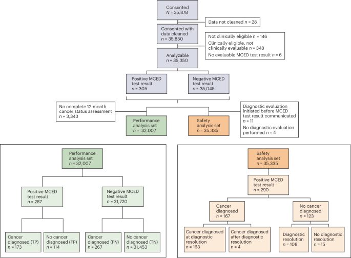
Analysis sets were prespecified in the statistical analysis plan. The analyzable set comprised consented, clinically eligible and clinically evaluable participants with an evaluable MCED test result (Fig. 1). The performance analysis set included analyzable participants with a completed 12-month cancer status assessment. The safety analysis set included analyzable participants who had one or more diagnostic evaluations performed only after communication of their MCED test result.
Study objectives and endpoints
Primary objectives were performance of the MCED test and its safety in terms of diagnostic testing triggered by the MCED test result (Supplementary Table 23). The primary performance endpoints included PPV, NPV, sensitivity, specificity, false-positive rate, positive likelihood ratio, negative likelihood ratio, overall CSO accuracy, observed cancer detection rate, number needed to screen to detect a cancer, MCED cancer detection rate, MCED cancer signal detection rate, accuracy of a CSO prediction by clinical CSO and precision of a CSO prediction by CSO prediction. The primary safety endpoint included the number and type of invasive procedures performed and the number and type of adverse events due to diagnostic testing in all participants with a positive MCED test result (safety measures detailed in Table 3).
All secondary endpoints are described in Supplementary Table 23. Secondary endpoints reported here included participant-reported anxiety assessed by State-Trait Anxiety Inventory (STAI) questionnaires pre-test (baseline), post-test, at diagnostic resolution (if applicable) and at 1 year (+30 days); utilization of standard-of-care cancer screening tests prior to study enrollment; the proportion of participants with cancer diagnosis at diagnostic resolution out of all participants who had a PET/CT only after initial negative diagnostic evaluation (no PET/CT during initial diagnostic evaluation); the proportion of participants with cancer diagnosis at diagnostic resolution out of all participants with initial negative diagnostic evaluation who had a diagnostic PET/CT at any time during diagnostic evaluation; the number and type of imaging procedures, number and type of invasive procedures, number and type of laboratory tests and time to diagnostic resolution; and primary test performance endpoints assessed in selected demographic and clinical subgroups classified on the basis of different categories of age, sex and smoking history.
Secondary endpoints not reported here include intention to follow standard-of-care cancer screening tests assessed pre-test, post-test and at 1 year (+30 days) and utilization of standard-of-care cancer screening tests during the first year and the second year (±30 days) after MCED test; the number and type of imaging procedures with radiation exposure and total per-participant radiation exposure during diagnostic evaluation; concordance between initial MCED test results and research blood draw results, including agreement in cancer signal detection and concordance of CSO1 prediction among participants with both test results; participant-reported outcomes and perceptions of the MCED test, assessed using participant-directed questionnaires, including health-related quality of life and other questionnaire-specific scores or item responses; primary test performance endpoints assessed in selected demographic and clinical subgroups classified on the basis of different categories of race, ethnicity, BMI, prior cancer history and genetic cancer predisposition; and MCED test positive rate among participants with potentially cross-reactive conditions.
All study objectives and endpoints were prespecified in the protocol and statistical analysis plan. The statistical analysis plan included analyses for two test versions: (1) MCED-V2, the test version used in the study for which results were returned to healthcare providers to inform diagnostic evaluations (that is, the study protocol version) and (2) a different MCED test version (referred to as MCED-I in the statistical analysis plan) that was analyzed to support an FDA premarket approval (PMA) submission. The results of the MCED-I test version were not returned during the PATHFINDER 2 study and, therefore, did not inform participants’ diagnostic evaluations. The analysis plan for MCED-I included composite success criteria and hypothesis testing (detailed in statistical analysis plan section 14.2.2.2), which were specific to the PMA analysis and, thus, were not included in the study protocol. The protocol and the present paper were focused on evaluating the safety and performance of MCED-V2. These analyses were designed to be descriptive, with no defined success criteria or formal hypothesis testing. Thus, the PMA-specific success criteria and hypothesis testing specified for MCED-I are not applicable to the MCED-V2 12-month analyses reported in this paper.
Performance
Performance of the MCED test, evaluated in analyzable participants with a completed 12-month cancer status assessment (performance analysis set), was assessed by multiple endpoints (see Glossary of Screening Terms for definitions and calculations). CSO prediction accuracy was calculated based on comparison of predicted CSO(s) with clinical CSO among participants with true-positive MCED test results. Twelve-month episode sensitivity was assessed by cancer type and within predefined cancer subgroups (Supplementary Table 22).
Safety
The safety analysis focused on analyzable participants with one or more diagnostic evaluations initiated only after MCED test result communication. Primary safety analyses evaluated the number and type of invasive procedures performed and adverse events occurring during the time of diagnostic testing triggered by a positive MCED test result. Invasive procedures were surgical or nonsurgical (for example, endoscopy and biopsy). Secondary analyses described laboratory tests, imaging, noninvasive procedures and adverse events in all consented participants with cleaned data.
Time to diagnostic resolution was calculated from the time the MCED test result was communicated to the participant or to the date of last contact for participants who did not achieve diagnostic resolution.
Participant-reported outcomes
Participant-reported anxiety resulting from MCED test use was assessed with the STAI38 based on questionnaires completed ≤2 months (61 days) after questionnaire release for pre-test, post-test and diagnostic resolution timepoints and ≤3 months (91 days) after questionnaire release for the 12-month timepoint. Questionnaires at the time of diagnostic resolution were administered only to participants with positive MCED test results.
Cancer detection method
The fold change in screen-detected cancers with the addition of MCED testing was calculated as follows: (number cancers detected by MCED screening + number of cancers detected by USPSTF-recommended screening) / number of cancers detected by USPSTF-recommended screening. MCED-detected cancers were defined as cancers in participants with true-positive MCED test results. There was no overlap in cases detected by the MCED test and USPSTF-recommended screening tests because only participants with true-positive MCED test results were counted as MCED detected, whereas participants with negative MCED test results and a screen-detected cancer during the 12-month follow-up period were counted as USPSTF screen detected.
Sample size and power
The sample size of this descriptive study was driven by the ability to enroll participants in the specific study subgroups based on age and sex to obtain information on diagnostic evaluation triggered by the MCED test result. The study was designed to enroll greater than or equal to 35,000 participants. Assuming that approximately 5% of participants would be excluded due to various clinical and assay evaluability reasons, approximately 33,250 analyzable participants were expected for analyses related to receiving MCED test results and associated diagnostic evaluation. Based on previous studies, MCED test specificity was expected to be in the range of 99−99.5%7.
A co-primary analysis objective focused on MCED test performance. The expected number of positive MCED test results, number of cancers diagnosed and test performance endpoints were estimated using microsimulations as previously described39. Under conservative assumptions about cancer incidence used in these microsimulations to account for a possible healthy volunteer effect (assuming underlying cancer incidence in the enrolled population was 65% of Surveillance, Epidemiology, and End Results (SEER) cancer incidence with the sex and age distribution described in the study protocol), approximately 370 cancers were expected to be diagnosed among 35,000 enrolled participants (or 33,250 analyzable participants) during 12 months of follow-up. The results of the microsimulations indicated that approximately 300−462 positive MCED test results were expected, with approximately 139 true-positive and 163−323 false-positive MCED test results. Based on these results, we expected to observe a PPV of approximately 30% and 46% if MCED test specificity is 99% and 99.5%, respectively. To illustrate the precision around these point estimates, an observed PPV of 30% based on 462 positive MCED test results would yield a two-sided Wilson 95% CI of 26.1−34.4%, and an observed PPV of 46% based on 300 positive MCED test results would yield a two-sided Wilson 95% CI of 40.4−51.7%. The other co-primary analysis objective focused on safety evaluation. The levels of precision for the primary safety Measure A (‘number of all participants with MCED positive test results and an invasive procedure divided by the total number of analyzable participants’) were estimated under six scenarios representing levels of false-positive rate (1.0% or 0.5%) and proportions of participants with invasive procedures ranging from 10% to 50%. Approximately 80% of participants with true-positive and 30% with false-positive MCED test results were estimated to have an invasive procedure based on data from the PATHFINDER study7. Based on the false-positive rate and the proportion of participants with false-positive MCED test results and invasive procedures, Measure A was estimated to range from approximately 0.4% to 0.8% across these scenarios. For reference, the proportion of participants with false-positive test results and invasive procedures among all screened participants for existing guideline-recommended noninvasive screening tests has been estimated as approximately 1.66% for breast cancer screening with mammography, 10.2% for colorectal cancer screening with a multi-target stool DNA test and 1.88% for lung cancer screening with low-dose CT13,40,41. The expected one-sided 95% upper confidence bound for Measure A based on all participants with positive MCED test results ranged from 0.45% to 0.92% across the scenarios prespecified in the protocol and was substantially lower than the reported point estimates for these existing screening tests among false-positive results. No formal comparison with existing screening tests was planned; for illustrative purposes only, the study would provide a high power (>99%) for such a comparison with even the lowest estimate reported above (1.66% for screening mammography) used as a threshold—that is, to test a null hypothesis that Measure A is above 1.66%.
Statistical analysis
No formal hypothesis testing or success criteria were defined for the endpoints reported in this paper; all endpoints were prespecified and analyzed descriptively. Based on microsimulations with conservative assumptions, greater than or equal to 370 cancers were expected with 35,000 participants screened, including 156 with true-positive MCED test results, which was considered sufficient for descriptive analyses of test performance and safety. Data lock date was 11 February 2026.
Performance estimates were not calculated for subgroups with fewer than five participants. Two-sided 95% CIs were constructed using the Wilson (score) method for performance endpoints not very near 0 or 1. Modified Wilson (score) CIs were used for endpoints near 0 or 1.
For categorical/binary variables, number and percentage of participants in each category were described. For continuous variables, number of participants and median with first and third quartiles (Q1, Q3) were reported.
The Kaplan−Meier method was used to estimate time to diagnostic resolution for participants with positive MCED tests.
Analyses were conducted using software R version 4.3.2.
Protocol and statistical analysis plan amendments
A complete list of amendments to the PATHFINDER 2 study protocol and statistical analysis plan is provided in Supplementary Table 24. Both documents are provided in full in the Supplementary Information.
Reporting summary
Further information on research design is available in the Nature Portfolio Reporting Summary linked to this article.
