


Patient recruitment and general considerations
This study recruited patients with various types of primary cancer, with the largest case series consisting of breast cancer patients. These patients require special consideration, as venous access to the ipsilateral limb is avoided following surgery with axillary lymphadenectomy. This necessitates continuous adaptation of the surgeon’s skills8.
In general, totally implantable venous access devices (TIVADs) provide a safe and comfortable route for the administration of cytotoxic drugs in patients with malignant neoplasms. Figure 2 presents a decision tree developed by the authors to summarize the key considerations during the surgical implantation of implantable vascular access devices. The schematic outlines the clinical and technical factors influencing procedural planning and frames the central question addressed by this study.
Fig. 2
Decisions in the implementation of implantable vascular access devices.
Although no premature port removal occurred in this study. The overall implantation success rate defined as successful device placement with adequate catheter tip positioning exceeded 96.6% patients: 97.8% in the technology-assisted approach (TA) group and 98.8% in the anatomical landmark-based approach (ARP) group. This is considered a high success rate compared with similar studies, which reported success rates of 91.9% with an ARP approach and nearly 100% among patients who received ultrasound-guided implantation9,10.
Operative time was stratified using a 30-minute cutoff based on institutional standards commonly applied in Colombian surgical practice for TIVAD implantation. Procedures exceeding this duration may reflect increased technical complexity or intraoperative difficulty. This pragmatic threshold was used to explore the potential association between prolonged operative time and postoperative complications rather than to define procedural quality.
Venous access selection
Certain conditions, such as burns or congenital and acquired morphological alterations, may impact the ability to identify anatomical landmarks11. Vascular access has been associated with complications, and these aspects have been evaluated in randomized controlled trials, in which the choice of vessel, laterality, as well as device caliber and length have been systematically analyzed12.
Meta-analyses have evaluated the risk of thrombosis associated with venous access, with no statistically significant differences found13,14. Other studies with lower statistical power have also failed to demonstrate the relevance of laterality in thrombosis risk15. The identification of a catheter associated with the innominate vein in the ARP group was an incidental finding during retrospective image review for catheter tip position assessment and does not reflect a routinely planned puncture technique for TIVAD implantation.
Surgical approach
The use of anatomical landmarks has been reported to achieve a success rate of up to 87%. However, the use of technology-assisted devices appears to improve this success rate, approaching 100%16. Historically, surgeons relied on the subclavian access route, which is based on anatomical landmarks, for training purposes. Nevertheless, ultrasound-guided venipuncture has gained popularity and has demonstrated sufficient evidence to support its use. In contrast, venous dissection approaches are becoming increasingly unnecessary17.
Venous dissection was employed in a limited number of cases as a secondary strategy after unsuccessful initial percutaneous access attempts, rather than as a planned first-line approach for TIVAD implantation. Given the retrospective nature of the data collection, detailed intraoperative decision-making processes and the precise anatomical rationale for vessel selection could not be fully captured, which may introduce bias in the interpretation of these findings.
Previous failed insertion attempts, particularly in emergency settings, are among the most common predictors of insertion-related complications. It has been reported that with a single successful catheterization attempt, the complication rate is 4.3%, whereas this increases to 24% with two or more attempts18,19.
The identification of a catheter associated with the innominate vein in the ARP group was an incidental finding during the retrospective image review for catheter tip position assessment and does not reflect a routinely planned puncture technique for TIVAD implantation.
Catheter tip positioning
Catheter tip location appears to be related to catheter dysfunction, manifesting as difficulty in aspiration, resistance, inflammation, and/or pain during infusion20. Additionally, catheter tip malposition is an independent risk factor for venous thrombosis in cancer patients, with a sevenfold increased risk when the catheter tip is located in the upper half of the superior vena cava21.
A noteworthy finding of this study was the heterogeneity observed in the interpretation of the final catheter tip location, as reported by both surgeons and radiologists. This finding suggests the need for additional studies to evaluate the degree of concordance in image interpretation.
Due to inconsistent documentation of the final catheter tip position across procedural reports, catheter tip location was retrospectively reassessed according to the Cadman classification by means of a systematic, patient-by-patient review of radiological studies. This assessment was performed by a radiologist to ensure data consistency and standardization.
Use of prophylactic antibiotics
As intravascular devices, catheters may be associated with bacteremia-related complications. However, retrospective studies have reported a relative risk (RR) of 0.97, indicating that there is no association between infection and the use of prophylactic antibiotics. However, this recommendation lacks the statistical power to be considered a standard22,23.
Our findings indicate that the use of prophylactic antibiotics during catheter insertion did not confer a protective effect against infectious complications. During the 90-day follow-up period, antibiotic prophylaxis use was associated with a significantly higher risk of complications compared to no prophylaxis (RR 2.56; 95% CI 1.20–5.44). It cannot be determined whether the observed association between antibiotic use and complications reflects a true causal relationship or is attributable to confounding, particularly given that antibiotics may have been administered as prophylaxis for concomitant procedures that themselves could have increased the risk of complications.
Recently, the Society of Interventional Radiology classified TIVAD placement as a clean procedure according to the National Academy of Sciences/National Research Council recommendations, suggesting that prophylaxis should not be routinely recommended, regardless of the technique used23,24.
Anesthesia management
The literature has not identified differences in terms of clinical outcomes, surgical time, blood loss, or radiation dose and duration. However, compared with local anesthesia, general anesthesia is associated with longer operating room times and increased costs25. In this cohort, the use of general anesthesia was not inherent to the TIVAD implantation technique but was instead dictated by the need to perform concomitant surgical procedures, including staging laparoscopies or oncologic resections. As such, anesthesia modality should be interpreted as a marker of overall surgical context rather than as a determinant of catheter implantation outcomes.
Complications
Complications have been classified into those related to catheter insertion, those associated with port implantation, and those categorized on the basis of the time of occurrence26. Table 6 was describes a summary tool to provide an overview of the temporal occurrence of procedure-related complications, facilitating interpretation as a descriptive synthesis rather than an independent analytical component.
Table 6 Complications associated with implantable vascular access devices.
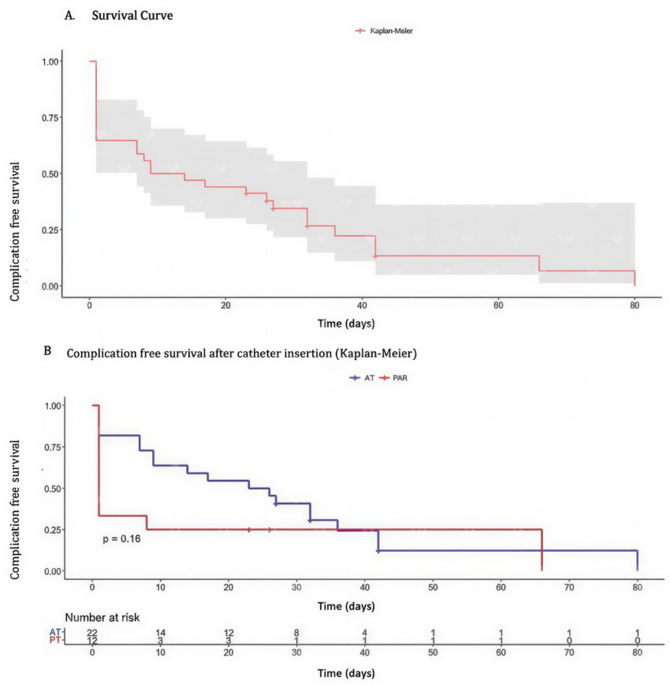
Recent publications report overall complication rates ranging from 2% to 14.4%, which is consistent with the data presented in this study10. In our study, the absence of statistically significant differences in complication rates between the two implantation techniques is consistent with findings reported in the existing literature. No significant differences were observed in the incidence of either early or late complications between the groups, supporting comparable safety profiles for both approaches. It is important to note that all procedures were performed electively and predominantly by surgeons with extensive professional experience, factors that may have contributed to the overall low complication rates observed in our cohort2.
Venous thrombosis is a multifactorial condition and appears to be secondary to fibrin deposits due to endothelial injury, leading to a reduction in the vessel lumen27. Some studies have demonstrated a benefit of using the jugular vein to reduce risk, although other authors have reported no significant differences28. In our study, the single case of deep vein thrombosis occurred in a female patient with advanced gastric cancer, a condition associated with an increased thrombotic risk. The catheter was placed via a left internal jugular approach using anatomical landmarks, and retrospective imaging review demonstrated suboptimal tip positioning, suggesting a multifactorial etiology involving both patient-related and procedural factors.
Cardiac arrhythmias are among the potentially life-threatening complications associated with catheter tip migration, occurring in up to 9% of cases. However, evidence suggests no statistically significant differences in vascular access when subclavian vein access is compared with internal jugular vein access29.
Hemothorax is less common and has been more commonly associated with venous access in the right internal jugular and subclavian veins, particularly following arterial puncture or laceration of intrathoracic veins. Vascular injuries are uncommon. The incidence of internal carotid artery puncture is 3%, whereas subclavian artery puncture is reported even less frequently26.
Although it has a very low incidence rate, pneumothorax is the most common complication in patients with anatomical landmark-based placement [ARP]. Several studies, including expert consensus, have indicated that technology-assisted TIVAD insertion completely eliminates the risk of pneumothorax and hemothorax. However, pneumothorax is a significant complication following TIVAD insertion and has an immediate clinical impact. Its reported incidence varies between 0.5% and 6% and is highly associated with the ARP approach and multiple puncture attempts30. In our study, the incidence of this complication was lower in the technology-assisted group compared with the anatomical landmark-based group; however, this difference did not reach statistical significance (P = 0.17). Although this observation suggests a possible trend, the low event rate limits the statistical power of the analysis, and therefore no definitive conclusions can be drawn regarding a protective effect of the technology-assisted approach.
Among the complications associated with port implantation, wound dehiscence and leakage of contents may be related to technical errors in wound closure, improper positioning and fixation of the reservoir, and patient immunocompromised status and malnutrition. However, maintaining skin integrity and ensuring a tension-free incision reduce the incidence of these complications26.
Major complications (as classified by the Clavien–Dindo system) and rare events such as gas embolism, hemothorax, pericardial tamponade, and brachial plexus injury were not observed in our cohort. Currently, there are no established quality control standards for TIVAD implantation techniques or specific guidelines for managing complications. Although our study results indicate that the complication rate does not seem to correlate with operator experience, especially since the procedures were performed by both surgeons and interventional radiologists, several studies suggest that the level of experience of the physician inserting the TIVAD is crucial. The likelihood of complications is halved when the procedure is performed by a physician with experience in 50 or more insertions compared with a physician with fewer than 50 implantations18,19. No cases of catheter fracture or reservoir malposition were identified in our cohort.
The occurrence of hematoma was identified retrospectively from clinical records, without standardized documentation regarding size or severity, which may introduce reporting bias. Furthermore, technology-assisted techniques may entail increased tissue manipulation during image acquisition and needle adjustment, potentially contributing to the observed distribution of this complication.
Medical specialty
The medical specialties currently involved in this study are limited to surgeons and interventional radiologists. However, the insertion procedure can also be performed by other specialties, including internal medicine, anesthesiology, and oncology. Nonetheless, the mastery of implantation techniques, complication management, and the appropriate use and maintenance of TIVADs remain inconsistent across different medical specialties. Furthermore, considering the low degree of participation of resident physicians reported in this study, there is a clear need to promote the development of these skills within specialized medical-surgical training programs.
The reduced involvement of resident physicians reflects the institutional characteristics of the participating centers, where formal training programs were not continuously available throughout the study period. Consequently, a proportion of procedures were performed exclusively by attending surgeons or radiologists, which should be considered when interpreting operator-related factors in this cohort.
Limitations
Despite the multicenter design of the present study, the relatively low number of complications may have limited the statistical power to detect meaningful differences between implantation techniques. This observation may be partly attributable to selection bias, as most procedures were elective and performed by experienced operators in high-volume centers, potentially resulting in a lower overall complication rate. Although matched-pair or propensity-based analyses could further control for confounding, the retrospective nature of the study and the limited number of outcome events precluded the implementation of a robust matching strategy without substantially compromising sample size. These limitations underscore the need for adequately powered prospective controlled studies to validate the present findings.
Although current guidelines recommend positioning the catheter tip at the cavoatrial junction or within the superior vena cava, optimal positioning was not achieved in all patients in this cohort. This finding is likely multifactorial and may reflect variability in intraoperative verification methods, institutional practices, and historical changes in positioning criteria over the extended study period. In addition, final catheter tip location was retrospectively reassessed using standardized radiological criteria rather than real-time intraoperative confirmation, which may have contributed to discrepancies between guideline-defined optimal positioning and observed anatomical locations. Importantly, these findings should not be interpreted solely as a surrogate for technical proficiency, but rather as a reflection of real-world practice in a multicenter retrospective setting.
