


Case description
A 30-year-old male with no comorbidities presented in January 2022 with progressive pain of the left upper arm and shoulder. MRI revealed a large proximal humeral lesion with intramedullary extension, cortical breakthrough, and an extensive soft tissue component, consistent with a high-burden primary tumor. The primary tumor was located in the proximal left humerus and measured 11 × 3.8 × 4.4 cm on baseline imaging. Initial staging by CT and 18F-FDG PET demonstrated multifocal osseous metastatic disease without pulmonary metastases, with documented skeletal involvement of the left humerus, C2, C4, T1, T10, T11, and the ilium. Skip metastases were also documented in the distal left humerus. The initial clinical stage was cT3 Nx M1b, corresponding to stage IVB according to UICC/AJCC. Histopathology demonstrated a small round blue cell tumor, and molecular profiling confirmed the presence of a type 1 EWSR1–FLI1 fusion, consistent with Ewing sarcoma1. Immunohistochemistry showed positivity for FLI1 and CD99, while EMA, desmin, S100, cytokeratin, smooth muscle actin, synaptophysin, and SSX were negative. Additional sequencing identified two stop-gain mutations in KMT2D, the clinical relevance of which remains uncertain. Despite the high radiographic tumor burden, serum lactate dehydrogenase (LDH) was not elevated at baseline, measuring 171 U/L on 26 January 2022, below the institutional upper limit of normal of 250 U/L. During longitudinal monitoring up to the February 2025 manuscript data cut-off, LDH remained within the institutional reference range, with a maximum value of 246 U/L recorded on 9 March 2022. After completion of chemotherapy and radiotherapy and shortly before vaccination initiation, LDH was 149 U/L on 23 November 2022. The patient underwent intensive multimodal therapy according to contemporary Ewing sarcoma treatment standards. Induction chemotherapy consisted of six cycles of VIDE between January and May 2022, comprising vincristine on day 1 and doxorubicin, etoposide phosphate, and ifosfamide on days 1–3. Cycles 1–4 were administered at full protocol dose for doxorubicin, etoposide phosphate, and ifosfamide: doxorubicin 20 mg/m²/day, etoposide phosphate 150 mg/m²/day, and ifosfamide 3000 mg/m²/day on days 1–3. Vincristine was administered at 2 mg on day 1 of each cycle, corresponding to the institutional absolute dose cap. After febrile neutropenia with pronounced mucositis and pancytopenia following VIDE cycle 4, doxorubicin, etoposide phosphate, and ifosfamide were reduced to 80% of the protocol dose from cycle 5 onward, corresponding to doxorubicin 16 mg/m²/day, etoposide phosphate 120 mg/m²/day, and ifosfamide 2400 mg/m²/day on days 1–3. The approximate delivered cumulative VIDE doses were vincristine 12 mg absolute dose, doxorubicin 336 mg/m², etoposide phosphate 2520 mg/m², and ifosfamide 50.4 g/m². Restaging after completion of induction chemotherapy demonstrated stable disease with local regression of the primary tumor. The patient subsequently received six cycles of VAI consolidation chemotherapy between June and October 2022, consisting of vincristine, dactinomycin, and ifosfamide. VAI cycle 1 was administered at full protocol dose for dactinomycin and ifosfamide, with dactinomycin 0.75 mg/m²/day and ifosfamide 3000 mg/m²/day on days 1–2. From VAI cycle 2 onward, dactinomycin and ifosfamide were reduced to 80% of the protocol dose because of prior febrile neutropenia and ongoing hematologic toxicity, corresponding to dactinomycin 0.6 mg/m²/day and ifosfamide 2400 mg/m²/day when administered. Vincristine was given at 2 mg in VAI cycles 1–4 and reduced to 1 mg in cycles 5–6. After three cycles of VAI, intra-articular resection of the proximal left humerus was performed on 18 August 2022, including resection of a 16.5 cm tumor-bearing bone segment and reconstruction with an inverse proximal humeral MUTARS endoprosthesis. The postoperative course was uncomplicated, with regular implant positioning on postoperative radiography. A formal histopathologic regression score, quantified tumor necrosis percentage, and resection-margin status were not available from the reviewed documentation. Three additional VAI cycles were then administered. During the final VAI cycle, administered from 17 to 18 October 2022, dactinomycin was omitted because of concurrent radiotherapy. Consolidative radiotherapy was delivered from 6 October to 17 November 2022 as part of multimodal local and metastatic-site control19. Intensity-modulated radiotherapy was applied to the primary tumor region of the left humerus with 50.4 Gy in 28 fractions of 1.8 Gy using 6 MV photons. Additional osseous metastatic sites were treated with 45 Gy in 1.8 Gy fractions, including lesions in the cervical and upper thoracic spine (C1–T2), thoracolumbar/pelvic target regions including T10–T11 and L1–L5, and the sternum. The sternal lesion was treated with photons up to 19.8 Gy followed by continuation with 8 MeV electrons to a cumulative dose of 45 Gy. Radiotherapy to the osseous metastatic sites was temporarily paused on selected treatment days because of pronounced pancytopenia and lymphocytopenia requiring transfusional support and G-CSF, while irradiation of the primary tumor region was not interrupted. Radiotherapy was completed on 17 November 2022. Acute radiotherapy-associated toxicity included grade 2 epitheliolysis in the left axillary region, mild erythema over the sternum, mild dysphagia, oral dryness, dysgeusia, appetite loss, mild weight loss, and transient diarrhea with intermittent blood admixture. The latter led to short inpatient evaluation after completion of radiotherapy. Colonoscopy showed nonspecific colitis without active bleeding. This episode occurred before initiation of peptide vaccination. Despite intensive multimodal therapy, prognosis at this stage remained poor, as long-term progression-free survival beyond two years is rarely achieved in patients with multifocal metastatic Ewing sarcoma20.
Peptide vaccination was initiated on 2 December 2022, 15 days after completion of radiotherapy. The priming phase consisted of four vaccinations within seven days, administered on 2, 5, 7, and 8 December 2022, followed by regular booster vaccinations. Most vaccinations were co-administered with subcutaneous sargramostim (GM-CSF, 83 µg) and topical imiquimod, as described in the “Methods” section and Supplementary Information. From October 2023 onward, a second vaccine batch was used. At the February 2025 manuscript data cut-off, the patient had received 23 vaccinations over 26 months, consisting of the initial priming phase followed by 19 booster vaccinations. Treatment was well tolerated, with only grade 1 local reactions (erythema, swelling, induration) reported. No systemic vaccine-associated adverse events or clinically relevant immune-mediated toxicities were observed.
Serial imaging demonstrated durable disease control during follow-up. Staging in January 2023 showed stable disease without new metastatic lesions. In March 2023, whole-body MRI no longer showed clearly recognizable osseous metastases, and CT of the thorax did not reveal new suspicious lesions. Subsequent imaging in July and October 2023 remained without evidence of new malignancy-suspicious disease. In January 2024, MRI of the left upper arm and whole-body MRI again showed no evidence of local recurrence or newly detectable metastatic lesions, and previously documented osseous metastases were no longer clearly demarcated morphologically. At clinical assessment in January 2024, the patient remained clinically stable with ECOG performance status 0 and Karnofsky performance status 90%. At the February 2025 manuscript data cut-off, more than three years after initial diagnosis and 26 months after initiation of peptide vaccination, the patient remained clinically well with ECOG performance status 0 and without evidence of disease progression. This duration of disease stability was notable in the context of adult-onset stage IVB Ewing sarcoma with multifocal osseous metastatic disease4,20.
Clinical outcome and safety
After completion of multimodal therapy, the patient had stable disease by radiographic criteria. Peptide vaccination was initiated on 2 December 2022, 15 days after completion of radiotherapy, with a priming phase of four vaccinations within seven days followed by 19 booster vaccinations over 26 months, resulting in 23 vaccinations in total by the February 2025 manuscript data cut-off. The full vaccination schedule is provided in the Supplementary Information. Vaccination was well tolerated throughout, with only grade 1 local skin reactions including erythema, swelling, and induration. No systemic vaccine-associated adverse events or clinically relevant immune-mediated toxicities were observed. LDH remained within the institutional reference range during longitudinal monitoring up to the February 2025 data cut-off, measuring 171 U/L at baseline and 149 U/L shortly before vaccination initiation, with a maximum value of 246 U/L. TSH measurements showed no persistent laboratory abnormality suggestive of immune-mediated thyroid disease. An isolated low TSH value had been recorded before vaccination initiation during multimodal therapy. In the absence of clinical or persistent laboratory evidence of thyroid dysfunction, thyroid autoantibody testing was not performed.
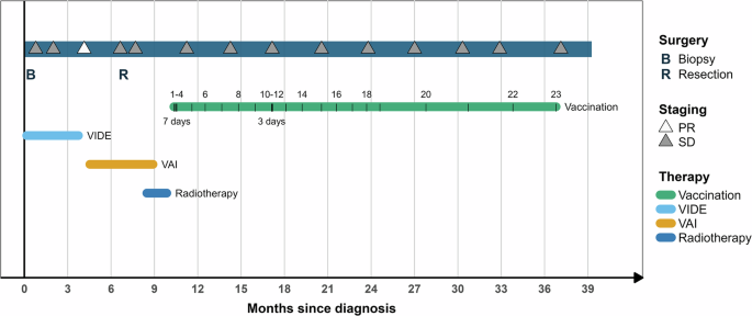
During follow-up, serial imaging with CT of the neck, chest, abdomen, and pelvis and MRI of the primary site demonstrated ongoing disease stability. Staging in January 2023 showed stable disease without new metastatic lesions. Whole-body MRI in March 2023 no longer showed clearly recognizable osseous metastases, and subsequent imaging in July, October, and January 2024 remained without evidence of local recurrence or newly detectable metastatic lesions. At the February 2025 manuscript data cut-off, more than three years after initial diagnosis and 26 months after initiation of peptide vaccination, the patient remained clinically well with ECOG performance status 0 and without evidence of recurrence or new metastatic lesions. This durable disease stability is notable in the context of adult-onset stage IVB Ewing sarcoma with multifocal osseous metastatic disease20. The clinical course and vaccination schedule are summarized in Fig. 1.
Fig. 1: Clinical course and treatment timeline.
Schematic representation of the patient’s clinical course from initial diagnosis (January 2022) to manuscript data cut-off (February 2025). The timeline depicts induction VIDE chemotherapy (six cycles), surgery of the proximal humerus with endoprosthesis implantation, adjuvant VAI chemotherapy (six cycles, three before and three after surgery), consolidative radiotherapy to the primary tumor region and osseous metastatic sites, and initiation of EWSR1–FLI1 peptide vaccination. Vaccination consisted of a priming cycle of four doses followed by 19 booster doses over 26 months. Disease status at each imaging assessment is indicated (PR, partial response; SD, stable disease). ECOG performance status remained 0 throughout follow-up. The patient experienced only grade 1 local injection-site reactions, without systemic vaccine-associated toxicities. The figure was created by the authors using the ggplot2 package, version 4.0.1, in R.
Vaccine-induced T-cell responses
Peripheral blood mononuclear cells were collected prior to the first vaccination and at seven longitudinal timepoints during treatment. Antigen-specific T-cell responses were assessed following in vitro peptide stimulation and intracellular cytokine staining (ICS). Responses were defined as polyfunctional CD4⁺ or CD8⁺ T-cells co-expressing at least two functional markers (CD154, IFN-γ, TNF-α, IL-2), with a stimulation index (SI) ≥ 2 considered positive14,15,16.
At baseline, no EWSR1–FLI1–specific responses were detectable. By month 7, polyfunctional CD4⁺ T-cell responses emerged against peptides E1 (0.4% of CD4⁺ T-cells, SI 2.1) and E4 (0.7%, SI 2.2). At month 17, responses had broadened to include peptides E1, E3, and E4, with frequencies ranging from 0.7–1.6% of CD4⁺ T-cells and stimulation indices consistently above 2. By month 26, polyfunctional responses were observed against all four peptides (E1–E4), with the strongest response directed against peptide E4 (17.7% of CD4⁺ T-cells, SI 82.7). Although a single CD8⁺ response was transiently detectable, the vaccine-induced immunity was dominated by durable CD4⁺ T-cell activity.
Representative flow-cytometry plots confirmed polyfunctional cytokine co-expression and validated the specificity of responses compared to mock-stimulated controls (Fig. 3). Longitudinal analysis demonstrated persistence of vaccine-induced CD4⁺ T-cell immunity for more than two years, consistent with the generation of durable immunological memory. Longitudinal kinetics are shown in Fig. 2, representative plots in Fig. 3.
Fig. 2: Longitudinal vaccine-induced CD4⁺ T-cell responses.
Polyfunctional CD4⁺ T-cell responses to the four EWSR1–FLI1 peptides (E1–E4) were assessed in peripheral blood by in vitro peptide stimulation and intracellular cytokine staining. Responses were defined as cells co-expressing ≥2 functional markers (CD154, IFN-γ, TNF-α, IL-2) with stimulation index (SI) ≥ 2. Graphs depict frequencies of polyfunctional CD4⁺ T-cells specific for each peptide across seven timepoints. No responses were detected prior to vaccination. By month 7, responses to peptides E1 and E4 emerged. By month 17, responses broadened to E1, E3, and E4. At month 26, polyfunctional CD4⁺ T-cell responses were detectable against all four peptides, with the strongest response to peptide E4. Data represent percentages of all CD4⁺ T-cells after subtraction of mock-stimulated controls. Raw values and gating details are provided in the Supplementary Information. The figure was created by the authors using GraphPad Prism, version 8.4.3 (GraphPad Software, Boston, Massachusetts, USA).
Fig. 3: Representative flow-cytometry plots of peptide-specific T-cells.
Flow-cytometry dot plots illustrating polyfunctional CD4⁺ T-cell responses prior to vaccination (V1) and after 22 vaccinations (V22). Cells were either mock-stimulated (negative control, upper row), stimulated with each of the four EWSR1–FLI1 peptides (E1–E4, lower row), or stimulated with CytoStim (Miltenyi Biotec) (positive control, PC). Shown are representative plots gated on CD4⁺ T-cells, with TNF on the x-axis and CD154 on the y-axis. Numbers indicate frequencies within all CD4⁺ T-cells. Robust vaccine-induced responses are evident after vaccination, while absent before treatment. Raw values and gating details are provided in the Supplementary Information. The figure was created by the authors using FlowJo™ Software, version 10.10.1 (BD Life Sciences).
