


Study design and trial oversight
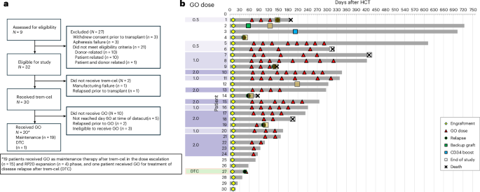
This research complies with all relevant ethical regulations, and the study protocol was approved by the Western IRB (WIRB/Copernicus Group), the NMDP IRB and the institutional review boards at Washington University; Miami Cancer Institute; the National Cancer Institute; Hôpital Maisonneuve-Rosemont; Hackensack University Medical Center; the University of California, San Diego; Memorial Sloan Kettering Cancer Center; the University of Kansas Cancer Center; the University of Minnesota; Winship Cancer Center; Emory University; Stanford University; and the University of Utah. All patients signed informed consent, and no compensation aside from study-required travel/lodging reimbusement was provided. Sex and gender were not considered in the study design. This was a first-in-human, phase 1/2a, open-label study in patients with AML or MDS at high risk of relapse who received an HLA-matched alloHCT with trem-cel followed by GO maintenance therapy (ClinicalTrials.gov identifier: NCT04849910, first submitted 21 April 2021; Extended Data Fig. 1). Trem-cel was manufactured as previously described and summarized in the ‘Trem-cell manufacturing’ section18. Patients underwent busulfan-based or total body irradiation (TBI)-based myeloablative conditioning prior to infusion of trem-cel on day 0. Supportive care was provided per local practice and study guidelines. T cell content of the graft was <1 × 10⁶ CD3+ cells per kg, and no post-transplant prophylaxis against GvHD was given. On day 60, patients achieving neutrophil engraftment were assessed for disease status and hematopoietic recovery by bone marrow biopsy; eligible patients initiated maintenance GO by infusion. Part 1 (GO dose escalation) followed a standard 3 + 3 design of 3−6 patients per dose cohort (0.5, 1.0 or 2.0 mg m−2 on day 1 of each 28-day cycle up to eight cycles) to identify maximum tolerated dose (MTD) and RP2D. Part 2 (GO dose expansion) assessed safety and preliminary efficacy of the RP2D. Patients who relapsed or were MRD+ could enter the DTC (Extended Data Fig. 1) to receive GO induction and consolidation.
The trial was approved by the institutional review board/independent ethics committee at each site and donor registry and was conducted in accordance with the principles of the Declaration of Helsinki and Good Clinical Practice guidelines. All patients and donors provided written informed consent. The study was designed and data were analyzed by the sponsor in collaboration with the investigators.
Eligibility
Inclusion/exclusion criteria (patients and donors)
Full patient eligibility
Inclusion criteria
-
1.
Patients must be ≥18 and ≤70 years of age.
-
2.
Ability to understand and willingness to sign the informed consent form or, if applicable, understanding and willingness of the patient’s caregiver or legally authorized representative prior to initiation of any study-related tests or procedures
-
3.
Patients must have confirmed diagnosis of AML or MDS according to World Health Organization (WHO) classification criteria.
-
4.
The patient’s AML or MDS must have evidence of CD33 expression (>0%) according to institutional flow cytometry criteria.
-
5.
Patients with AML must have one of the following groups of features that are known to be a risk factor for leukemia relapse:
-
Bone marrow in morphological remission (<5% blasts) with adverse-risk disease-related genetics at presentation (according to ELN guidelines40), or
-
Intermediate-risk genetics in morphologic remission (<5% blasts) with other recognized high-risk criteria, such as MRD+ after therapy, or
-
Bone marrow with evidence of persistent leukemia 5–10% blasts after induction/salvage therapy. Patients with bone marrow blast count >10% may participate with Sponsor Medical Monitor approval (note: these patients may have disease-related genetics of any risk criteria at presentation), or
-
Any patient in second or greater remission
-
a.
Patients with MDS must have all of the following:
-
Previous or current Revised International Prognostic Scoring System (IPSS-R) score of High or Very High risk
-
Previous or current MDS-IB1 or MDS-IB2 per the 2022 WHO criteria
-
6.
Patients must be a candidate (per investigator opinion) for HLA-matched alloHCT using a myeloablative conditioning regimen (see protocol for allowed regimens).
-
7.
Patients must have a related or unrelated stem cell donor that is an 8/8 match for HLA-A, HLA-B, HLA-C and HLA-DRB1, as assessed by high-resolution DNA-based typing. The selection of related donors is per institutional standards and, for unrelated donors, is per registry standards.
-
8.
Patients exceeding a weight of 120 kg (265 lbs) may participate with Sponsor Medical Monitor approval.
-
9.
Patients must have adequate performance status and organ function as defined below:
-
Performance status: Karnofsky score of ≥70
-
Cardiac: left ventricular ejection fraction ≥45% by echocardiogram or radionuclide scan (that is, multiple-gated acquisition (MUGA) scan)
-
Pulmonary: diffusing capacity of lung for carbon monoxide ≥66% (adjusted for hemoglobin levels) and forced vital capacity (FVC) and forced expiratory volume in 1 second (FEV1) ≥66%
-
Renal: estimated glomerular filtration rate >60 ml min−1 as assessed by Cockcroft−Gault formula and actual body weight
-
Hepatic: total bilirubin <1.5× upper limit of normal (ULN) or, if ≥1.5× ULN, direct bilirubin
-
10.
Female patients of childbearing potential must test negative for pregnancy at screening, not be pregnant, nor breastfeeding, nor expecting to conceive within the projected duration of the trial. Sexually active women, unless surgically sterile, must be willing to use a highly effective method of birth control up to 180 days after the last dose of study drug (VOR33 or Mylotarg). Examples of birth control methods are as follows: implants, injectables, combined oral contraceptives, intrauterine devices (IUDs) or vasectomized partner.
-
11.
Up to 90 days after the last dose of study drug (VOR33 or Mylotarg), male patients with partners of childbearing potential must either be vasectomized or agree to use a condom, in addition to having their partners use another method of contraception, resulting in a highly effective method of birth control such as implants, injectables, combined oral contraceptives or IUDs. Male patients should not have sexual intercourse with females who are either pregnant or lactating without double-barrier contraception. Male patients must not donate sperm during the course of the study.
-
12.
Currently not requiring systemic corticosteroid therapy (10 mg or less of prednisone or equivalent doses of other systemic steroids are allowed)
Exclusion criteria
-
1.
Patients with TT genotype (SNP rs12459419, a population variation missing Mylotarg-binding epitope on CD33)
-
2.
Prior autologous or allogeneic stem cell transplantation
-
3.
Presence of the following disease-related genetics: t(15; 17)(q22; q21) or t(9; 22)(q34; q11) or other evidence of acute promyelocytic leukemia or chronic myeloid leukemia
-
4.
Prior treatment with Mylotarg (GO) in the past 3.5 months, except with Sponsor Medical Monitor approval, or any prior Mylotarg-related SOS/VOD
-
5.
Active central nervous system leukemia
-
6.
Patients diagnosed with Gilbert’s syndrome
-
7.
Uncontrolled bacterial, viral or fungal infections or known HIV, hepatitis B or hepatitis C infection. (Patients cured of hepatitis C for ≥3 years prior to enrollment are permitted.)
-
8.
Known sensitivity to components of CliniMACS Prodigy CD34 System or VOR33, including iron dextran or murine proteins
-
9.
Have a history of other active malignancy(ies) unless the patient has been disease free for at least 2 years and is deemed by the treating investigator to be at low risk for recurrence of that malignancy or the only prior cancer is curatively treated cervical cancer in situ or basal cell or squamous cell carcinoma of the skin
-
10.
Women who are pregnant or who are lactating
-
11.
Received any investigational drug (that is, an agent that has not received regulatory approval for any indication) within the 30 days prior to the start of conditioning, unless approved by the Sponsor Medical Monitor
-
12.
History of cardiovascular disease including, but not limited to, myocardial infarction, unstable angina, stroke or transient ischemic attack within the 6 months prior to the initiation of study or congestive heart failure (New York Heart Association class III−IV)
-
13.
Any serious underlying medical or psychiatric condition that would increase the risk associated with study participation, in the opinion of the investigator or the Sponsor Medical Monitor
Donor eligibility
Inclusion criteria (donor)
-
1.
Must be ≥18 years of age
-
2.
Must be 8/8 HLA-matched to the patient (HLA-A, HLA-B, HLA-C and HLA-DRB1)
-
3.
Willing to receive injections of granulocyte colony-stimulating factor (G-CSF) and plerixafor to stimulate the peripheral blood circulation of HSCs and donate hematopoietic progenitor cells by apheresis for up to two consecutive days
-
4.
Willing and able to donate within the United States. Related donors for patients in Canada may donate at clinical trial site(s) in Canada.
-
5.
Meet the donor requirements under 21 CFR 1271 and related August 2007 guidance regarding the Eligibility Determination for Donors of Human Cells, Tissues, and Cellular and Tissue-Based Products. Donors considered ‘ineligible but suitable’ (per CFR § 630.20) may still be considered for donation by the investigator with Sponsor Medical Monitor approval.
-
6.
Meet additional criteria for donation as per registry or clinical trial site-specific requirements
-
7.
Willing and able to comply with the protocol procedures, in the opinion of the investigator or registry
-
8.
Willing and able to provide informed consent
Exclusion criteria (donor)
-
1.
For female donors of childbearing potential: pregnancy or uninterruptible breastfeeding. Pregnancy is an absolute contraindication under this protocol. Women who are breastfeeding must be willing and able to interrupt breastfeeding during G-CSF (filgrastim) and plerixafor administration and for 2 days after the final dose.
-
2.
Sensitivity to filgrastim or to Escherichia coli-derived recombinant protein products
-
3.
History of autoimmune disorders, including rheumatic diseases and thyroid disorders (although donors with a history of thyroid disease who have undergone successful therapy may be suitable). Exemptions for mild or limited disease may be granted after discussion with the investigator and the Sponsor Medical Monitor.
-
4.
History of deep vein thrombosis or pulmonary embolism
-
5.
History of iritis or episcleritis
-
6.
Platelet count <150,000 per µl at baseline evaluation
-
7.
Current treatment with lithium. Drug interactions between filgrastim and lithium, which may potentiate the release of neutrophils, have not been fully evaluated.
-
8.
Positive hemoglobin solubility (for example, SickleDex or equivalent) test
-
9.
Donors receiving experimental therapy or investigational agents within 30 days of the start of mobilization. FDA-approved therapies under investigation for alternative uses are permitted.
-
10.
Donors deemed not medically suitable per the registry or the clinical trial site or in the opinion of the Sponsor Medical Monitor
-
11.
Donors who do not meet the eligibility requirements per the registry or clinical trial site-specific procedures, unless otherwise approved to donate by the Sponsor Medical Monitor and the patient’s physician.
Donor procedures
Donors were consented after patients consented for the study. Coordination of the unrelated donor process was managed by NMDP Biotherapies and Gift of Life Marrow Registry. Clinical trial sites identified a related donor or initiated a database search for an unrelated donor prior to approaching the patient for consent in this study. The registry or the clinical trial site that consented the donor confirmed that the donor is medically suitable to donate. Donors underwent dual mobilization procedure of G-CSF/plerixafor. After mobilization, up to two apheresis procedures were performed. Collection of apheresis product was obtained sufficient to manufacture trem-cel and provide a minimally manipulated backup graft, both with a minimum of 3 × 10⁶ viable CD34+ cells per kg. Apheresis product was shipped fresh to a manufacturing facility, and cryopreserved trem-cel and backup graft were shipped to the clinical sites.
Trem-cel manufacturing
All cell manipulation procedures were performed in accordance with current Good Manufacturing Practices (GMPs) following process-specific procedures and batch records. A detailed description of the gRNA guide selection, off-target analysis using karyotyping and GUIDE-seq, editing efficiency, InDel distribution, colony-forming unit (CFU) analysis and manufacturing process was previously published18. In brief, the gRNA is manufactured as a 100-mer sequence: [5′-c*c*u* CAC UAG ACU UGA CCC ACG UUU UAG AGC UAG AAA UAG CAA GUU AAA AUA AGG CUA GUC CGU UAU CAA CUU GAA AAA GUG GCA CCG AGU CGG UG C u*u*u* U-3′; * indicates phosphorothioate linkage; lowercase nucleotides indicate 2′O-Me-rU or 2′O-Me-rC (Ac)]. The trans-activating CRISPR RNA (tracrRNA) sequence and chemical modification were previously published by Hendel et al.41. This gRNA targets exon 3 of the human CD33 gene, with the target sequence being 5′-CCTCACTAGACTTGACCCAC-3′. In summary, a minimally manipulated backup graft was removed from the apheresis material for cryopreservation, and the remaining cells were used for trem-cel manufacturing. The donor apheresis material was washed to remove platelets and plasma. The cells underwent positive selection for CD34+ cells using magnetic beads on a CliniMACS Prodigy system (Miltenyi Biotec). After selection, CD34+ cells were resuspended in MaxCyte electroporation buffer (MaxCyte), combined with a CD33-targeted ribonucleoprotein (RNP) comprising a single precomplexed gRNA and Cas9 protein, and electroporated in a MaxCyte GTx device using the CL1.1 assembly. The Cas9 nuclease is derived from Streptococcus pyogenes and manufactured in the form of sNLS-SpCas9-sNLS nuclease, which is commercially available for purchase from Aldevron. Cells were then resuspended in SCGM media (Cellgenix) containing stem cell factor (SCF), Fms-related tyrosine kinase 3 ligand (FLT-3L) and thrombopoietin (TPO) in a cell culture bag and cultured for up to 3 days. The cells were centrifuged and washed to reduce cellular debris and cytokines. The cells were then resuspended in the final formulation buffer, transferred to an infusion bag and cryopreserved in a controlled rate freezer and stored in liquid nitrogen. Trem-cel and the backup graft were shipped to the clinical sites cryopreserved and thawed per institutional method. A frozen aliquot of trem-cel was thawed, and a viable cell count provided the final predicted CD34+ dose calculation based on the weight of the patient.
Conditioning regimens
Targeted busulfan/fludarabine/melphalan/antithymocyte globulin (allowable for all patients) or TBI/cyclophosphamide/thiotepa antithymocyte globulin (option for patients under age 60) were as previously described19. Busulfan pharmacokinetics were performed per institutional standards.
GO dosing
Maintenance GO
At approximately day 60 after HCT, all patients who had successful neutrophil engraftment had a bone marrow biopsy to assess disease status and hematopoietic recovery. Patients required ≥1,000 cells per µl CD33− absolute neutrophil count (ANC) and platelet count ≥50,000 per µl and were required to be in complete remission prior to receiving maintenance GO at the current dose level. Patients who had evidence of frank leukemia/MDS relapse (defined as either presence of circulating blasts or bone marrow with >5% leukemic blasts) at any time after trem-cel infusion and recovery of peripheral counts were moved to the DTC.
For all cohorts, dose escalation was executed using a 3 + 3 design. The DLT period was 28 days. The cohort 1 dose was 0.5 mg m−2; for cohort 2, it was 1 mg m−2; and for cohort 3, it was 2 mg m−2. Subsequent doses of GO were given a minimum of 28 days between dosing for up to eight cycles. A DEC reviewed pharmacokinetic and toxicity data prior to escalation between cohorts or with cohort expansion. Dose adjustments after the initial GO dose were guided by the FDA package insert. If a patient was ready to begin GO but the prior patient had not cleared their DLT period, they were allowed to backfill into the previous cohort. The DEC was convened to review all available data prior to the expansion of the 2 mg m−2 dose and, after the sixth patient at that dose completed their DLT period, to confirm RP2D and the initiation of the expansion RP2D cohort.
DTC
Patients who relapsed were eligible to enter the DTC. Patients were dosed with GO on days 1, 4 and 7 for 1–2 induction cycles every 28 days followed by consolidation dosing on day 1 of up to eight 28-day cycles. Dosing was at 0.5 mg m−2 per dose and escalated in a 3 + 3 strategy as with the maintenance cohorts up to 2 mg m−2.
Stopping rules
Stopping rules included the following: increased incidence of patients with primary or secondary graft failure; elevated incidence of grade 3 or higher acute GvHD; non-relapse mortality ≤100 days and >100 days after HCT; and elevated incidence of SOS/VOD in patients receiving GO in comparison to an unedited, standard-of-care, CD34-selected HCT. Triggering of a stopping rule required review of the relevant patient data by the Data and Safety Monitoring Board and a safety determination before further dosing of trem-cel and/or GO could proceed.
Endpoints
Primary endpoint
-
1.
Cumulative incidence of patients who achieve neutrophil engraftment (first day of three consecutive days of ANC ≥500 cells per µl) by day 28
Secondary endpoints
-
1.
Overall safety on use of VOR33 as a stem cell source in HCT:
-
a.
Time to neutrophil engraftment after HCT from day 0, calculated as the first day of three consecutive laboratory values obtained on separate days where the ANC is ≥500 cells per µl
-
b.
Time to platelet recovery. Platelet recovery is defined as the first day of a sustained platelet count ≥20,000 per µl with no platelet transfusion in the preceding 7 days. The first day of sustained platelet count above this threshold will be designated the day of platelet engraftment.
-
c.
Incidence of acute GvHD grade (G) G2−G4 and G3−G4 over the course of the trial
-
d.
Incidence of chronic GvHD (all and moderate/severe) assessed at months 12 and 24 after HCT
-
e.
Incidence of primary and secondary graft failure measured after HCT. Primary graft failure is defined as no neutrophil recovery to ≥500 cells per µl by day 28 after transplant and bone marrow aplasia. Secondary graft failure is defined as initial neutrophil engraftment by day 28 followed by subsequent decline in ANC to <500 cells per µl with bone marrow aplasia, unresponsive to growth factor therapy, but cannot be explained by disease relapse, infections or medications.
-
2.
Safety and tolerability of VOR33 and Mylotarg administered in the maintenance setting after HCT:
-
a.
Incidence, severity and causal relationship of adverse events, TEAEs and SAEs grade 3 or higher, graded according to Common Terminology of Adverse Events (CTCAE) version 5.0; SOS/VOD was graded per European Society for Blood and Marrow Transplantation (EBMT) criteria.
-
b.
Incidence of DLTs as a function of dose to determine the MTD and RP2D of Mylotarg
-
c.
All treatment-related toxicities due to Mylotarg assessed from cycle 1, day 1 up to 30 days after last Mylotarg administration
-
d.
Incidence of SOS/VOD after receiving at least one dose of Mylotarg administration
-
a.
-
3.
Long-term safety of CD33− hematopoiesis:
-
a.
Incidence of TRM assessed at day 100 and at months 12 and 24 after HCT
-
a.
-
4.
Percentage of CD33-− myeloid cells in the blood and marrow:
-
a.
Percent donor myeloid chimerism and CD33− myeloid cells in peripheral blood at days 28, 60, 100 and 180 and at months 12 and 24
-
a.
-
5.
RFS, overall survival, MRD and cumulative incidence of relapse (CIR):
-
a.
RFS measured from the time of HCT to the date of disease relapse (bone marrow blasts ≥5% or reappearance of blasts in the blood or development of extramedullary disease) or the date of death from any cause, whichever comes first. Median RFS and other parameters related to the Kaplan–Meier analysis will be reported if applicable.
-
b.
Overall survival is defined as the time from HCT to the date of death from any cause. Median overall survival and other parameters related to the Kaplan–Meier analysis will be reported if applicable.
-
c.
Presence of MRD assessed at screening through 24 months/end of study
-
d.
CIR is measured from the time of HCT to the date of disease relapse.
-
a.
-
6.
Pharmacokinetic parameters for Mylotarg in patients who received VOR33:
-
a.
Maximal concentration (Cmax), time to maximal concentration (Tmax), AUC, volume of distribution and clearance of Mylotarg
-
a.
Exploratory endpoints
-
1.
Early-onset and late-onset viral, bacterial and fungal infections:
-
a.
Incidence of G2–G5 and G3–G5 viral, bacterial and fungal infections that occur up to 100 days after receiving VOR33
-
b.
Incidence of G2–G5 and G3–G5 viral, bacterial and fungal infections that occur 100 days after receiving VOR33
-
a.
-
2.
Biomarkers for predicting persistence of VOR33 editing and long-term engraftment:
-
a.
Percentage of CD33 editing at days 28, 60, 100 and 180 and at months 12 and 24
-
b.
Characterization of HSPCs from bone marrow and peripheral blood using assays including CFU and flow cytometry
-
a.
-
3.
Immune reconstitution:
-
a.
Absolute counts of T cell subsets, B cells and NK cells in the peripheral blood at days 60, 100 and 180 and at months 12 and 24. Assay of T cell function and repertoire.
-
b.
Percent donor chimerism of T, B and NK subsets
-
a.
-
4.
Donor characteristics:
-
a.
Donor safety adverse events after administration of plerixafor and filgrastim for mobilization. Review donor characteristics across all patients for trends. Determine if they have any impact on VOR33 manufacturing, patient outcome and/or safety.
-
a.
-
5.
Expression and activity of drug efflux transporters
DTC only
Primary endpoint
-
1.
Safety as a function of dose to determine the RP2D of induction and consolidation Mylotarg in patients with R/R or MRD+ AML/MDS after alloHCT with VOR33
Secondary endpoints
-
1.
Pharmacokinetic parameters for induction and consolidation Mylotarg, including Cmax, Tmax, AUC, volume of distribution and clearance
-
2.
Percentage of patients who achieve morphologic remission (defined as <5% bone marrow blasts).
-
3.
Percentage of patients who achieve response by ELN criteria for AML/MDS
-
4.
Percentage of patients who achieve MRD negativity by flow cytometry (<0.1%) after Mylotarg Induction course
-
5.
RFS after Mylotarg induction and consolidation course in patients achieving morphologic remission or MRD negativity
-
6.
Overall survival after Mylotarg induction and consolidation course
Exploratory endpoints
-
1.
Percentage of CD33− myeloid and monocyte populations prior to and after treatment with induction and consolidation Mylotarg
-
2.
Percentage of CD33+ AML/MDS cells prior to and after treatment with induction Mylotarg
-
3.
Biomarkers for predicting persistence of VOR33 editing and long-term engraftment:
-
a.
Percentage of CD33 editing at days 28, 60, 100 and 180 and at months 12 and 24
-
b.
Characterization of HSPCs from bone marrow and peripheral blood using assays including CFU and flow cytometry
-
a.
-
4.
CD33 antigen density of AML/MDS cells prior to and after treatment with induction Mylotarg
-
5.
Expression and activity of drug efflux transporters
Endpoints for the DTC were not assessed as only one patient was enrolled and had not completed treatment at time of the data cut. Due to the relatively short follow-up time of the study at termination, incidence of chronic GvHD was assessed over the course of the trial instead of 12-month and 24-month post-HCT timepoints, and TRM was assessed to day 100 and over the course of the trial after day 100 (instead of 12-month and 24-month timepoints). Within the exploratory endpoints, data on characterization of HSPCs, assay of T function, donor characteristics and expression and activity of drug efflux transporters were not analyzed.
Study assessments
The ‘evaluable’ population comprised any patient receiving trem-cel and was used for analysis of engraftment, time to engraftment and platelet recovery, GvHD, chimerism, gene editing, immune reconstitution, RFS, overall survival and adverse event reporting. The ‘efficacy’ population comprised patients also receiving at least one cycle of GO and was used for determining GO pharmacokinetics and hematologic toxicity/shift tables. The primary endpoint was cumulative incidence of neutrophil engraftment by day 28, defined as the first day of three consecutive days of ANC ≥500 cells per µl. Key secondary objectives were time to neutrophil engraftment and platelet recovery; incidence and severity of GvHD; the safety, tolerability, MTD and RP2D of GO administered after trem-cel; the percentage of CD33− cells and donor myeloid chimerism; RFS; overall survival; and the pharmacokinetic profile for GO after trem-cel.
Patient enrollment
Patient enrollment of the part 1 dose-escalation portion of the trial was within the range prespecified in the protocol (9−18 evaluable patients). The part 2 RP2D expansion cohort was planned for 12 patients and was not fully enrolled due to the early termination of the study. At the time of the data cut, four RP2D patients were evaluable, one patient was in the DTC and five patients had not yet been eligible to receive GO at the RP2D.
Engraftment definitions
Primary graft failure was defined as no neutrophil recovery to ≥500 per µl by day 28 after transplant and bone marrow aplasia. Secondary graft failure was defined as initial neutrophil engraftment by day 28 followed by subsequent decline in ANC to <500 per µl with bone marrow aplasia, unresponsive to growth factor therapy, but could not be explained by disease relapse, infections or medications.
Neutrophil engraftment and platelet recovery
Neutrophil engraftment was monitored by daily ANC from infusion to engraftment. Platelet count was monitored until recovery was achieved, defined as the first day of a sustained platelet count ≥20,000 per µl with no platelet transfusion in the preceding 7 days.
GvHD
Grading of acute GvHD was based on the Mount Sinai Acute GvHD International Consortium scale42 and grading of chronic GvHD according to the National Institutes of Health Consensus Criteria43. Adverse events were monitored until study end and graded according to CTCAE version 5.0.
Donor chimerism and CD33 editing
Donor chimerism (performed by C3i) and CD33 editing (performed by Microsyth) were evaluated in specific linages from peripheral blood at each timepoint. In brief, peripheral blood cells were immunostained with anti-human CD45, CD3, CD19, CD16, CD56, CD14 and CD15 and fluorescence-activated cell sorted (FACS) for T, B, myeloid and NK cells. Chimerism was determined with a short tandem repeat (STR) multiplex assay that targeted 15 tetranucleotide repeat loci and the amelogenin gender-determining marker in a single polymerase chain reaction (PCR) amplification. All 13 of the required loci for the Combined DNA Index System (CODIS) used in forensic individual identification are part of the 16-locus multiplex of the AmpFLSTR Identifiler Plus PCR Amplification Kit (Thermo Fisher Scientific). After amplification, the single PCR reaction was analyzed on a SeqStudio 24 Flex Genetic Analyzer (Thermo Fisher Scientific), and the resulting 15-locus STR and amelogenin marker profile of the patient was followed over time, with comparative analysis to the donor and recipient pretransplant profiles carried out with ChimeRMarker Automated Chimerism Analysis software version 3.1.8 (SoftGenetics).
For CD33 editing analysis, genomic DNA isolated from the FACS-sorted T, B, NK and myeloid cells was PCR amplified at the CD33-targeted site18 using the Nextera two-step PCR protocol, followed by deep sequencing of the amplicons using the MiSeq system (Illumina). Small InDels generated by CRISPR–Cas9 editing for each amplicon were analyzed using a bioinformatics analysis pipeline based on CRISPResso2, and the frequencies of all InDels were combined and reported as percent editing.
Bone marrow and peripheral blood AML assessments were done by flow cytometry (Hematologics and Vor Biopharma). CD33 negativity in myeloid populations was also assessed using this method. In brief, samples were immunostained with antibody cocktails containing HLA-DR, CD7, CD11b, CD13, CD14, CD16, CD19, CD33, CD34, CD36, CD38, CD45, CD56, CD64, CD117 and CD123 for 20 minutes at room temperature in the dark. Red blood cells were lysed with the use of 3.5 ml of buffered NHCl (0.83%) at 37 °C for 5 minutes, followed by centrifugation at 300g. The cells were washed with 3 ml of PBS containing 2% fetal calf serum and resuspended to 0.5 ml in 1% paraformaldehyde for analysis on a FACSCalibur flow cytometer (BD Biosciences). A total of 200,000 events were collected for each tube. The flow cytometers were standardized and calibrated with the use of RCP-5 and RFP-5 beads (Spherotech). Patient sample collection for this analysis was performed up to cycle 4 on previous versions of the study protocol but later amended to collect up to cycle 8. For the histogram in Extended Data Fig. 3, the proportion of monocytes expressing CD33 was based on cells expressing high levels of CD14. CD33 expression on myeloid cells was based on CD45 versus log side scatter comparing autofluorescence to cells stained with CD33.
Immune profiling
Immune profiling was determined by mass cytometry based on Maxpar Direct Immune Profiling Assay (Fluidigm). In brief, cells were stained with a 30-marker antibody panel and a viability stain, acquired on a Helios mass cytometer and analyzed in FlowJo software versions 9 and 10 (BD Biosciences).
GO pharmacokinetic assessment
Pharmacokinetics of GO was characterized by measurements of plasma hP67.6, the CD33-targeted humanized antibody portion of GO44. Plasma hP67.6 concentration was measured 2, 3, 4, 6, 72 and 168 hours after GO administration on cycle day 1 using a validated sandwich ELISA. In brief, 96-well plates were coated with CD33 antigen, and bound hP67.6 was detected using an HRP-conjugated anti-human IgG4 detection antibody and tetramethylbenzidine peroxidase substrate. Absorbance was read at 450 nm, and the optical density of GO standards was fitted to a four-parameter logistic curve. The lower limit of quantification for this validated assay was 10 ng ml−1. Data collection was performed by Immunologix, and pharmacokinetic parameters were calculated using the actual sampling time. Phoenix analysis software (Certara) was used to calculate pharmacokinetic parameters according to a two-compartment model with nonlinear clearance.
T cell/B cell receptor repertoire assay
Whole blood from patients was sampled on day 60, day 100, day 180 and month 12 (if available) after trem-cel HCT and collected into PAXgene Blood RNA Tubes. T cell receptor/B cell receptor repertoire was identified at iRepertoir using a proprietary multiplex amplification strategy that captures the VDJ rearrangements from immune-specific RNA with the use of unique molecular indices. The resulting libraries generated from input RNA were sequenced using Illumina next-generation sequencing platforms at a read depth of approximately 5 million and a read length of 300-bp paired-end reads.
Unique CDR3 regions of B cells (IgH) or T cells (TCRβ) were used to identify clonotypes. To determine the clonotype origin for samples at any particular timepoint after trem-cel HCT, unedited donor leukapheresis starting material (prior to the trem-cel manufacturing process) and a patient screening sample (prior to transplant) were also processed similarly and used as a reference. To determine the origin of clonotypes, the following criteria were used. For B cells, if a clonotype was found only in patient screening sample or donor leukapheresis starting material, it was assigned as ‘residual recipient’ or ‘donor contaminant’ (carried over into drug product during the trem-cel manufacturing process), respectively. A B cell clonotype was assigned as a ‘donor de novo’ clone when it was exclusively found in timepoints after HCT but not in either donor leukapheresis starting material or patient screening sample. A B cell clonotype was assigned as unknown if none of the above criteria was fulfilled. For T cells, a clonotype was assigned as ‘residual recipient’ or ‘donor contaminant’, similar to B cells. In addition, a T cell clonotype was assumed to be ‘donor de novo’ (that is, trem-cel HSPC derived) only when it was found in none of the following: donor leukapheresis starting material, patient screening, day 60 and day 100 (it is assumed that T cell reconstitution from a CD34-selected graft cannot occur prior to day 60).
Neutrophil oxidative burst assessment
At day 28 after HCT, neutrophil function was assessed by a clinical neutrophil oxidative burst assay that measured the functional ability of neutrophils to produce reactive oxygen species. In brief, neutrophils were stimulated to induce an oxidative burst, and the activity was quantified via flow cytometry. Assays were performed by commercial laboratories and result in a ‘normal’ or ‘abnormal’ readout compared to a control specimen.
Statistics and reproducibility
The ’evaluable’ analysis population comprised all patients who received trem-cel, and no patients were excluded. As a phase 1/2, non-randomized safety study, no statistical method was used to determine sample size. The investigators were not blinded to allocation during experiments and outcome assessment. Descriptive statistics are reported for all variables. Time-to-event endpoints were estimated using Kaplan–Meier methods. RFS was defined as the time from trem-cel infusion to the date of disease relapse (bone marrow blasts ≥5% or reappearance of blasts in the blood or development of extramedullary disease) or the date of death from any cause, whichever came first. Overall survival was defined as the time from trem-cel infusion to the date of death from any cause. Median duration of follow-up was calculated using a reverse Kaplan–Meier curve. Patients were excluded from immune reconstitution, CD33 negativity and chimerism analysis for any timepoint after receiving the backup graft (n = 2).
Reporting summary
Further information on research design is available in the Nature Portfolio Reporting Summary linked to this article.
