


In silico biological activity screen identifies AML-targeting compounds
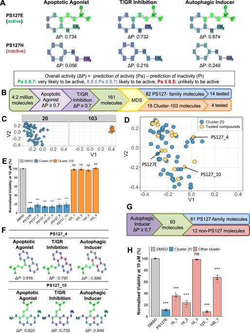
Previous research in our lab identified PS127 as a novel activator of mitophagy that selectively killed AML cells [21]. A small-scale structural similarity search and structure-activity relationship analysis identified ~15 analogs of PS127, 12 of which showed cytotoxicity against AML cells in vitro [12]. To identify potential mechanisms of action for these molecules, we employed the cheminformatic tool Prediction of Activity Spectra for Substances (PASS) [25]. PASS uses a training set of drug-like compounds with known biological activities to predict the activities of query molecules. PASS analysis predicted high probability for two activities, apoptotic agonism and thioredoxin reductase/glutathione reductase (T/GR) inhibition, for all 12 active PS127-series molecules [12]. Analogs inactive in vitro (such as PS127H) had low Pa (probability of activity) scores for these activities (Fig. 1A). These results suggested that one or both of these activities may be involved in selective, PS127-family-mediated killing of AML cells.
Fig. 1: In silico screening and hit validation.
A Representative Prediction of Activity Spectra for Substances (PASS) outputs of active and inactive PS127 molecules on relevant biological activities. B The in silico screening pipeline to uncover additional active novel small molecules. C The top two clusters from multidimensional scaling (MDS) analysis. D Scatter plot of cluster 20 (PS127-family), showing all PS127 molecules (blue) and tested compounds (yellow). Three molecules with the strongest cytotoxic activity are labelled. E Cytotoxicity of representative molecules from cluster 20 (n = 5) and cluster 103 (n = 5) at 10 μM on MOLM-13. F PASS results of active PS127-family compounds that helped identify key third predicted activity: autophagic induction. G Flow chart of a downstream in silico screening with the third PASS-predicted activity, an autophagy inducer. H Cytotoxicity comparison of compounds with three PASS-predicted activities. Representative compounds from the non-PS127-family (n = 5) were tested at 10 μM in MOLM-13. Bar graphs display the average of biological replicates, each of which is represented as data points. Error bars indicate the standard error of the mean (SEM). One-way ANOVAs followed by Dunnett’s post hoc tests were used to assess statistical significance. T/GR = Thioredoxin/Glutathione reductase. *** – p < 0.001; ns: not significant (p > 0.05).
To identify additional molecules with these predicted functions, an in silico screen of over 4.2 million compounds was performed. To increase stringency and reduce the number of false positive hits, the screening criteria were refined. Compounds were considered hits only if ΔP (the difference between Pa and Pi, Probability of inactivity) was at least 0.7 for each of the desired activities (Fig. 1B). This screen yielded 161 potential hits. Using structural similarity analysis and multidimensional scaling, the molecules were grouped into 41 clusters (Supplementary Fig. 1). The majority of these compounds were within two clusters, referred to herein as cluster 20 and cluster 103 (Fig. 1C).
Cluster 20 contained 82 compounds, including multiple previously-identified PS127-family hits, including PS127E and PS127B. This cluster is hereafter referred to as the PS127-family. A subset of five compounds from this cluster was selected and tested as representatives of the broad chemical space of the PS127 family (Fig. 1D). Compound cytotoxicity was tested in MOLM-13 cells at 10 µM for 72 h (Fig. 1E). All PS127-family compounds tested showed high cytotoxicity (Table 1 and Supplementary Table 3).
Table 1 CC50 value of PS127-family compound in MOLM-13, PBMCs, and H9C2.
Cluster 103 consisted of 18 molecules, four of which were selected and tested as above (Fig. 1E). Despite being predicted to have apoptotic agonism and T/GR inhibition, none of the compounds exhibited activity in vitro. Tanimoto-based structural analysis indicated that all compounds in this cluster had substantial structural similarity to each other (Tanimoto coefficient ≥0.75), whereas compounds from the PS127-family cluster formed distinct sub-clusters (Supplementary Fig. 2 and Supplementary Table 4). For example, PS127_10 and PS127E were more structurally similar to each other (Tanimoto coefficient 0.833) than they were to PS127_4 or PS127_6, which were more closely related to each other (Tanimoto coefficient 0.758).
To refine our understanding of the functional differences between these clusters of compounds, PASS software was used to analyze each cluster. This process revealed several additional activities present in active PS127-family molecules that were absent from inactive compounds (Fig. 1A, F). The most intriguing of these was autophagic induction, a feature we have previously linked to mitochondrial health and AML drug sensitivity [12, 26]. We hypothesized that this third activity was required for cytotoxicity and may differentiate active and inactive compounds in our group of 161 hits. This criterion excluded all the compounds in cluster 103 and reduced the number of hits to 93 (Fig. 1G). Of these 93 molecules, 81 belonged to the PS127-family cluster, while the remaining 12 were structurally distinct from this group. Our observation that this approach removed all of Cluster 103 and narrowed our pool of hits suggested that the three activities were most directly responsible for the observed cytotoxicity. To test this hypothesis, five of the 12 non-PS127-family compounds were selected and tested for cytotoxicity in MOLM-13 cells. Four of these five significantly reduced AML cell viability (Fig. 1H). Three compounds, 125_1, 16_2, and 148_1, were structurally unrelated to each other and the PS127-family (Supplementary Fig. 2), but were cytotoxic to AML cells (Fig. 1H and Supplementary Table 5), suggesting that these compounds represent a valuable, mechanism-based toolkit for predicting cytotoxicity. These findings also provided proof of principle for the utility of using cheminformatic screening to identify compounds with desired molecular activities.
PS127 family has selective cytotoxicity against AML cell lines
After initial cytotoxicity screening indicated that PS127-family molecules were likely to be active, 14 molecules were chosen to represent the chemical space of the PS127 family. To evaluate potency, dose-response curves were generated to determine the concentration that would induce 50% cytotoxicity (CC50) in MOLM-13 cells. Twelve of the 14 PS127-family molecules displayed CC50 under 8 µM, with four molecules (PS127_4, PS127_9, PS127_10, and PS127E) displaying sub-micromolar CC50 values (Table 1 and Supplementary Fig. 3).
To evaluate whether this cytotoxicity was specific to leukemic cells, CC50 values were also determined for healthy donor peripheral blood mononuclear cells (PBMCs). Each compound showed at least two-fold higher CC50 values in PBMCs compared to MOLM-13. Notably, CC50 values of lead PS127-family compounds in PBMCs were at least 12- and 8-fold higher than in MOLM-13 and MV4;11, respectively, indicating consistent compound potency across AML cell lines harboring FLT3-ITD mutations.
Previous research indicated that PS127 (the founding member of the compound family) likely functions by stimulating autophagic recycling of mitochondria [12]. We considered the possibility that these compounds may exhibit greater toxicity to cells with increased mitochondrial dependence, such as cardiomyocytes [27]. To test this, CC50 values were also calculated for H9C2 cardiomyocytes for PS127-family compounds with sub-micromolar CC50 values in MOLM-13 cells. Observed CC50 values were ~5 to 19-fold higher for cardiomyocytes than AML cells, suggesting that cytotoxicity was specific to leukemic cells due to their unique metabolic adaptations (Table 1).
These compounds were also evaluated in a panel of other AML cell lines, including MOLM-14, MV4;11, NB4, and OCI-AML2 (Table 2), which have a variety of genetic lesions frequently found in AML patients. Each of the prioritized PS127-molecules was active against other AML cell lines, a result crucial for targeting the heterogeneous nature of AML. Notably, PS127E and PS127_10 displayed similar activity in large-scale dose response analysis in MOLM-13 and MV4;11 and much lower toxicity to PBMCs (Supplementary Fig. 3). Based on their strong activity in AML cell lines, PS127_4, PS127_10, and PS127E were selected for further evaluation.
Table 2 CC50 value of prioritized PS127-family compounds in other AML cell lines.
PS127-family compounds are synergistic with current commercial chemotherapeutics
Due to the prevalence of drug resistance in AML, most modern chemotherapeutics are administered in combinations. The synergy of our top compounds (greater-than-additive effect) was tested in combination with commonly-used AML chemotherapeutics [28], including the first-line treatments doxorubicin (DOX), cytarabine (ara-C), venetoclax (VEN) [29,30,31], and midostaurin (MID), a targeted therapy used in AML patients with FLT3 mutations [32, 33]. Synergy was evaluated by assessing cell viability after single and combinatorial treatments using a Bliss independence model, with a score above 10 considered to indicate synergy [28]. The prioritized candidates, PS127_4, PS127_10, and PS127E, showed some synergy with DOX and ara-C but had the strongest synergy with MID in MOLM-13 (Fig. 2A–D and Supplementary Fig. 4).
Fig. 2: Synergistic effect of PS127-compounds and commercial chemotherapeutics.
A Average maximum synergy score ( ± standard error of mean [SEM]) of PS127-family compounds and commercial chemotherapeutics: midostaurin (MID), doxorubicin (DOX), and ara-C (n = 3). B Representative synergy plot of PS127_4 and MID in MOLM-13 cells (left) or peripheral blood mononuclear cells (PBMCs) (right). C Representative synergy plot of PS127_4 and DOX in MOLM-13 cells (left) or PBMCs (right). D Average maximum synergy score ( ± SEM) of PS127-family compounds and various FLT3 inhibitors (MID, quizartinib [QUIZ], sorafenib [SORA], gilteritinib [GIL]) (n = 3). E Comparison of MV4;11 and PBMCs viability upon monotherapy or combined treatments with PS127_4 and MID. All bar graphs represent mean ± SEM from three biological replicates (n = 3). Black asterisks indicate comparison of AML cells vs. healthy PBMCs under the same combinatorial treatment conditions. Pink asterisks indicate significantly lower survival under combinatorial treatment compared to single PS127-family compound treatment; Blue asterisks indicate significantly lower survival under combinatorial treatment compared to a single MID treatment. A black dot represents the maximum synergy point in leukemic cells and its corresponding condition in PBMCs. Statistical significance was assessed via Student’s t-test. F Representative synergy plot of PS127_4 with MID in MV4;11 (left) and PBMCs (right). G Representative synergy plot of PS127_4 with QUIZ in MV4;11 (left) and PBMCs (right). H Average maximum synergy score ( ± SEM) of PS127-family compounds with venetoclax (VEN) in AML cells (n = 3). I Representative synergy plot of PS127_4 and VEN in MOLM-13 cells (left) or PBMCs (right). *** – p < 0.001; ns: not significant (p > 0.05).
We also evaluated the synergistic potential of these compounds in healthy PBMCs under the same conditions. In these cells, no synergistic activity was observed (Fig. 2B, C and Supplementary Fig. 4). We found that the survival in the presence of drug combinations resulting in maximum synergy was significantly lower in AML cells than in PBMCs, with survival in the latter remaining >80% for these tested combinations (Fig. 2E and Supplementary Fig. 5). This reinforced that drug combination cytotoxicity was specific to leukemic cells and is unlikely to have overt side effects on healthy cells.
Since PS127-family compounds showed the greatest synergy with MID, we evaluated these combinations in two additional AML cell lines: MV4;11 and OCI-AML2. MV4;11, like MOLM-13, carries an FLT3-ITD mutation, while OCI-AML2 lacks known FLT3 mutations and maintains detectable amounts of FLT3 [34]. As anticipated, top PS127-compounds showed significant synergy with MID in MV4;11 (Fig. 2F and Supplementary Fig. 6). The combination of PS127_4 and MID produced the strongest synergy, with the average maximum Bliss score being above 60 (Fig. 2D, F). This combination did not significantly affect PBMC cell viability (Fig. 2E). The difference in combined PS127_4 and MID treatment survival between MV4;11 and PBMCs was approximately 7-fold (13% vs 95%). Notably, prioritized PS127-compounds displayed a range of synergy with MID in OCI-AML2, despite the lack of FLT3 mutation in this cell line (Fig. 2D and Supplementary Fig. 6C–E). MID is also known to target S6 phosphorylation and sphingosine kinase-1, which may contribute to its interaction with PS127-family compounds in OCI-AML2 cells [34, 35]. Importantly, while PS127E had comparable synergy with MID in MV4;11 and OCI-AML2, synergy scores for PS127_4 and PS127_10 were approximately two-fold lower in the latter cell line, indicating complex relationships between compound effects and cancer genotype.
Due to the strong synergy with MID, we evaluated the synergistic potential of PS127E, PS127_4, and PS127_10 with other FLT3 inhibitors, quizartinib (QUIZ), sorafenib (SORA), and gilteritinib (GIL). We found that these compounds had strong synergy with QUIZ and SORA (Fig. 2D, G and Supplementary Fig. 7A–H) and ranged from moderate to strong synergy with GIL (Fig. 2D and Supplementary Fig. 7I–L) in both AML cell lines. These data indicate that our PS127-family compounds will likely be active in AML patient cells, regardless of whether they carry wild-type or mutant FLT3 alleles.
Furthermore, we evaluated the synergistic potential of lead compounds in combination with venetoclax (VEN), an established standard-of-care agent for newly-diagnosed and refractory/relapsed AML [36]. All leads demonstrated strong synergy in MOLM-13 cells (Fig. 2H, I and Supplementary Fig. 8). Importantly, PS127_4 showed the strongest synergy with VEN in leukemic cells while remaining antagonistic in PBMCs (Fig. 2I). Similar synergistic activity was observed in FLT3-ITD MV4;11 cells. These data are consistent with a dual-action mechanism wherein PS127-mediated cellular stress sensitizes cells to VEN-induced apoptosis, highlighting the potential for these combinations in broader clinical applications.
In vitro validation of PASS activity predictions
To elucidate the mechanisms used by the prioritized compounds to selectively induce leukemic cell death, we first verified the PASS-predicted activities used for screening: apoptotic agonism, T/GR inhibition, and autophagic induction.
To evaluate compounds’ ability to induce apoptosis, flow cytometry experiments were performed using annexin V-FITC/ Hoechst/PI staining. Flow cytometry analysis demonstrated that 24-h treatment with PS127E, PS127_4, or PS127_10 significantly induced apoptosis in leukemic cells, as indicated by annexin V-positive staining (Fig. 3A, B), suggesting that apoptosis is a primary mechanism of cell death.
Fig. 3: Validating predicted activities of PS127-treatment in MOLM-13.
A Representative flow cytometry analysis of apoptotic cells induced by PS127E (8 μM, blue) or DMSO (red) for 24 h. Apoptotic assay was determined using flow cytometry with Annexin V-FITC/Propidium Iodide (PI)/Hoechst staining. B Quantification of cell populations upon PS127-molecule treatment compared to DMSO following annexin V-FITC/PI staining (n = 4). Apoptotic cells were detected by annexin V-FITC+/PI- and annexin V-FITC+/PI+, while non-apoptotic dead cells were indicated by annexin V-FITC-/PI+ and live cells were annexin V-FITC-/PI-. Bar graphs show the average percentage of cell population, and error bars display the standard error of the mean (SEM). Significance levels represent the comparison of PS127-induced apoptotic cells to DMSO. C Average antagonist score ( ± SEM) of PS127-molecules with various autophagic inhibitors (n = 3). D Representative antagonism landscape of PS127E and wortmannin (left) or chloroquine (CQ) (right) treatment. E The effect of PS127-molecules on thioredoxin reductase (TrxR) activity (n = 3). F Changes of reduced glutathione (GSH) to oxidized glutathione (GSSG) ratio upon treatment of PS127-molecules (n = 4). G Quantification of glutathione reductase (GR) enzymatic activity upon PS127-molecule treatment in MOLM-13 and peripheral blood mononuclear cells (PBMCs). H Fold change (FC) of dead transfected MOLM-13 cells after GSR knockdown relative to scrambled silencing RNA (siRNA). Biological replicates are shown as data points. One-way ANOVAs followed by Dunnett’s post hoc tests were used to assess statistical significance. * – p < 0.05; ** – p < 0.01; *** – p < 0.001; ns: not significant (p > 0.05).
Next, the dependence of PS127-family compound activity on autophagic stimulation was evaluated. MOLM-13 cells were treated for 24 h with PS127-family molecules and either early-stage (wortmannin and 3-methyladenine [3-MA]) or late-stage (chloroquine [CQ] and hydroxychloroquine [HCQ]) inhibitors of autophagy [37,38,39]. The addition of autophagic inhibitors to PS127-family-treated cells resulted in strong antagonism, suggesting that functional autophagy was required for PS127-induced cytotoxicity (Fig. 3C, D and Supplementary Fig. 9).
Finally, we evaluated the predicted inhibitory effect of PS127-family molecules on enzymes responsible for redox metabolism, specifically TrxR or GR. None of the prioritized PS127-family compounds had a significant effect on the activity of TrxR in MOLM-13 (Fig. 3E), suggesting that none of them interacted with TrxR.
In contrast, compound-mediated effects on glutathione metabolism were observed. The GSH pool maintains ROS homeostasis in the cell and reduces oxidative stress [40, 41]. GSH is oxidized to GSSG, which is then reduced back to GSH by GR, restoring its potential to serve as an antioxidant. The steady-state ratio of GSH to GSSG represents the current oxidative burden of the cells, but can also serve as a readout of GR activity. Cells treated with PS127-family compounds exhibited significantly decreased GSH-to-GSSG ratios (Fig. 3F), suggesting that the compounds may be disrupting the function of the enzyme.
To test the specificity of this activity in vitro, MOLM-13 or PBMC cells were treated with PS127-family compounds for 24 h and then lysed. GR activity in cell-free lysates was quantified and normalized to total protein concentration, in accordance with the GR activity assay. Lead compounds significantly inhibited GR activity in leukemic cells, but not in PBMCs (Fig. 3G), likely reflecting increased basal GR activity in leukemic cells. By inhibiting GR activity, lead compounds prevented recycling GSSG to GSH, thereby increasing oxidative stress and cytotoxicity.
To confirm the importance of GR for survival in MOLM-13 cells, siRNAs were used to knock down GSR or GAPDH (the latter served as a positive control). Disruption of GSR resulted in considerable cytotoxicity in MOLM-13 cells compared to scrambled siRNA (Fig. 3H). This corroborates our observations with chemical inhibition and reinforces the conclusion that GR activity is critical for the survival of MOLM-13 cells. Unfortunately, a limited number of viable cells were obtained after GSR knockdown, which precluded the determination of CC50 values in GSR knockdown cells.
PS127-compounds induce redox-dependent cytotoxicity by interfering with glutathione metabolism
To further validate the mechanism of compound activity on redox metabolism, PS127E-treated cells were supplemented with exogenous GSH, GSSG, or the GSH precursor, N-acetyl-cysteine (NAC). GSH and NAC completely rescued the viability of PS127E-treated cells (Fig. 4A). In contrast, supplementation with GSSG did not provide any rescue. As neither GSH nor GSSG is toxic to MOLM-13 (Supplementary Fig. 10A), we hypothesized that this difference is directly related to their distinct functions in redox metabolism.
Fig. 4: The effect of PS127-induced cytotoxicity on redox metabolism and reactive oxygen species (ROS) levels.
A Redox metabolite-mediated rescue of cell viability upon PS127E treatment (n = 3) for 72 h. B A representative histogram of DHE+ population in the presence of redox metabolites. C Representative histogram of MitoSOX Red+ population in the presence of redox metabolites. Peaks with dashed lines indicate control treatment, while solid lines represent treatment with PS127-molecule. D Total ROS quantification using median fluorescence intensity (MFI) of DHE in the presence of redox metabolites in the presence and absence of PS127E (n = 3). E Mitochondrial ROS quantification using MFI of MitoSOX Red in the presence of redox metabolites and presence and absence of PS127E (n = 3). ROS level was indicated by the MFI of the ROS staining dye. F Melting temperature shift of human glutathione reductase (GR) in the presence of PS127-family compounds. PS127_4 (n = 4), PS127_9 (n = 3), PS127_10 (n = 3), and 2-AAPA (n = 3) significantly changed the melting temperature of GR compared to DMSO (n = 7), indicating compound-enzyme binding. PS127H (n = 3) did not change the melting temperature. PS127H = previously identified inactive PS127 analog, 2-AAPA = GR inhibitor control. G Raw fluorescence of Sypro Orange for technical replicates of the differential scanning fluorometry (DSF) to determine dissociation constant (Kd) between PS127_10 and GR (left). Melting temperature of GR with PS127_10 at 0, 2.5, 5, 10, 20, and 40 µM (right). One-way ANOVAs followed by Dunnett’s post hoc tests were used to assess statistical significance. GSH = reduced glutathione, GSSG = oxidized glutathione, NAC = N-acetyl cysteine, a.u. = arbitrary unit. ** – p < 0.01; *** – p < 0.001; ns: not significant (p > 0.05).
To verify that the increase in viability conferred by GSH or NAC was not simply a consequence of providing antioxidants to MOLM-13 cells, MOLM-13 cells were treated with PS127-family compounds and then supplemented with other known antioxidants that act independently of GR. Reduced ascorbate, which does not affect glutathione peroxidase or GR activity [40, 42], was tested at the highest concentration that could be used without causing significant cytotoxicity on its own (Supplementary Fig. 10C). Even at this concentration, the ascorbate did not significantly affect cell death (Fig. 4A). Supplementation with α-tocopherol, another antioxidant [43, 44], also failed to significantly improve survival of PS127E-treated cells (Supplementary Fig. 10B and Supplementary Fig. 11A, B). The combination of ascorbate and α-tocopherol generates enhanced ROS scavenging activity [45]. Interestingly, this combination still did not rescue cells from PS127-induced cytotoxicity (Supplementary Fig. 11B). Likewise, the cytotoxicity of PS127_4 and PS127_10 was inhibited by GSH but remained unaffected by GSSG or α-tocopherol, ascorbate, or their combination (Supplementary Fig. 11A, B). These results reinforced the likelihood that PS127-family molecules specifically compromise the glutathione pathway, likely by preventing GR from reducing GSSG, and that this effect cannot be compensated for by other antioxidative pathways.
GSH serves as a scavenger for intracellular and mitochondrial ROS, its depletion would be expected to increase both total and mitochondrial ROS levels [46]. To examine the relationship between ROS levels and cytotoxicity, total and mitochondrial ROS were measured in cells treated with PS127-family molecules using dihydroethidium (DHE) and MitoSOX Red staining, respectively. PS127-family compound treatment increased both total and mitochondrial ROS levels (Fig. 4B–E and Supplementary Fig. 11C–H). Consistent with the viability rescue data, only supplementation with GSH, but not with GSSG or α-tocopherol, restored total and mitochondrial ROS levels in PS127-family-treated cells to approximately basal levels.
PS127-family compounds interfere with glutathione reductase
This evidence strongly suggests that GR is the target of the PS127-family compounds. To directly prove physical interaction between the compounds and GR, we used differential scanning fluorimetry (DSF). This technique measures the melting temperature of a target protein in the presence or absence of small molecules that may interact with the protein [47]. Changes to the melting temperature of the protein indicate stabilization of the protein, likely due to interaction between the enzyme and the small molecule.
Incubation of GR with active PS127-family compounds (PS127_4, PS127_10, or PS127E) significantly increased its melting temperature, suggesting that the compounds interact with the enzyme (Fig. 4F). In contrast, PS127H, an inactive PS127-like compound, did not significantly alter the melting temperature of GR, indicating that the lack of activity is likely due to a lack of interaction between GR and PS127H. Consistent with these findings, 2-AAPA, a known inhibitor of GR [48], produced a comparable increase in melting temperature.
To further verify this interaction, a thermal shift binding assay was performed between PS127_10 and purified GR, which demonstrated a concentration-dependent increase in melting temperature (Fig. 4G). Using these data, a binding constant (Kd) for the experimental conditions was calculated to be 0.204 µM [95% CI: 0.160–0.247 µM], indicating high-affinity binding of PS127_10 to GR under the experimental conditions used (2.5 µM GR). These data indicate that active PS127-family molecules bind to GR, inhibiting GSSG reduction, resulting in depletion of cellular GSH pools.
A non-PS127-family compound, 125_1, shares PS127-family compounds’ properties
As described above, the PS127 hits tested were predicted to, and were confirmed to, have a high probability of apoptotic agonism, T/GR inhibition, and autophagic induction. These effects are likely to have driven the observed cytotoxicity to MOLM cells. The screen that identified several of these hits was structure-agnostic, so other compounds with all three activities may also be cytotoxic.
To test this prediction, we procured compound 125_1, which is structurally unrelated to the PS127-family (Tanimoto coefficient < 0.7, Supplementary Table 4) but was predicted to have all three activities. Compound 125_1 exhibited a low CC50, 0.41 ± 0.04 µM in MOLM-13, which was over 30-fold lower than its CC50 for PBMCs (15.68 ± 0.75 µM) or cardiomyocytes ( > 16 µM). Like PS127-family compounds, 125_1 induced apoptotic cell death, was antagonistic with 3-MA, and its cytotoxicity was inhibited by GSH, but not by GSSG or α-tocopherol (Fig. 5A–C). Compound 125_1 also increased total and mitochondrial ROS levels, an effect attenuated by GSH (Fig. 5D–G). Finally, compound 125_1 reduced GR activity (Fig. 5H), likely by directly binding to GR, as shown by the protein’s increased melting temperature in thermal shift assay (Fig. 5I). Compound 125_1 also showed synergy with MID in MOLM-13, like PS127-family molecules (Fig. 5J). Overall, these data indicate that 125_1 and the PS127 family share the same mechanistic properties, despite structural dissimilarity.
Fig. 5: Evaluation of Prediction of Activity Spectra for Substances (PASS)-predicted activities for 125_1 and the impact of PS127-family compounds and 125_1 on mitochondrial bioenergetics.
A Quantification of apoptotic cells upon 125_1 treatment in MOLM-13, indicated by annexin V/Propidium Iodide staining (n = 4). B Representative antagonistic interactions between 3-methyladenine (3-MA) and 125_1. C Cytotoxicity upon 125_1 treatment was rescued by reduced glutathione (GSH) (n = 8). D DHE+ population representative histogram upon 125_1 treatment in the presence or absence of redox metabolites. E) MitoSOX Red+ population representative histogram upon 125_1 treatment in the presence or in the absence of redox metabolites. Peaks with dashed lines indicate control treatment or the absence of PS127-molecule, while solid lines represent treatment with PS127-molecule. F Quantification of total ROS in the presence of redox metabolites (n = 3) based on median fluorescence intensity (MFI) of DHE. G Quantification of MitoSOX Red MFI in the presence of redox metabolites (n = 3). H Quantification of glutathione reductase (GR) activity in MOLM-13 treated with 125_1 (n = 3). I Melting temperature shift of human GR upon 125_1 treatment in MOLM-13 (n = 5). J Representative synergy plot of compound 125_1 and midostaurin (MID) in MOLM-13 (left) and peripheral blood mononuclear cells (PBMCs) (right). K Oxygen consumption rate (OCR) measurement of MOLM-13 upon 3-h compound treatment. PS127E (n = 4), PS127_4 (n = 5), PS127_10 (n = 4), or 125_1 (n = 3) impaired the routine OCR compared to DMSO (n = 7). OCR was normalized to the number of live million cells per chamber volume. L The steady state adenosine triphosphate (ATP) level in MOLM-13 after 3-h exposure to the compound (n = 7). M The steady state ATP level in PBMCs after 3-h exposure to the compound (n = 3). All AML cells and PBMCs had > 90% viability before cell lysis occurred to measure total ATP. One-way ANOVAs followed by Dunnett’s post hoc tests were used to assess the statistical significance of apoptotic cells, ROS and ATP levels, and oxygen consumption. Significance level of differential scanning fluorometry assay was determined using Student’s t-test. a.u. = arbitrary unit, RLU = Relative Luminescence Unit. ** – p < 0.01; *** – p < 0.001; ns: not significant (p > 0.05).
Prioritized PS127-family compounds interfere with mitochondrial bioenergetics
As noted above, PS127-family compounds compromised redox homeostasis and increased mitochondrial ROS, suggesting that the compounds may be affecting mitochondrial function, particularly respiration. AML cells, especially LSCs, have low coupling efficiency and therefore rely heavily on OXPHOS to support their increased demand for ATP [12, 49, 50]. We previously reported that other compounds that impair respiration and ATP production exhibit selective cytotoxicity [12].
To test whether PS127-family compounds similarly impaired mitochondrial respiration, oxygen consumption was measured in MOLM-13 cells that were exposed to PS127_4, PS127_10, PS127E, or 125_1 for 3 h. Each of the compounds impaired routine respiration, as shown by reduced OCR (Fig. 5K). Consistent with previous results [12], all lead compounds significantly reduced ATP production in MOLM-13, but not in PBMCs (Fig. 5L, M).
PS127-family activity in AML patient samples
Patient-derived samples were used to validate PS127-family compound activity in a more clinical context. In a panel of wild-type and FLT3-mutant samples, we evaluated the ability of compounds to induce cytotoxicity, synergy between PS127_10 and MID or DOX, oxygen consumption rate, and rescue by exogenous GSH. Due to limited cell numbers in patient-derived samples, limited experiments could be performed.
In line with the heterogeneity of AML in patients, the OCR of the primary samples ranged from ~5 to ~30 pmol/s per million viable cells; MOLM-13 and PBMC OCR values were~18 and ~3 pmol/s per million viable cells, respectively (Fig. 6A, F). Additionally, GSH supplementation rescued AML patient cells treated with PS127E or compound 125_1, regardless of FLT3 mutation status (Fig. 6B, C). A range of effects, from additivity to strong synergy, were seen in various patient-derived cells when PS127_10 was combined with MID or DOX (Fig. 6D–F). FLT3 mutation status did not appear to correlate with the degree of synergy.
Fig. 6: Screen hits are effective in AML patient samples.
A Oxygen consumption rate (OCR) of FLT3-mutated and wild-type AML patient cell samples, peripheral blood mononuclear cells (PBMCs), and MOLM-13 in the 0.5 mL chamber. OCR was normalized to the number of live million cells per mL [pmol·s–1 per million cells (Mx–1)]. Error bars represent standard error of mean (SEM) from two technical replicates. Bars without error bars represent one replicate due to limited sample size. B Viability rescues of PS127E- or 125_1-treated wild-type AML patient cells (n = 10) by 500 µM reduced glutathione (GSH) supplementation. C Viability rescues of PS127E- and 125_1-treated FLT3-mutated patient cells (n = 7) by 500 µM GSH supplementation. D Representative synergy plot of FLT3-mutated (AML-1) (left), wild-type FLT3 (AML-16) (middle), and PBMCs (right) of PS127_10 and midostraurin (MID). E Representative synergy plot of FLT3-mutated (AML-1) (left), wild-type FLT3 (AML-17) (middle), and PBMCs (right) of PS127_10 and doxorubicin (DOX). Blue and black dots represent the maximum FLT3-mutated synergy point and its corresponding condition in PBMCs. Black dots correspond to maximum wild-type FLT3 synergy point and its corresponding PBMC condition. F Summary table of OCR and max synergy value with PS127_10 and MID or DOX in AML patient cells. ND = not determined. G The effect of 500 µM GSH supplementation on CD34+ cells treated with lead compounds (n = 3). H Representative synergy plot of PS127_10 and MID in CD34+ cells. I Hypothesized mechanistic model of active PS127-family compounds inhibiting GR, causing an increase in ROS, leading to lethal mitophagy. Leukemic metabolism without (left) and with (right) PS127-compound treatment is shown. ATP = adenosine triphosphate, OXPHOS = oxidative phosphorylation, GSH = reduced glutathione, GSSG = oxidized glutathione, NAC = N-acetylcysteine, Gln = glutamine, Glu = glutamate, Cys = cysteine, CAC = citric acid cycle, α-KG = α-Ketoglutarate, GPX = glutathione peroxidase. *** – p < 0.001; ns: not significant (p > 0.05).
Activity of lead compounds in human bone marrow-derived CD34+ progenitors
To further evaluate the therapeutic potential of these compounds, lead compounds were tested on human bone marrow-derived CD34+ cells, which are related to the CD34+CD38- population that gives rise to LSCs [51,52,53], enabling evaluation of potential off-target effects on healthy hematopoietic progenitors. PS127E and PS127_10 exhibited significantly higher median CC50 values, 14.73 and 39.15 μM, respectively, than were seen for AML cells. Unlike MOLM-13 cells, GSH supplementation had a negligible effect on the viability of the CD34+ HSCs (Fig. 6G), likely due to the low cytotoxic effect of the compound, indicating that the redox status of these cells has not been compromised. PS127_10 showed a moderate synergistic effect with MID and no synergy with DOX in CD34+ progenitor cells (Fig. 6H), markedly weaker than the synergistic interaction observed in MOLM-13 cells, suggesting that healthy cells lack the specific redox and signalling susceptibilities that are exploited in drug cooperation and further supporting the conclusion that treatment with these compounds will specifically target cancerous cells with minimal impact on HSCs.
