


Aged mice show increased breast cancer metastasis
To examine the impact of aging on breast tumor growth and metastasis, syngeneic murine breast cancer cell lines were orthotopically implanted into young 3-month-old C57BL/6J mice, age analogous to a young 20-year-old patient, and into 21-month-old hosts, comparable in age to humans aged 65. Two TNBC lines from the MMTV-PyMT mouse model (Py8119 and AT3)36,37 and a lung-trophic, metastatic subline of the luminal B E0771 cell line (E0771-LMB)38,39 were each injected into groups of young and old mice (n = 5 per group).
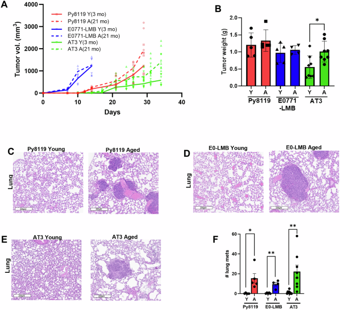
Primary tumor incidence and latency in young and old hosts were similar in all 3 mouse models assayed Fig. 1A). AT3 breast cancers grew significantly faster in aged versus young mice, (Fig. 1A, p < 0.0001) and reached greater sizes (Fig. 1B, p < 0.05); Py8119 cancers showed modestly increased growth (Fig. 1A, p < 0.05); and E0771-LMB cancer growth rates and final tumor weights were similar in young and aged hosts (Fig. 1A, B). Histological analysis of primary breast cancers is shown in Supplementary Fig. 1. Cancer metastasis to the lungs was consistently higher in aged compared with young hosts across all breast cancer models examined (Fig. 1C–F). Py8119 tumors showed a significant increase in metastatic burden in aged mice (p < 0.05), with similar effects observed for E0771-LMB (p < 0.01) and AT3 tumors (p < 0.01) (Fig. 1C–F). As expected, none of the non-tumor-bearing mice (young or aged) developed lung metastasis (Supplementary Fig. 2).
Fig. 1: Aged mouse models of breast cancer show increased metastasis.The alternative text for this image may have been generated using AI.
A Py8119, E0771-LMB, and AT3 cells were injected into the MFP of young (3 months) and aged (21 months) female C57BL/6J mice. Tumor size was measured every 3 days, and B tumors were weighed at the end of the experiment. Data are from 5 to 8 mice per group. H&E analysis on lung metastasis from C Py8119 young and aged mice, D E0771-LMB young and aged mice and E AT3 young and aged mice. F Number of lung metastasis observed by H&E analysis in young (Y) and aged (A) mice for Py8119, E0771-LMB, and AT3 cells. Values shown are mean ± SEM. Statistical analysis: one or two-way ANOVA with Dunnett’s multiple comparisons test. *p < 0.05; **p < 0.01; and ****p < 0.0001.
Breast cancer metastasis in aged mice is impaired by host RAGE knockout
RAGE and its ligands promote breast cancer metastasis through tumor intrinsic and host inflammatory mechanisms31,32,35. We tested if host RAGE activation drives aging-promoted metastasis by implanting Py8119, AT3, and E0771-LMB tumors into young (Y) and aged (A) wild-type (RAGE+/+) and RAGE knockout (RAGE−/−) C57BL/6J mice. Primary orthotopic Py8119 tumors exhibited faster tumor growth in aged RAGE+/+ compared to young RAGE+/+ mice (Fig. 2A and Supplementary Fig. 3A). This effect was RAGE-dependent, as Py8119 mammary cancers in aged RAGE−/− mice did not show growth advantage compared to that in both young RAGE–/– and young RAGE+/+ mice (Fig. 2A and Supplementary Fig. 3B–H). Similar results were seen with the AT3 model, with faster tumor growth limited to aged RAGE+/+ hosts and comparable growth rates across all young mice regardless of genotype (Fig. 2B and Supplementary Fig. 4A–H). Primary tumor growth in E0771-LMB injected mice was similar in aged RAGE+/+ and aged RAGE−/− hosts (Supplementary Fig. 4I). However, in aged hosts, RAGE knockout almost completely eliminated the greater lung metastasis of PY8119 cells seen in aged RAGE+/+ mice (Fig. 2C, D) compared to young RAGE+/+ mice. Lung metastases from primary PY8119 cancers were not observed in young mice, regardless of RAGE+/+ or RAGE−/− host genotype (Fig. 2C, D). Similarly, aged RAGE−/− mice bearing AT3 and E0771-LMB tumors demonstrated significantly fewer lung metastatic lesions than their aged RAGE+/+ counterparts (Fig. 2E–H).
Fig. 2: Host RAGE signaling is a drives aging-associated breast cancer metastasis.The alternative text for this image may have been generated using AI.
Tumor growth curves for Py8119 (A) and AT3 (B) cells implanted orthotopically into the mammary fat pad of young (3-month-old) or aged (21-month-old) female C57BL/6J RAGE⁺/⁺ or RAGE⁻/⁻ mice (n = 9–12 per group). Representative H&E-stained lung sections and quantification of spontaneous lung metastases are shown for Py8119 (C, D), AT3 (E, F), and E0771-LMB (G, H) tumor-bearing mice from young and aged RAGE⁺/⁺ or RAGE⁻/⁻ hosts, with metastatic burden quantified as the total number of metastatic lesions per lung. I, J Show experimental metastasis following tail vein injection. Py8119 cells were injected intravenously via the tail vein into young (3-month-old) or aged (21-month-old) RAGE⁺/⁺ and RAGE⁻/⁻ mice, and metastasis was quantified as the percent metastatic area of the lung (n = 6–9 per group). Values represent mean ± SEM. Statistical analysis was performed using one- or two-way ANOVA with Dunnett’s multiple comparisons test. *p < 0.05; **p < 0.01; and ****p < 0.0001.
RAGE promotes all stages of metastasis, whereas aging acts mainly at early metastatic stages
Experimental tail vein metastasis models bypass the early steps of local invasion and intravasation and directly assess the ability of tumor cells to survive in circulation, extravasate, and colonize distant organs40. To determine which stages of the metastatic process are influenced by aging and RAGE signaling, Py8119 tumor cells were intravenously injected into young and aged RAGE+/+ and RAGE−/− C57BL/6J mice. Quantification of lung metastases (Fig. 2I, J) showed no significant difference in metastatic burden between aged RAGE+/+ mice and young RAGE+/+ mice. In contrast, aged RAGE−/− mice showed significantly fewer lung metastases than either young (p < 0.05) or aged (p < 0.01) RAGE+/+ hosts (Fig. 2I, J). Based on metastatic patterns in the orthotopic and tail vein models, these data indicate that aging enhances metastasis at early stages of dissemination, whereas RAGE is required for both early- and late-stage metastasis.
S100A8/9 and AGEs accumulate in the tumor microenvironment and metastatic niche of aged hosts
Effects of age on RAGE ligand expression were next evaluated in orthotopic Py8119 tumors from young and aged RAGE+/+ and RAGE–/– C57BL/6J mice, focusing on RAGE-ligands S100A8/9 and AGEs. Histological analysis and western blot showed higher S100A8 levels in cancers of aged compared to young wild-type mice (Fig. 3A–C and Supplementary Fig. 5), and this aging-related increase was attenuated in cancers of aged RAGE−/− mice (Fig. 3A–C). Similarly, tumor AGE levels showed a similar pattern, with greater expression in aged than young RAGE+/+ mice and decreased in aged RAGE−/− mice (Fig. 3D). Immunofluorescence analysis of lung sections from orthotopically Py8119 tumor-bearing mice showed an age- and RAGE-dependent pattern of pulmonary S100A9 accumulation (Fig. 2E, F). Aged RAGE+/+ hosts showed a higher number of S100A9⁺ cells in the lungs compared with young RAGE+/+ mice, whereas aged RAGE–/– mice did not exhibit any age-related increase in pulmonary S100A9⁺ cells (Fig. 2E, F).
Fig. 3: Aging increases RAGE ligand S100A8/9 and AGEs in tumors and metastatic tissues.The alternative text for this image may have been generated using AI.
IHC analysis of S100A8 expression in tumors from young and aged RAGE⁺/⁺ and aged RAGE⁻/⁻ mice is shown in representative images (A) and quantified as S100A8⁺ cells per area (B). C Western blot of S100A8 protein levels in tumor lysates from young and aged RAGE+/+ and aged RAGE−/− mice. Beta-actin is shown as loading control (n = 3 tumor samples per condition). D IHC analysis of AGE expression in tumors from young and aged RAGE⁺/⁺ and aged RAGE−/− mice is shown in representative images. IF analysis of S100A9 expression in lungs from young and aged RAGE+/+ and aged RAGE−/− mice is shown in representative images (E) and quantified as S100A9⁺ cells per area (F). Values shown are mean ± SEM. Statistical analysis was performed using one-way ANOVA with Dunnett’s multiple comparisons test. *p < 0.05; **p < 0.01.
To identify the cellular source of S100A8/9+ within tumors, co-immunofluorescence was performed on orthotopic Py8119 tumors. S100A9 showed strong co-localization with Ly6G+ granulocytic myeloid cells (Fig. 4A), suggesting neutrophil and/or MDSCs are the primary S100A8/9 expression cells in the tumor. In contrast, S100A9 did not co-localize with pan-cytokeratin, indicating that tumor cells themselves are not the main source of S100A9 (Fig. 4B). These data identify neutrophils and MDSCs as the primary producers of S100A8/9 in the aged tumor microenvironment. Together, these findings indicate that aging permits greater accumulation of the RAGE ligands S100A8/9 and AGEs in both tumors and in the lungs of tumor-bearing mice, linking increased MDSC-derived RAGE ligands to the pro-metastatic phenotype observed in aged hosts.
Fig. 4: Aging promotes RAGE-dependent accumulation and tumor association of S100A9⁺ myeloid cells.The alternative text for this image may have been generated using AI.
A Representative co-immunofluorescence images of S100A9 (red) and Ly6G (green) in tumors from young (3-month) and aged (21-month) RAGE⁺/⁺ mice and aged RAGE−/− mice (n = 3–4 mice per group). B Representative co-immunofluorescence images of S100A9 (red) and pan-cytokeratin (green) in tumors from young and aged RAGE⁺/⁺ mice and aged RAGE−/− mice (n = 3–4 mice per group). Scale bars = 100 μm.
Aging promotes pro-metastatic gene expression in the tumor
To investigate how aging affects tumor progression and metastasis, and to define the role of RAGE in these processes, genome-wide expression profiling was performed on orthotopic Py8119 tumors from young and aged RAGE+/+ and RAGE–/– mice. This analysis revealed age- and RAGE-dependent changes in oncogenic pathways affecting both tumor cells and the tumor microenvironment. Bioinformatic pathway analysis revealed that aging is associated with the upregulation of tumor- and metastasis-promoting gene programs in RAGE+/+ tumors in aged hosts compared to their younger counterparts (Fig. 5A, B). Specifically, tumors in aged compared to young RAGE+/+ mice showed upregulation of pathways associated with EMT, angiogenesis, hypoxia, and ECM production and organization, and cell migration and adhesion (Fig. 5A, B). In addition, tumors from aged RAGE+/+ hosts displayed upregulation of immune and inflammatory pathways, including inflammatory response signatures, IL6/JAK/STAT3 signaling, and cytokine-mediated communication, relative to tumors from young hosts (Fig. 5A, B).
Fig. 5: RAGE-dependent transcriptional and cytokine alterations define a prometastatic and inflammatory tumor and systemic environment in aged mice.The alternative text for this image may have been generated using AI.
A Hallmark pathways enriched in tumors from aged (21-month) RAGE⁺/⁺ mice compared with young (3-month) RAGE⁺/⁺ mice, based on differential expression from bulk tumor RNA-seq. B Gene Ontology-biological process (GO-BP), WikiPathway, and KEGG pathways enriched in aged RAGE⁺/⁺ versus young RAGE⁺/⁺ tumors from RNA-seq. C Enriched GO-BP, hallmark, KEGG, and WikiPathway terms identified among RAGE-dependent aging-associated genes (genes altered with age in RAGE⁺/⁺ tumors but unchanged between young and aged RAGE−/− tumors). D Heat map of cytokine array results from tumors of young RAGE⁺/⁺, aged RAGE⁺/⁺, and aged RAGE−/− mice. E Heat map of cytokine array results from serum collected from young RAGE⁺/⁺, aged RAGE⁺/⁺, and aged RAGE−/− tumor-bearing mice. Serum was pooled from 3 mice per group. Quantification of CCL cytokine changes in F tumor lysates and G serum from young RAGE⁺/⁺, aged RAGE⁺/⁺, and aged RAGE−/− mice. Data show mean normalized cytokine intensities (relative pixel density). H Western blot analysis of ERK1/2 phosphorylation in tumor lysates from young RAGE⁺/⁺, aged RAGE⁺/⁺, and aged RAGE−/− mice. Total ERK1/2 and β-actin are shown as loading controls. All studies on animal samples included n = 3 mice per group for molecular analyses.
To determine which of these aging-associated transcriptional programs specifically require RAGE, we compared age-related gene expression changes in RAGE+/+ tumors with those in RAGE–/– tumors. RAGE-dependent aging signatures were defined as genes that changed with aging in RAGE+/+ tumors but remained unchanged between aged and young RAGE–/– tumors. We identified a subset of RAGE-dependent genes specifically upregulated in tumors from aged hosts that are enriched in pathways associated with prometastatic signaling (ECM organization, EMT, angiogenesis, and relaxin pathway) and proinflammatory gene programs (IL-6/STAT3 signaling, cytokine–cytokine receptor interactions, decreased T-cell apoptosis) (Fig. 5C). Together, these data suggest that aging promotes multiple prometastatic processes within the tumor and its microenvironment, and that RAGE is required for the induction of these inflammatory and tumor-promoting pathways in aged hosts.
Host RAGE drives a proinflammatory, prometastatic tumor microenvironment and contributes to systemic inflammation during aging
To further evaluate RAGE-dependent inflammatory pathways altered in aged tumor-bearing animals, cytokine arrays were performed on both primary tumors and serum from orthotopic Py8119 tumor-bearing mice. Aging was associated with increased expression of 25 tumor-derived cytokines, 19 of which were reduced in tumors from aged RAGE–/– hosts (Fig. 5D, Supplementary Figs. 6 and 7). Four cytokines were reduced in tumors in aged RAGE+/+ tumors relative to young RAGE+/+ or aged RAGE−/−. These cytokines elevated in aged RAGE+/+ mice included factors known to promote myeloid recruitment, inflammatory signaling, and tumor progression, many of which participate in IL-6/JAK/STAT3, TNF-α/NFκB, and cytokine–cytokine receptor networks (Supplementary Fig. 7C).
To determine whether these RAGE-dependent cytokine changes in the tumors were also reflected systemically, we profiled serum cytokines from the same groups of tumor-bearing mice. Thirty-eight cytokines differed significantly between groups; 23 were increased in aged compared with young RAGE+/+ mice and were reduced in aged RAGE–/– hosts (Fig. 5E, Supplementary Figs. 8 and 9). Among the cytokines upregulated in both tumors and serum with age, CCL6, CCL11, CCL17, and CCL22 were the most strongly aging- and RAGE-dependent. These chemokines markedly increased in both tumor lysates and serum of aged compared to young RAGE+/+ mice, and reduced in aged RAGE−/− hosts (Fig. 5F, G, Supplementary Figs. 8 and 9). These findings identify a RAGE-dependent activation of CCL chemokine networks that coordinate local and systemic inflammation and are predicted to enhance myeloid cell recruitment and suppress antitumor immunity, providing a mechanistic link between aging, RAGE signaling, and pro-metastatic cytokine induction.
RAGE signaling mediates tumor progression and metastasis in part through activation of mitogen-activated protein kinase (MAPK) pathways31,41,42. Consistent with this mechanism, ERK1/2 phosphorylation was increased in tumors from aged compared with young RAGE+/+ hosts, with little change in total ERK1/2 levels, and this activation was attenuated in tumors from aged RAGE–/– mice (Fig. 5H and Supplementary Fig 10). We next assessed whether serum factors altered by aging and RAGE status influence tumor cell invasion. Transwell invasion assays were performed using Py8119 and 4T1 cells with serum from young RAGE+/+, aged RAGE+/+, and aged RAGE–/– tumor-bearing mice as chemoattractants. Serum from aged RAGE+/+ mice significantly increased Matrigel invasion of both Py8119 and 4T1 cells compared with serum from young RAGE+/+ hosts, indicating that circulating factors in aged mice promote tumor cell invasiveness (Fig. 6A, B). In contrast, serum from aged RAGE−/− mice stimulated substantially lower Py8119 and 4T1 cell invasion (Fig. 6A, B), indicating that host RAGE is required for the circulating factors that drive this invasive response.
Fig. 6: Effects of aged host serum and RAGE-associated pathway inhibition on tumor-cell invasion and migration.The alternative text for this image may have been generated using AI.
Transwell invasion assays using Py8119 (A) and 4T1 (B) breast cancer cells were performed using 1% serum pooled from three mice per group (young RAGE⁺/⁺, aged RAGE⁺/⁺, or aged RAGE−/−) as the chemoattractant, and invasion was quantified after 24 h. Transwell assays assessing migration (C) and invasion (D) of Py8119 cells toward 1% serum pooled from aged RAGE⁺/⁺ mice (n = 3) were performed in the presence of the RAGE inhibitor TTP488 (1 μM), the S100A8/9 inhibitor paquinimod (25 μM), the CCR1 inhibitor BX471 (10 μM), or the CCR2 inhibitor BMS-CCR2-22 (10 μM). Data represent mean ± SEM from four independent biological experiments. Statistical analysis: one-way ANOVA with Dunnett’s multiple-comparisons test. *p < 0.05; **p < 0.01; ***p < 0.001; and ****p < 0.0001.
Based on the clear age and RAGE-dependent pro-invasive effects observed in Fig. 6A, B, subsequent migration and invasion experiments were performed using serum from aged RAGE⁺/⁺ mice. We next performed migration and invasion assays with pharmacologic inhibition of RAGE or its ligand S100A8/9. Inhibition of RAGE signaling with TTP488 (1 µM)32 or blockade of S100A8/9 with paquinimod (25 µM)43, significantly reduced both migration and invasion of Py8119 cells toward serum from aged RAGE+/+ mice (Fig. 6C, D). These results indicate that tumor cell-intrinsic RAGE signaling and activation by S100A8/9 are required for maximal migration and invasion towards the cytokine-rich serum from aged RAGE+/+ mice. We next examined chemokine pathways that might contribute to this RAGE-dependent, age-driven invasive response. CCL6 was the most consistently elevated aging and RAGE-dependent chemokine across both tumor lysates and serum of aged RAGE+/+ mice (Fig. 5F, G, Supplementary Figs. 7 and 9). Although CCL12 was RAGE- and age-dependent only in tumor lysates, it was further evaluated since it is the murine homolog of human CCL244, which mediates CCR2-dependent myeloid recruitment and supports lung metastasis45. Inhibition of CCR1 (receptor for CCL6) with BX471 (10 µM) or CCR2 (receptor for CCL12) with BMS-CCR2 22 (10 µM), each reduced tumor cell invasion, with CCR1 blockade producing the strongest suppression (Fig. 6C, D). These findings indicate that RAGE-dependent activation of CCL6/CCR1 and CCL12/CCR2 signaling contributes to the enhanced invasion induced by aged serum, linking S100A8/9-RAGE signaling and myeloid chemokine pathways to aging-associated increases in tumor cell invasiveness.
RAGE (AGER) expression and RAGE-regulated aging signatures define pro-inflammatory and prometastatic programs in human primary breast cancer
Expression of RAGE and its ligands, S100A8/9, in human breast cancer are associated with poor patient outcome31,35,46,47. To determine if RAGE expression is more associated with adverse human breast cancer outcomes as a function of aging, AGER (encoding RAGE) expression was analyzed in 1107 primary breast cancers from TCGA. Elevated AGER expression was significantly associated with shorter progression-free survival (PFS) across all subjects (HR = 1.6, 95% CI = 1.0–2.5; p = 0.031) (Fig. 7A). Notably, the poor prognostic effect of high AGER expression was greater in women older than 55 years of age at breast cancer diagnosis (HR = 2.3, 95% CI = 1.2–4.6; p = 0.016) (Fig. 7B). Thus, increased AGER expression predicts poor prognosis, with a stronger association in older patients.
Fig. 7: RAGE expression and aging- and RAGE-associated transcriptional signatures are associated with progression-free survival in human breast cancers.The alternative text for this image may have been generated using AI.
Kaplan–Meier analysis of progression-free survival (PFS) in the TCGA breast cancer cohort stratified by AGER expression (AGER high vs. AGER low), in A the overall cohort and in B patients ≥ 55 years. PFS in all TCGA breast cancers stratified by enrichment of the murine AGED signature (high vs. low) in C the overall cohort and in D patients ≥ 65 years. PFS of tumors with high AGER expression stratified by AGED signature enrichment in E the overall cohort and in F patients ≥ 65 years. G Correlation between AGED-signature expression and RAGE-signature expression across TCGA breast cancers (n = 1093). Linear regression R² and p-value are shown. Hazard ratios (HR), confidence intervals, log-rank p-values, and sample sizes (n) are indicated on each plot.
Next, aging- and RAGE-associated transcriptional programs identified in murine tumors were evaluated in relation to clinical patient outcome. Two signatures were derived from the murine RNAseq dataset (Fig. 5A–C and Supplementary Table 1); an “AGED” signature (genes up-regulated in tumors from aged vs. young RAGE⁺/⁺ mice) and a “RAGE” signature (genes up-regulated in aged RAGE⁺/⁺ vs. aged RAGE−/− tumors), both defined using FC ≥ 1.5 vs. control and q ≤ 0.1. The human orthologs of these signatures were then used to evaluate their prognostic relevance in 1093 primary breast cancers. Across all TCGA breast cancer cases, enrichment of the murine AGED signature showed a nonsignificant trend toward shorter PFS (HR = 1.3, 95% CI = 0.9–1.8; p = 0.17; n = 1109) (Fig. 7C), that became significantly associated with worse PFS among women age ≥65 years at breast cancer diagnosis (HR = 1.9, 95% CI = 1.0–3.6; p = 0.036; n = 323) (Fig. 7D).
This AGED signature association with poor breast cancer outcome was even stronger in BC that also showed high RAGE signature expression in women over age 65 (HR = 3.7, 95% CI = 1.2–10.9; p = 0.013; n = 176) (Fig. 7F), but not in breast cancer from all age groups (Fig. 7E). Expression of the AGED and RAGE signatures were strongly correlated in human breast cancer from TCGA (R² = 0.64, p = 2.49 × 10−²⁴⁵) (Fig. 7G). In addition, cancers with High AGED signature enrichment frequently also showed High RAGE signature expression (odds ratio = 19.48, p = 0, Supplementary Fig. 11A). Furthermore, cancers enriched for WikiPathways high AGE/RAGE signature frequently also expressed high AGE signature (odds ratio = 4.61, p = 6.71e-33, Supplementary Fig. 11B) and high RAGE signature (odds ratio = 4.82, p = 3.41e-35, Supplementary Fig. 11C).
AGED and RAGE signatures define profiles of proinflammatory DEGS in primary human malignant breast epithelial cells on sRNA seq
To evaluate these relationships at the cellular level, scRNAseq data for >15,000 cancer-epithelial (CA EP) cells from 26 primary human breast cancers48 were examined for our AGED and RAGE signature expression. Cancer cells with enrichment of the mouse-derived AGED signature frequently also show enrichment of the RAGE signatures (Fig. 8A right, odds = 1.47, p = 9.41e-51, Supplementary Fig 11D) and both are highly correlated in human breast cancer cells (Fig. 8B). An analysis of genes differentially expressed in primary CA EP cells enriched for the AGED- signatures yielded 526 genes overexpressed were defined as AGED DEGs (Supplementary Table 2) (q < 0.05, FC ≥ 1.5 vs. low). Similarly, a 253 RAGE DEG gene set (Supplementary Table 3) was overexpressed in CA EP cells with high RAGE signature expression (q < 0.05, FC ≥ 1.5 vs. low). Expression of the curated WIKI AGE/RAGE pathway49 in TCGA breast cancers was strongly correlated with each of the AGED DEG and the RAGE DEG groups strongly with, further validating their biological specificity (Fig. 7D, E).
Fig. 8: Aging- and RAGE-driven gene signatures overlap in breast cancer epithelial cells and enrich for related inflammatory and ECM pathways.The alternative text for this image may have been generated using AI.
A UMAP visualization of single-cell RNA-seq data from 26 primary breast cancer scRNA seq data (Wu et al., Nat Gen, 2021)48, showing cancer epithelial (CA-EP) cells (red) and normal epithelial cells (blue). The Venn diagram shows the overlap of AGED and RAGE signature in CA-EP cells. B Correlations of AGED and RAGE signature DEGs expressions in TCGA. Correlation of AGED-signature (C) and RAGE-signature (D) DEG) with the curated WIKI AGE/RAGE pathway in TCGA. E Pathway enrichment analysis of AGED-signature DEGs in CA-EP cells (GO biological processes, KEGG, and hallmark). F Pathway enrichment analysis of RAGE-signature DEGs in CA-EP cells. G Kaplan–Meier progression-free survival (PFS) analysis of TCGA breast cancers stratified by AGED-signature DEG expression. H PFS analysis for tumors with high RAGE-signature DEG expression, stratified by high vs. low AGED-signature DEG expression.
Pathway analysis revealed that the 526 AGED DEGs significantly associated with pro-metastatic pathways, including immune cell modulation, PD1/PDL1, proinflammatory, ECM, and VEGF pathways (Fig. 7E). The RAGE DEGS were also associated with similar pathways (Fig. 7F). Moreover, high AGED DEGs expression was associated with shorter PFS in breast cancer from TCGA (Fig. 7G), and tumors with combined high AGED and high RAGE DEGs had the poorest outcomes (Fig. 7H). Together, these data indicate that in older individuals with breast cancer, intratumor RAGE overexpression amplifies aging-associated transcriptional programs, linking age-dependent inflammation to promote metastatic progression.
