


Reagents and antibodies
Rosiglitazone was purchased from Sigma-Aldrich (St. Louis, MO, USA). The primary antibodies, namely anti-poly [ADP-ribose] polymerase 1 (PARP1), anti-β-Actin, anti-Perforin, anti-Granzyme B, anti-phospho-AKT, and anti-phospho-p44/42 MAPK (extracellular signal-regulated kinase; ERK1/2) and the secondary antibodies, including anti-mouse and anti-rabbit IgG horseradish peroxidase (HRP), were procured from Cell Signaling Technology (Danvers, MA, USA). Allophycocyanin (APC)-conjugated anti-human NKp30, APC-conjugated anti-human NKp44, and APC-conjugated anti-human NKp46 antibodies were acquired from BioLegend (San Diego, CA, USA).
HTS
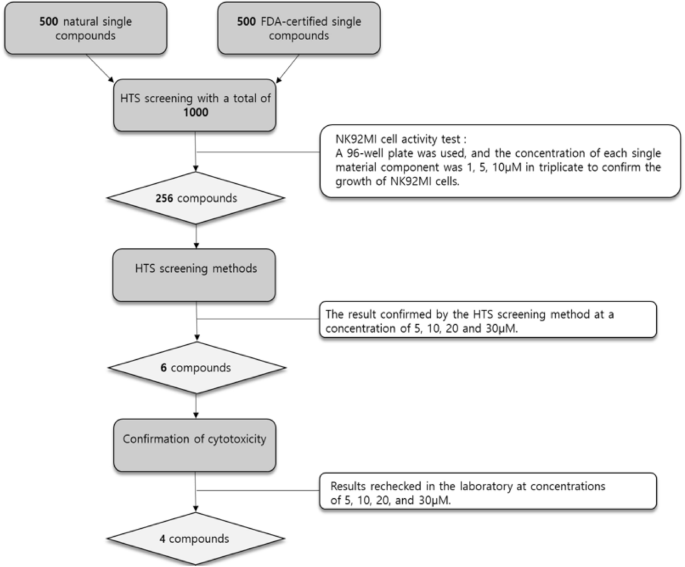
A total of 1,000 compounds, including 500 natural single-component compounds from the natural product library of the Korea Institute of Science and Technology and 500 FDA-approved single-component drugs, were screened using NK-92MI cells. High-throughput screening was performed using the Integrated Natural Products High-throughput/Content Screening System (iHTac-LAB). Each compound was tested at concentrations of 1, 5, and 10 μM in 96-well plates containing 100 μL of cell suspension containing NK-92MI cells and incubated for 24 h. Experiments were performed in triplicate. Cell viability was measured to exclude cytotoxic compounds, and compounds that maintained ≥80% cell viability compared to the control were selected as candidate compounds. Selected compounds were further evaluated at concentrations of 5, 10, 20, and 30 μM using the same screening system.
Cell culture
Human pancreatic cancer cell lines (Panc-1 and BxPC-3) were graciously provided by Dr. Kim (Asan Medical Center, Seoul, Korea). NK-92MI cells were purchased from ATCC. Pancreatic cancer cells were cultured in Dulbecco’s Modified Eagle Medium (DMEM; Gendepot, USA) supplemented with 10% (v/v) fetal bovine serum (FBS; Gibco, USA) and 20 μg/mL penicillin/streptomycin (Lonza, Switzerland). NK-92MI cells were cultured in an alpha modification of Eagle’s minimum essential medium (α-MEM; Gibco, USA) supplemented with 2 mM l-glutamine, 0.2 mM inositol, 0.02 mM folic acid, 0.01 mM 2-mercaptoethanol, 12.5% FBS, and 12.5% horse serum (Gibco, USA). All cell lines were maintained at 37℃ in a 5% CO2 incubator.
Cell viability assay
Cells were seeded in 96-well plates at a density of 1 × 10⁴ cells per well and then cultured at 37℃. Cell viability was analyzed using an EZ-Cytox assay kit (DoGen Bio, Seoul, South Korea). EZ-Cytox solution (10 μL) was incorporated into each well, and the plates were incubated for 2 h. Absorbance was measured at 450 nm using a microplate reader (Beckman Coulter, Brea, CA, USA).
Flow cytometric analysis
The surface natural cytotoxic receptors (NCR) were analyzed via flow cytometric analysis using APC-conjugated anti-human NKp30, anti-human NKp44, and anti-human NKp46 antibodies. NK-92MI cells were inoculated into 6-well plates at 3.0 × 10⁵ cells per well and cultured overnight. Following a 24-h incubation with rosiglitazone, the cells were washed with ice-cold PBS, incubated with 5 μL of conjugated antibodies for 30 min at 4 °C, washed twice in PBS, and analyzed directly using a flow cytometer (Beckman Coulter, Brea, CA, USA). Flow cytometry data were analyzed by calculating mean fluorescence intensity (MFI), and values were normalized to the control group. All experiments were performed in at least three independent biological replicates (n = 3).
Western blot
The cells were lysed in a radioimmunoprecipitation assay buffer (50 mM Tris, 150 mM NaCl, 1% Triton X-100, 0.1% sodium dodecyl sulfate, SDS, and 1% sodium deoxycholate; pH 7.4) supplemented with a protease and phosphatase inhibitor cocktail (Sigma-Aldrich, St. Louis, MO, USA). Protein concentrations were quantified using the bicinchoninic acid assay. Equal aliquots of proteins were resolved using SDS-polyacrylamide gel electrophoresis and transferred onto polyvinylidene fluoride membranes. The membranes were blocked with TBS containing 0.2% Tween 20 and 5% skim milk, incubated overnight with primary antibodies at 4℃, and incubated with HRP-conjugated secondary antibodies. Subsequently, the protein signals were visualized using an electrochemiluminescence solution on a Chemidoc (Amersham Image Quant 800).
IVIS assay
BxPC-3-Luc cells were seeded in 24-well plates at a density of 1 × 10⁵ cells per well and cultured at 37 ℃. NK-92MI cells were treated with 30 µM rosiglitazone for 24 h. Then, BxPC-3-Luc with NK-92MI were co-cultured for 24 h. To confirm fluorescence, D-luciferin (100 ng/ml) was added to co-cultured cells, incubated in the dark room for 10 min, and bioluminescence was measured using an IVIS imaging system. Although IVIS is primarily designed for in vivo imaging, it was used here as a relative indicator of viable luciferase-expressing cells under in vitro conditions.
Enzyme-linked immunosorbent assay (ELISA)
ELISA was performed to evaluate the expression of TNF-α and IFN-γ in rosiglitazone-treated NK-92MI cells. All relevant reagents were procured from the R&D Systems (Minneapolis, MN, USA). Culture plates and reagents were brought to room temperature (22 ℃), following which 50 μL each of assay diluent RD1F and the samples or standards were aliquoted to each well and incubated for 2 h at room temperature. Subsequently, the wells were washed, 200 μL of detection antibody solution was incorporated into each well for 2 h at room temperature, the wells were washed for color development, and 200 μL of substrate solution was added to each well for 30 min at room temperature. Following the conclusion of the incubation period, the reaction was terminated by introducing 50 μL of stop solution to each well. The absorbance was computed at 450 nm using a microplate reader.
Apoptosis assay
Panc-1 cells were inoculated into 6-well plates at 1.5 × 10⁵ cells per well and cultured overnight. The cells were administered with either 30 µM of rosiglitazone, NK-92MI, or both combined. Thereafter, an apoptosis assay was performed in compliance with the kit protocol. Cells were harvested using trypsin-EDTA and centrifuged at 1000 ×g for 5 min. The supernatant was collected, washed with ice-cold PBS, and centrifuged at 1000 ×g for 5 min. Following centrifugation, the cells were resuspended in 500 µL of 1X binding buffer. Subsequently, the cells were incubated with Annexin V stain (5 µg/mL) at room temperature in a dark room for 15 min, the mixture was centrifuged at 1000 ×g for 5 min, and the supernatant removed. Next, 500 µL of 1X binding buffer and 10 µL of propidium iodide were homogenized with the supernatant, and cell apoptosis was immediately analyzed using flow cytometer (Beckman Coulter, Brea, CA, USA) to determine the percentage of viable or apoptotic cells.
Statistical analysis
Data are presented as mean ± standard deviation (SD). Experiments were performed in triplicate. Statistical significance was determined using Student’s t-test for comparisons between two groups or one-way analysis of variance (ANOVA) for multiple group comparisons. A p-value < 0.05 was considered statistically significant.
