


Cell culture and treatment
Breast cancer cell lines MDA-MB-231 (ATCC HTB-26, Cellosaurus: CVCL_0062) and BT549 (ATCC HTB-122, Cellosaurus: CVCL_1092) were purchased from Shanghai Cell Bank of Chinese Academy of Sciences (Shanghai, China) and maintained in DMEM (Hyclone, USA) that contains 10% fetal bovine serum (FBS; BI, China) and 1% streptomycin/penicillin (Sigma, USA) in a 37 °C incubator filled with 5% CO2.
Cell transfection and treatment
The siRNA targets the USP25 gene, and the negative control siRNA (Sequences: sense, 5′-UUCUCCGAACGUGUCACGUdTdT-3′; antisense, 5′-ACGUGACACGUUCGGAGAAdTdT-3′) were purchased from RiboBio (Guangzhou, China). The C1ql4 overexpression vectors were constructed using pcDNA3.1 as a backbone. The siRNAs and overexpression vectors were transfected into cancer cells using Lipofectamine RNAiMAX reagent (Invitrogen, USA) and PEI reagent (Sigma, USA), respectively. The cells were transfected for 48 h and treated with the cycloheximide (CHX, 200 ng/mL) for 24 h to suppress protein synthesis.
Quantitative real-time PCR
Total RNA was extracted from cells using Trizol reagent (Thermo, USA) and reverse transcribed to cDNA by using First-strand synthesis kit (Transgene, China). RNA level was measured using a SYBR Green Supermix (Transgene) and detected using the Real-Time PCR Detection System (Bio-Rad, USA). The β-Actin level was set as internal reference for normalization. USP25 primers (5′–3′): forward, 5′‑GTTCTATGGCAGATTCCTGGCTG‑3′; reverse, 5′‑GCAGCTTCTAGGCACTCATGCA‑3′.
Cell viability
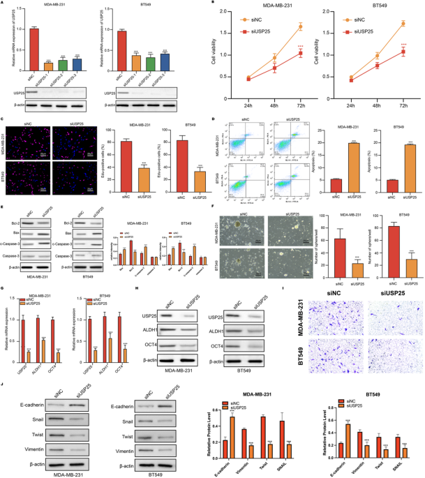
Cell viability and proliferation were measured by cell counting kit 8 (CCK-8) and 5-ethynyl-2′-deoxyuridine (EDU) assay. For the CCK-8 assay, cells were incubated in 96-well plates and hatched for 24, 48, and 72 h. After reaction with CCK-8 reagent (Beyotime) for 2 h, the absorbance values at 450 nm were detected by a microplate spectrometer (Thermo, USA). For Edu assay, cells were fixed and permeabilized, and incubated with Edu reagent (Beyotime) and DAPI reagent (Sigma, USA). The images were captured using fluorescence microscopy (Leica, Germany).
Apoptosis detection
Cell apoptosis was assessed by using flow cytometry. The cells were incubated with propidium iodide (PI) and Annexin V (Beyotime, China) for 30 min and analyzed with the flow cytometer (BD Biosciences, USA).
Western blotting assay
Total proteins were harvested after homogenizing the cells with RIPA lysis buffer (SolarBio, China) and centrifuged at 12,000 g for 10 min. The proteins were separated with SDS-PAGE and transferred to a PVDF membrane (Millipore, Germany). Followed by blocking in skimmed milk. Protein bands were then probed with primary anti-USP25, anti-Bcl-2, anti-Bax, anti-Caspase-3, anti-ALDH1, anti-OCT4, anti-C1ql4, anti-β-actin overnight at 4 °C. Next day, the blots were incubated with horseradish peroxidase (HRP)-conjugated secondary anti-mouse or anti-rabbit antibodies and reacted with ECL reagent (Millipore). Images were visualized in an imaging system (Tanon, China).
Sphere formation assay
Cells were suspended as single cells in serum-free DMEM/F12 medium (BI) that added with 20 ng/mL b-FGF (Sigma), 20 ng/mL EGF (Sigma), and 2% B27 (Sigma). Then, 5,000 cells were seeded into each well of the ultra-low attachment 96-well plate (Corning, USA) and incubated for 10 days at the 37 °C incubator. The images of mamo spheres were taken under a microscope (Leica, Germany).
Immunoprecipitation assays
The interaction of USP25 and C1ql4 was analyzed by co-immunoprecipitation (Co-IP) in the MDA-MB-231 and BT549 cells transfected with flag-USP25. Co-IPs were conducted by Pierce Co-Immunoprecipitation Kit (Fisher Scientific, Schwerte, Germany) according to the manufacturer’s instructions.
Xenograft model
Animal experiments were authorized by the Ethics Committee of the Affiliated Hospital of Chengde Medical University. Female athymic nude mice (4–6 weeks old; 18–22 g; SiPeiFu, China) were housed under specific pathogen-free (SPF) conditions. In brief, MDA-MB-231 cells transfected by USP25 siRNAs, siNC, and C1ql4 overexpression vectors and collected in saline with 6 × 10⁵ cells/100 µl. Then each mouse was hypodermically inoculated with 100 µl cell suspension in the right fat pad (n = 5 mice for each group). The width and length of the tumor were measured using vernier calliper every 5 days for 30 days. The tumor volume was calculated as width (mm)2 × length (mm)/2. All mice were finally sacrificed, and the tumors were weighed, photographed, and stored for analysis.
Immunohistochemistry (IHC) analysis
Tumor tissues were fixed in 10% neutral formalin (Sigma) for 3 days, dehydrated in serial ethanol, embedded with paraffine, and sliced into 5 μm thickness samples. The tissue samples were de-paraffinized and rehydrated. The endogenous peroxidase was blocked using 3% H2O2 at room temperature for 10 min and goat serum for 30 min. The tissue samples were then probed with anti-Ki-67 (Abcam, ab16667), anti-USP25 (Abcam, ab246948), and anti-C1ql4 (Affinity, DF9409) overnight at 4 °C. The samples were then incubated with HRP-conjugated secondary antibodies for 1 h. The samples were counterstained with hematoxylin, dehydrated, and mounted with a neutral balsam. Images were captured with a microscope (Leica).
Clinical samples
A total of 50 patients diagnosed with breast cancer and received surgical operation were recruited in this study and have signed informed consents. The tumor tissues and adjacent non-tumor tissues were resected during surgery and immediately frozen in liquid nitrogen, then stored at -80 °C refrigerator for subsequent experiments.
Statistics
Data are shown as mean ± standard deviation (SD). Statistical analyses are analyzed by using GraphPad Prism 7 software. Comparison between two groups and multiple groups was assessed using the unpaired two-tailed Student’s t test and one- or two-way analysis of variance followed by Tukey’s correction. A p-value less than 0.05 was regarded as statistically significant.
Animals and ethical statement
All methods were performed in accordance with the relevant guidelines and regulations. The animal study is reported in accordance with the ARRIVE guidelines. Throughout the experiment, the health status of the mice was continuously monitored to prevent any unintended harm. Upon completion of all experimental procedures, the animals were humanely euthanized via cervical dislocation under isoflurane anesthesia, with tumor size strictly maintained below 13 mm in accordance with ethical guidelines. The carcasses were subsequently stored in a designated temporary freezer at the animal experiment center and later collected for standardized, environmentally safe disposal.
