


Design and identification of TCRm Bi-NbTE
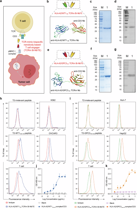
To enable the redirection of T cell against intracellular tumor antigens in a WT1+ or GPC3+ and HLA-A2+ restricted manner, we engineered a novel TCRm Bi-NbTE antibody. Specifically, an anti-CD3ε nanobody previously screened and characterized was fused via a flexible Gly4Ser linker to a TCRm nanobody targeting either the HLA-A2/WT1126-134 complexes or HLA-A2/GPC3144-152 complexes, both identified in our earlier studies (Fig. 1a). A negative control construct (Irrelevant NbTE) was generated by replacing the TCRm nanobody with an irrelevant nanobody derived from the human antibody germline repertoire that recognizes a nontumor target. Protein homology modeling of the designed TCRm Bi-NbTE constructs was performed using the SWISS model (https://swissmodel.expasy.org), yielding a predicted spatial arrangement of the molecules (Fig. 1b, e). While the TCR Bi-NbTE format is distinct from previously reported TCRm bispecific antibodies,29 ImmTACs30 and TCRm BiTE molecules,23 it aligns with the modularity of the nanobody-based bispecific T cell engager format (Supplementary Fig. S1a). Heterologous expression in E. coli BL21(DE3) revealed that the HLA-A2/WT1126 TCRm Bi-NbTE exhibited maximal yield under optimal induction with 0.5 mM IPTG at 37 °C for 6 h (Supplementary Fig. S2a, c, d), whereas the HLA-A2/GPC3144 TCRm Bi-NbTE achieved optimal expression under induction conditions with 0.5 mM IPTG at 16 °C for 16 h (Supplementary Fig. S2b, e, f). Both TCRm Bi-NbTE proteins were efficiently purified from inclusion bodies by a Ni2+–NTA affinity chromatography (Supplementary Fig. S3a, b). Purity was confirmed to exceed 90% by SDS–PAGE and SEC–HPLC analysis (Fig. 1c, f, Supplementary Fig. S3c, d). Western blotting with anti-His tag antibodies further verified molecular identities with expected immunoreactive band sizes (Fig. 1d, g). Overall, these results demonstrate the successful generation and purification of functional HLA-A2/WT1126 TCRm Bi-NbTE and HLA-A2/GPC3144 TCRm Bi-NbTE proteins, establishing a robust foundation for subsequent binding and functional evaluation. This innovative TCRm Bi-NbTE design extends the BiTE therapeutic repertoire for targeting intracellular tumor antigens.
Fig. 1
Generation and characterization of TCRm Bi-NbTE. a Schematic diagram of the TCRm Bi-NbTE molecule, consisting of a TCRm nanobody targeting pMHC complex and a CD3ε nanobody engaging T cells. Predicted structural models of (b) HLA-A2/WT1126 TCRm Bi-NbTE and (e) HLA-A2/GPC3144 TCRm Bi-NbTE were generated using the SWISS-MODEL model (https://swissmodel.expasy.org). SDS–PAGE analysis of purified (c) HLA-A2/WT1126 TCRm Bi-NbTE and (f) HLA-A2/GPC3144 TCRm Bi-NbTE was visualized by Coomassie blue staining. Lanes: M, marker; 1, purified protein. Western blot validation of purified (d) HLA-A2/WT1126 TCRm Bi-NbTE and (g) HLA-A2/GPC3144 TCRm Bi-NbTE using an anti-His tag antibody. Lanes: M, marker; 1, purified protein. h Binding analysis of HLA-A2/WT1126 TCRm Bi-NbTE to HLA-A2+/WT1+ OVCAR3 cells, HLA-A2-/WT1+ K562 cells, T2 cells pulsed with WT1126-134 peptides or irrelevant peptides, and primary human T cells assessed by flow cytometry. i Binding analysis of HLA-A2/GPC3144 TCRm Bi-NbTE to HLA-A2+/GPC3+ HepG2 cells, HLA-A2-/GPC3+ Huh-7 cells, T2 cells pulsed with GPC3144-152 peptides or irrelevant peptides, and primary human T cells assessed by flow cytometry. j, k Sandwich ELISA demonstrating that TCRm Bi-NbTEs simultaneously engage CD3 and their respective peptide-MHC complexes without steric interference
Binding specificity of TCRm Bi-NbTE
To systematically characterize the binding specificity of TCRm Bi-NbTE, we performed flow cytometry-based binding assays using both antigen-positive and antigen-negative cell lines. The HLA-A2/WT1126 TCRm Bi-NbTE specifically bound to OVCAR3 (HLA-A2+/WT1+) and T2-WT1126-134 cells (HLA-A2+/WT1+), as well as primary human T cells, with no detectable binding to K562 cells (HLA-A2-/WT1+), T2-irrelevant peptide cells or unpulsed (Fig. 1h, Supplementary Fig. S4a-c). Similarly, the HLA-A2/GPC3144 TCRm Bi-NbTE exhibited specific binding to HepG2 (HLA-A2+/GPC3+) and T2-GPC3144-152 (HLA-A2+/GPC3+) cells, as well as primary human T cells, while no binding was observed for Huh-7 (HLA-A2-/GPC3+), T2-irrelevant peptide cells or unpulsed (Fig. 1i, Supplementary Fig. S4a, d, e). Importantly, competitive inhibition assays confirmed the binding specificity of TCRm Bi-NbTE. Preincubation of T cells with soluble recombinant hCD3ε protein significantly reduced TCRm Bi-NbTE binding (Supplementary Fig. S4c, e). Furthermore, binding affinity of the TCRm Bi-NbTE was determined by biolayer interferometry (BLI). The HLA-A2/WT1126 TCRm Bi-NbTE displayed equilibrium dissociation constants (KD) of 2.49 × 10⁻⁸ M for recombinant CD3ε and 5.39 × 10⁻⁷ M for the HLA-A2/WT1126-134 complex protein (Supplementary Fig. S5a, b). Similarly, the HLA-A2/GPC3144 TCRm Bi-NbTE showed 1.18 × 10⁻⁸ M for CD3ε and 1.01 × 10⁻⁷ M for the HLA-A2/GPC3144-152 complex protein (Supplementary Fig. S5d, e). No measurable binding was detected toward mimetic peptides (Supplementary Fig. S5c, f). Moreover, a sandwich enzyme‑linked immunosorbent assay (ELISA) results showed that TCRm Bi-NbTE can simultaneously bind to two target antigens, indicating no steric interference between epitopes (Fig. 1j, k). These results establish TCRm Bi-NbTE as a dual-specific and HLA-A2/peptide-restricted T cell engager capable of simultaneously recognizing CD3 and pMHC complexes.
To further assess potential off-target interactions, we evaluated its cross-reactivity using the sCRAP algorithm, which predicts structural similarities between target epitopes and human proteome-derived peptides presented on HLA-A*02:01.31 Based on these predictions, four top-ranked homologous peptides were identified and selected for experimental validation.26 Flow cytometry analysis revealed that no detectable binding by either TCRm Bi-NbTE to T2 cells pulsed with these predicted cross-reactive peptides (Supplementary Fig. S6a, b). Alanine-scanning further identified the critical contact residues mediating recognition. Specifically, substitutions at Arg1, Asn5, Ala6, and Leu9 significantly diminished HLA-A2/WT1126 TCRm Bi-NbTE binding, indicating these residues as critical contribution to recognition. Similarly, residues Val2, Gly3, Phe5, and Val9 are particularly important for recognition by HLA-A2/GPC3144 TCRm Bi-NbTE (Supplementary Fig. S6c, d). Taken together, these data confirm high specificity and dual antigen recognition of TCRm Bi-NbTE, supporting its therapeutic potential against intracellular tumor antigens in an HLA-restricted manner.
TCRm Bi-NbTE triggers T cell activation, proliferation, and effector function
To assess the functional responses of TCRm Bi-NbTE-mediated T cell engagement, we analyzed T cell activation, proliferation and cytokine production when co-cultures with OVCAR3 (HLA-A2+/WT1+) or HepG2 (HLA-A2+/GPC3+) cells. Strikingly, compared to Irrelevant NbTE or blank controls (ctr), both TCRm Bi-NbTE induced robust T cell activation, evidenced by significantly upregulated surface expression of CD25 (Fig. 2a, e, Supplementary Fig. S7a, c) and CD69 (Fig. 2b, f, Supplementary Fig. S7a, c). Concurrently, degranulation activity, measured by CD107a expression increased in TCRm Bi-NbTE-mediated T cells compared to Irrelevant NbTE or blank ctr (Fig. 2c, g, Supplementary Fig. S7a, c). Meanwhile, no T cell activation was observed when TCRm Bi-NbTE was applied to both HLA-A2⁺/antigen⁻ tumor cell lines (ARH77, SW480) or normal CD34+ cells from an HLA-A2+ healthy donor (Supplementary Fig. S9a-c), further supporting that TCRm Bi-NbTE triggering potent antigen-dependent T cell responses.
Fig. 2
TCRm Bi-NbTE induced T cell activation and effector function. Upregulation of (a, e) CD25, (b, f) CD69, and (c, g) CD107a on T cells after co-culture with OVCAR3 or HepG2 cells in the presence of TCRm Bi-NbTE analyzed by flow cytometry. d, h T cell proliferation following co-cultured with OVCAR3 cells or HepG2 cells in the presence of respective TCRm Bi-NbTE measured by flow cytometry and presented as division percentage. T cell subpopulation analysis after stimulation with (i) HLA-A2/WT1126 TCRm Bi-NbTE or (m) HLA-A2/GPC3144 TCRm Bi-NbTE, defined by CCR7 and CD45RA expression. j, n IL-2 and k, o IFN-γ secretion levels in co-cultures of T cells with OVCAR3 cells or HepG2 cells in the presence of TCRm Bi-NbTE or at equimolar controls. ELISPOT assays quantifying IFN-γ-secreting T cells induced by (l) HLA-A2/WT1126 TCRm Bi-NbTE or (p) HLA-A2/GPC3144 TCRm Bi-NbTE. Data are representative of three independent experiments. **P < 0.01; ***P < 0.001; ****P < 0.0001
Another key hallmark of activated T cell is their proliferative capacity. Flow cytometry analysis demonstrated that TCRm Bi-NbTE mediated substantial T cell proliferation in pMHC+ tumor co-cultures, whereas Irrelevant NbTE or blank ctr exhibited minimal cell division (Fig. 2d, h, Supplementary Fig S7a, c). Notably, TCRm Bi-NbTE also promoted differentiation into central memory T cells (TCMs, CD45RA−/CCR7+), increasing their proportion compared to controls (Fig. 2i, m, Supplementary Fig. S7b, d). This phenotype is associated with heightened proliferative potential, lymphoid homing and rapid effector differentiation upon antigen re-encounter, suggesting enhanced potential for sustained antitumor immunity.
Consistent with this activated state, TCRm Bi-NbTE engagement triggered a potent antigen-specific cytokine response. Secretion of key effector cytokines, including IL-2 and IFN-γ, was significantly elevated compared to Irrelevant NbTE in the presence of pMHC+ tumor cells (Fig. 2j, k, n, o). Enzyme-linked immunospot (ELISPOT) analysis revealed an increase in IFN-γ+ T cell frequencies in TCRm Bi-NbTE-treated groups (Fig. 2l, p). Taken together, these data demonstrate that TCRm Bi-NbTE selectively activate and expand T cells in an antigen-dependent manner, driving potent effector function, align with emerging evidence highlighting the therapeutic value of antigen-restricted T cell engagers in cancer immunotherapy.
TCRm Bi-NbTE mediates specific cytotoxicity against pMHC+ tumor cells in vitro
To evaluate the antigen-specific cytotoxic potential of TCRm Bi-NbTE in vitro, we performed flow cytometry-based cytotoxicity assays. Expression analysis of public datasets confirmed variable WT1 and GPC3 levels across tumor tissues and tumor lines (Supplementary Fig. S8a-g). Co-culture of T cells with various HLA-A2+/GPC3+ cells (T2-GPC3144-152, HepG2, and primary hepatocellular carcinoma (HCC) cells) or HLA-A2+/WT1+ cells (T2-WT1126-134, OVCAR3, and OCIAML3 cells) at an effector-to-target (E:T) ratio of 10:1 revealed that both TCRm Bi-NbTE induced potent, dose-dependent T cell cytotoxicity (Fig. 3a, d, e, h, i, m). This cytotoxicity was significantly superior to that mediated by the Irrelevant NbTE or T cells alone (Fig. 3b, f). Strikingly, no appreciable lysis was observed against pMHC− cells (T2-irrelevant peptides, T2 cells, K562, ARH77, Huh-7, SW480 or Hela cells) at different concentrations (Fig. 3a, e, j–l, n–p), confirming its tumor cell lysis in a strictly antigen-specific and dose-dependent manner.
Fig. 3
TCRm Bi-NbTE mediated T cell-specific cytotoxicity against pMHC+ complex target cells in vitro. Dose-dependent cytotoxicity of T2 cells pulsed with (a) WT1126-134 peptide or (e) GPC3144-152 peptide in the presence of various concentrations of TCRm Bi-NbTE, and specific lysis of T2 cells pulsed with (b) WT1126-134 peptide or (f) GPC3144-152 peptide at E:T ratio of 10:1 compared to T2 cells pulsed with irrelevant peptide, or unpulsed. E:T ratio-dependent cytotoxicity against (c) OVCAR3 and (g) HepG2 cells. Dose–response cytotoxicity of (d) OVCAR3, (i) OCIAML3, (j) K562, (k) ARH77 and (l) Hela cells. Dose–response cytotoxicity of (h) HepG2, (m) primary HCC, (n) Huh-7, (o) SW480 and (p) Hela cells. Data are representative of three independent experiments. ****P < 0.0001; ns not significant
Moreover, results demonstrated that TCRm Bi-NbTE-mediated T cell cytotoxicity was dependent on the E:T ratio. TCRm Bi-NbTE induced the maximal lysis of pMHC+ cells HepG2 or OVCAR3 at an E:T ratio of 10:1, and reduced but significant lysis at a ratio of 5:1 (Fig. 3c, g). As expected, cytotoxicity occurred only in the presence of T cells, with no lysis observed in their absence, underscoring that the cytotoxicity was entirely T cell-dependent (Supplementary Fig. S9d, e). Besides, TCRm Bi-NbTE was significantly more effective than its anti-TCRm Nb counterpart, highlighting the critical role of CD3 engagement in driving efficient killing. Collectively, these data establish that TCRm Bi-NbTE selectively lyse pMHC+ tumors cells by inducing T cell-mediated cytotoxicity in an HLA-A2+/WT1+ or HLA-A2+/GPC3+ restricted manner. This precise antigen specificity and potent cytotoxic capacity highlight TCRm Bi-NbTE as a promising therapeutic strategy for targeting intracellular tumor antigens with minimal off-target effects.
TCRm Bi-NbTE exhibits robust antitumor efficacy in humanized mouse models
To evaluate the therapeutic potential of TCRm Bi-NbTE in vivo, we established cell-derived xenograft (CDX) models using HepG2-Luc and OVCAR3 tumor cells, respectively. NOD/SCID mice bearing subcutaneous tumors were randomized into treatment groups and received daily intravenous injections of TCRm Bi-NbTE or control molecules for 6 days following adoptive transfer of human peripheral blood mononuclear cells (PBMCs) (Fig. 4a, g). TCRm Bi-NbTE treatment significantly suppressed tumor growth compared to both control groups, as corroborated by tumor size measurement and in vivo bioluminescent imaging assessment (Fig. 4b, h, j), as well as conferred a significant survival benefit with TCRm Bi-NbTE compared to Irrelevant NbTE or PBS groups (Fig. 4c, i), providing robust evidence for the therapeutic efficacy of the TCRm Bi-NbTE in preclinical models.
Fig. 4
Antitumor efficacy of TCRm Bi-NbTE in multiple xenograft mouse models. Schematic diagram of the experimental timeline. NOD/SCID mice (n = 5) were subcutaneously implanted with (a) OVCAR3 tumor cells, (d) PDX tumors, or (g) HepG2-Luc tumor cells, injected intravenously with human PBMCs and were treated daily TCRm Bi-NbTE or control antibodies for six consecutive days. (b, e) Tumor growth curves. (c, f, i) Kaplan–Meier survival curves. h Quantitative analysis of luminescence intensity, and (j) representative in vivo bioluminescence imaging of HepG2-Luc tumors. Quantification of (k) Ki-67-positive proliferating cells, and (l) apoptotic cells in resected tumor tissues from the OVCAR3 or HepG2-Luc model after TCRm Bi-NbTE treatment. *P < 0.05; **P < 0.01; ***P < 0.001; ****P < 0.0001
To further evaluate its translational potential, we established a patient-derived xenograft (PDX) model using primary human HCC tissue grafted into NOD/SCID mice (Fig. 4d). TCRm Bi-NbTE treatment again significantly inhibited tumor growth and extended survival compared to controls (Fig. 4e, f). Importantly, no significant weight loss (Supplementary Fig. S10a, b) or signs of treatment-related toxicity, as indicated by stable levels of alanine aminotransferase (ALT), aspartate aminotransferase (AST), and pro-inflammatory cytokines including IL-6 or IL-1β (Supplementary Fig. S11a-e). Collectively, these results demonstrate that TCRm Bi-NbTE effectively suppresses tumor progression and improves survival in multiple mouse xenograft models, validating its potent antitumor activity.
We then performed histological analysis of harvested HepG2-Luc or OVCAR3 xenograft tumors. TCRm Bi-NbTE-treated tumors exhibited a significant increase in apoptosis cells and concurrent decrease in Ki-67+ proliferating cells compared to Irrelevant NbTE and PBS controls (Fig. 4k, l). Additionally, flow cytometry analysis revealed increased numbers of CD3+ T cells within the tumors and spleens of TCRm Bi-NbTE-treated mice (Fig. 5a, b, d, e, Supplementary Fig. S12a-b), with sustained peripheral CD3+ T cell counts observed within two weeks post-injection (Fig. 5c, f, Supplementary Fig. S12a-b).
Fig. 5
TCRm Bi-NbTE promotes intratumoral T cell functionality by boosting anti-tumor responses. Flow cytometry analysis showing CD3+ T cell infiltration in (a, d) tumor tissues, (b, e) spleens, and (c, f) peripheral blood from the OVCAR3 or HepG2-Luc model after TCRm Bi-NbTE treatment. g, j Heatmaps depicting differential expression of immune-related genes in tumor tissues from TCRm Bi-NbTE-treated versus Irrelevant NbTE-treated mice. h, k Relative expression of selected key genes associated with T cell activation, proliferation, exhaustion and cytotoxicity in the respective groups. i, l GO enrichment analysis showing significantly enriched pathways in tumor tissues from TCRm Bi-NbTE-treated versus Irrelevant NbTE-treated mice. **P < 0.01; ***P < 0.001; ****P < 0.0001
Transcriptomic profiling of tumor tissues provided further mechanistic insight. TCRm Bi-NbTE treatment upregulated a broad signature of T cell activity, including lineage markers (CD3E, CD8A, CD4), activation markers (IL2RA, CD69), cytotoxic effectors (GzmB, GzmA, PRF1, FASLG). A moderate increased expression of exhaustion markers (PDCD1, CTLA4, HAVCR2, LAG3, TIGIT) when compared to the control groups, consistent with sustained activation signaling accompanying robust effector responses (Fig. 5g, h, j, k). Furthermore, gene ontology (GO) enrichment analysis between the TCRm Bi-NbTE and Irrelevant NbTE groups revealed significant enrichment of pathways related to T cell proliferation, activation, mediated cytotoxicity and immunity, and proinflammatory cytokine signaling (Fig. 5i, l). Taken together, these results demonstrate that TCRm Bi-NbTE mediates robust antitumor efficacy in multiple preclinical models, mediating tumor regression through antigen-specific T cell recruitment, activation, and cytotoxic effector functions (Fig. 6a–c), positioning TCRm Bi-NbTE as a robust and scalable therapeutic platform for expanding T cell-engaging immunotherapy to intracellular tumor targets.
Fig. 6
Schematic overview of the TCRm Bi-NbTE platform and its therapeutic mechanism. a Workflow outlining the generation of specific anti-TCRm nanobodies and an anti-CD3ε nanobody, and their subsequent engineering into the TCRm Bi-NbTE construct. b Proposed working model of the TCRm Bi-NbTE mediated T cell–tumor cell engagement (Left), and therapeutic characterization of TCRm Bi-NbTE molecule targeting the HLA-A2/WT1126-134 complexes or HLA-A2/GPC3144-152 complexes (Right), redirecting T cell cytotoxicity independent of endogenous TCR specificity. c Summary diagram depicting that TCRm Bi-NbTE promotes potent antitumor efficacy in multiple mouse xenograft models, validating this modular platform as a novel strategy for targeting intracellular antigens. Generated by Adobe Illustrator software
