


Sociodemographic and clinical differences based on tumor HPV status
The study cohort comprised 58 OPC patients and 1 CUP patient. Patients were grouped according to tumor HPV status, with 39 HPV-driven and 20 HPV-negative patients. The majority of HPV-OPC was solely positive for HPV16 (n = 33, 84.6%), while two tumors were HPV16/HPV33 double positive. Three OPC cases were positive for HPV33, HPV35 and HPV58, respectively. One patient was positive for HPV69, which is not a recognized high-risk HPV type. This patient was excluded in the subsequent dPCR analysis.
Patients with HPV-negative OPC had a higher prevalence of alcohol consumption (55% vs. 13%) and smoking (90% vs. 49%) compared to patients with HPV-OPC (Table 1). More than 90% of patients with HPV-negative OPC presented with advanced disease (stage III or IV), in comparison to 26% of patients with HPV-OPC.
Table 1 Baseline characteristics in OPC/CUP patients by tumor HPV status.
HPV-negative patients had an overall higher ECOG status, and thus an overall worse performance status.
The HPV-driven and HPV-negative groups did not differ in age (68 vs. 69), gender (64% vs. 70% male) nor tumor location, with the most frequent subsite in both groups being the tonsils (44% and 45%, respectively).
Clinical follow-up and outcomes
OPC patients were under clinical follow-up for a median of 1.2 years (range 20 days – 2.9 years) and outcomes were recorded. Seven patients died during the follow-up period, all of whom had HPV-negative OPC (35%, 95% CI 15–59%) with advanced disease (clinical stage III or IV) (Table 2). Deaths occurred at a median of 7 months (range 20 days – 1.9 years) after diagnosis. Similarly, recurrent disease was more common in patients with HPV-negative OPC (n = 6; 43%, 95% CI 18–71%) compared to those with HPV-OPC (n = 3; 8.8%, 95% CI 2–24%) (Table 2). OPC recurrence was diagnosed at a median of 15 months (range 6–20 months) after diagnosis among patients with available timing information (n = 8).
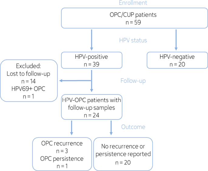
All OPC patients provided blood samples at diagnosis, while follow-up samples were analyzed for 24 of 39 HPV-OPC patients (Fig. 1). Fourteen HPV-OPC patients did not provide follow-up samples: Nine only gave consent to analyze samples at diagnosis due to the pandemic, two withdrew consent to collection of follow-up samples, and three were lost to follow-up because radiotherapy was performed as an outpatient procedure at a different facility. One additional HPV-OPC patient was excluded from further analyses due to not testing positive for a high-risk HPV type included in the test panel.
Table 2 Clinical outcomes in OPC/CUP patients by tumor HPV status.Fig. 1The alternative text for this image may have been generated using AI.
Flowchart of study participants.
Concordance of HPV cfDNA in blood and HPV status in tissue at time of diagnosis
The presence and quantity of HPV cfDNA at time of diagnosis in the blood plasma of patients with HPV-OPC (n = 38) and HPV-negative OPC (n = 20) were investigated using a multiplex dPCR assay detecting the eight high-risk HPV types HPV16, 18, 31, 33, 35, 45, 52 and 58. All included plasma samples contained sufficient human beta-globin with a median of 2,190 copies/ml (range 548–685,777 copies/ml).
HPV cfDNA was present in 36 of 38 HPV-OPC patients at time of diagnosis, indicating a sensitivity of 95% (95% CI 82–99%, Fig. 2A). HPV cfDNA was not detected in two patients, one with HPV16-driven OPC and one with HPV33-driven OPC. Both patients had stage I OPC with minor lymph node involvement (N1).
Conversely, HPV cfDNA was not detected in 19 of 20 patients with HPV-negative OPC, resulting in a specificity of 95% (95% CI 75–100%). The single HPV cfDNA false positive patient had 8.5 copies/ml HPV16 cfDNA but was negative for p16 IHC and HPV16 E6 antibodies.
HPV cfDNA concentration in patients with HPV-OPC who tested positive for HPV cfDNA at time of diagnosis ranged from 3 to 104,009 copies/ml (median 560 copies/ml) for HPV16 and from 7 to 226 copies/ml (median 26 copies/ml) for HPV33, HPV35 or HPV58. A higher HPV cfDNA concentration was not statistically significantly correlated with any tumor parameters, although a positive trend for clinical, tumor and nodal stage was observed.
HPV genotypes identified by the dPCR assay matched the tumor HPV status in all cases. HPV-type specific sensitivity was 97% for HPV16 (33/34, 95% CI 85–100%), 50% for HPV33 (1/2, 95% CI 9–99%), 100% for HPV58 (1/1, 95% CI 0.025–100%) and 100% for HPV35 (1/1, 95% CI 0.025–100%) (Fig. 2B–D). Due to limited multiplexing capacity, only four channels were available to test for the eight HPV types included, and channel-wise specificity was 92% for HPV16 (22/24, 95% CI 73–100%), 98% for HPV33/52/58 (64/65, 95% CI 92–100%), 100% for HPV31/35 (67/67, 95% CI 95–100%) and 100% for HPV18/45 (68/68, 95% CI 95–100%).
Fig. 2The alternative text for this image may have been generated using AI.
HPV cfDNA levels in blood plasma of 58 patients at time of diagnosis in relation to tumor HPV status. (A) Sum of HPV copies across all HPV types, and copies of (B) HPV16, (C) HPV33/58 and (D) HPV35 detected per sample. The HPV cfDNA status determined by dPCR was compared to the tumor HPV status. Mean copy numbers for HPV16 and HPV33/35/58 from two dPCR replicates per ml plasma volume are shown. Samples that were reproducibly tested positive or negative across both duplicates were classified as positive (red) or negative (blue), respectively, while samples tested positive in only one replicate were labeled as inconclusive (grey). Values of 0 were replaced with a value of 0.1 (here labeled as 0) to allow the display on a logarithmic scale. 58 out of 59 study participants are shown here, since samples from one HPV69-positive patient were excluded from dPCR analysis.
Monitoring HPV cfDNA levels supports early diagnosis of recurrence
Twenty-four HPV-OPC patients who were positive for HPV cfDNA at baseline were followed up for a median of 1.5 years (range 8.4 months – 2.4 years) after diagnosis, and data on the course of disease and final outcomes of the patients were collected. Ninety-one plasma samples from a median of 3 follow-up visits per patient (range 1–8) were collected after diagnosis and tested for HPV cfDNA. The first follow-up sample was taken at a median of 41.5 days (range 14–273 days) after diagnosis. Therapy started at a median of 21 days (range 1–37 days) after diagnosis and lasted for a median of 70 days (range 14–190 days) across all treatment modalities.
Four patients within the HPV-OPC group experienced OPC recurrence or persistence during the follow-up period (Figs. 3 and 4). One patient (patient 1) presented with local recurrence at 15 months after diagnosis, while another patient (patient 2) was diagnosed with pulmonary metastases approximately 17 months after diagnosis. Both patients had a positive HPV cfDNA status at time of diagnosis (3853 and 1969 copies/ml, respectively), which were cleared upon completion of CRT. HPV cfDNA levels increased again prior to the clinical diagnosis of recurrence in both patients. HPV16 cfDNA concentration of patient 1 increased to 82 copies/ml three months before recurrence and further to 494 copies/ml two weeks after recurrence had been diagnosed. In patient 2, a positive HPV cfDNA status with 4 copies/ml was detected again seven months after end of therapy, and remained at this concentration for another three months. The first plasma samples consistently positive for HPV cfDNA post-treatment were collected approximately 3 and 8 months prior to the clinical diagnosis of recurrence in patient 1 and 2, respectively. The third patient (patient 3) presented with locoregional recurrence 7 months after collection of the last blood sample, which had negative HPV cfDNA status. One patient (patient 4) had persistent disease at end of therapy confirmed by imaging and biopsy, but their HPV cfDNA status remained negative after treatment.
Out of 24 patients with positive HPV cfDNA status at diagnosis and any available follow-up blood samples, 23 were HPV cfDNA-negative after treatment completion (Figs. 3 and 4). One patient (patient 22) had one positive HPV cfDNA result (10.4 copies/ml) shortly after end of therapy, despite not suffering a recurrence during the follow-up period, and further follow-up samples were negative. After completion of treatment this patient still had residual tumor mass, but a biopsy revealed no viable tumor cells. The other 19 patients had neither positive HPV cfDNA test results post-treatment nor recurrent or persistent disease reported.
To evaluate test performance in post-treatment surveillance, 59 HPV cfDNA test results from 24 HPV-OPC patients with follow-up were evaluated on a per-test basis. The 59 corresponding samples were taken after end of treatment and prior to a potential recurrence or persistence. If disease recurrence or persistence was diagnosed within 12 months of a positive HPV cfDNA result, the test was considered true positive. Conversely, if no recurrence or persistence was diagnosed within 12 months of a negative HPV cfDNA test result, the result was considered true negative. HPV cfDNA detection after completion of therapy had a positive predictive value (PPV) of 75% (3/4, 95% CI 19–99%) for recurrence of HPV-OPC within one year. Specificity was 98% (49/50, 95% CI 89–100%), as one test result during follow-up was false positive. The negative predictive value (NPV) for recurrence within one year was 89% (49/55, 95% CI 78–96%) and sensitivity was 33% (3/9, 95% CI 7–70%), both due to six false negative or inconclusive results across patients with OPC recurrence or persistence. Patient 1 and 4 each also had one sample collected shortly after their respective OPC recurrence or persistence, with a positive HPV cfDNA test result for patient 1 and an inconclusive HPV cfDNA test result for patient 4.
HPV cfDNA kinetics were compared between patients treated with RT or CRT (Fig. 4A) and those treated by surgery (with or without adjuvant treatment, Fig. 4B). As HPV cfDNA levels change rapidly after initiation of treatment, only patients with samples positive for HPV cfDNA at diagnosis and available follow-up samples within the first 4 months after diagnosis were included in this comparison (n = 20). Patients treated surgically (n = 5) had cleared their HPV cfDNA at the first follow-up sample after a mean of 37 days (range 14–61 days). Patients treated with RT or CRT alone (n = 15) had on average one positive sample after start of treatment (range 0–4), with the last positive sample at a mean of 34 days (range 0–100 days) after diagnosis. The first negative test result for HPV cfDNA occurred at a mean of 68 days (range 36–118 days) after diagnosis. Among patients treated with RT or CRT alone, a gradual decline of HPV cfDNA concentration was observed in some cases (Fig. 4A, patients 2 and 13). In two patients HPV cfDNA levels initially increased after start of treatment (Fig. 4A, patients 6 and 9), but their HPV cfDNA was eventually cleared.
Fig. 3The alternative text for this image may have been generated using AI.
HPV cfDNA status in 24 HPV-OPC patients during the follow-up period. HPV cfDNA status for any HPV type determined by dPCR across all patient plasma samples over the course of disease. Samples that were tested positive or negative across both duplicates for any HPV type were classified as positive (red) or negative (blue), respectively. Samples that tested positive in only one replicate for any HPV type were labeled as inconclusive (grey), even if they were negative for the tumor-driving HPV type (e.g. for patient 4). Grey lines indicate the clinical follow-up period. Brown bars mark the therapy period. Patient outcomes (persistence, recurrence) are marked at the corresponding date on the timeline.
Fig. 4The alternative text for this image may have been generated using AI.
HPV cfDNA concentrations in 24 HPV-OPC patients during and after treatment. HPV cfDNA status for the respective indicated HPV type determined by dPCR across all patient plasma samples over the course of disease. For each subplot, a patient ID and the HPV type are indicated (HPV16: grey, HPV33/58: green, HPV35: yellow). Mean copy number of HPV16, HPV33/58 or HPV35 based on dPCR duplicates per ml plasma volume is shown. Samples that were tested positive or negative across both duplicates for the indicated HPV type were classified as positive (red) or negative (blue), respectively, while samples tested positive in only one replicate were labeled as inconclusive (grey). Therapy period (dashed lines) and time of disease recurrence or persistence (solid lines) are marked. Values of 0 were set to 0.1 (here labeled as 0) to allow display on a logarithmic scale. (A) Patients that did not receive surgery (RT/CRT only). (B) Patients that received surgery (with or without adjuvant RT/CRT).
