


Study design and patient inclusion
This study was conducted at University Hospital Augsburg over a two-year period from December 2018 to November 2020. 40 Patients were enrolled as part of a prospective colorectal cancer (CRC) cohort, for which previous investigations have also been published4,15,21. The present evaluation represents a post hoc analysis of this cohort. All included patients underwent surgical treatment without receiving neoadjuvant therapy. Surgery was performed according to established guidelines for UICC tumor stages I–III22.
To minimize confounding factors influencing immune function, patients with acute or chronic infections, inherited or acquired immunodeficiencies, autoimmune disorders, or concomitant malignancies were excluded from the study. Patients with stage IV disease that was pretherapeutically inapparent were likewise excluded from evaluation.
A total of 27 age matched healthy individuals served as the reference cohort. These participants were blood donors recruited from the blood bank of the University Hospital Augsburg (Bavaria, Germany). Patient demographic and tumor characteristics are depicted in Table 1.
Ethical considerations
The study was approved by the ethics committee of Ludwig Maximilian University of Munich, the relevant ethics committee for our institution (Reference: Project No. 18–726) and conducted in accordance with the principles of the Declaration of Helsinki at University Hospital Augsburg, Germany. Prior to study inclusion, all patients provided written informed consent.
Definition of SARIFA
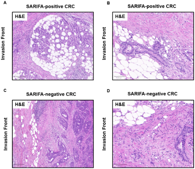
SARIFA status was established for this patient cohort for a previous publication for which we now present updated and additional data15. SARIFA status was assessed across all available tumor slide sections, prioritizing those with the greatest depth of invasion. Following our previous publication on SARIFA, SARIFA was defined as a region at the tumor invasion front (IF) where at least one tumor gland, or a cluster of at least five tumor cells, is directly adjacent to adipocytes, with no intervening inflammatory infiltrate or desmoplastic stromal response on standard hematoxylin and eosin (H&E) staining7. The stromal reaction could include features such as collagen deposition, reactive histiocytes, or fibroblastic proliferation. The presence of even a single SARIFA, such as a solitary tumor gland surrounded by adipose tissue, was sufficient to classify the case as SARIFA positive. Experienced pathologists (NGR; BM) blinded to clinical data, clinical course, and other tumor characteristics conducted all tumor evaluations. In rare cases where SARIFA status was unclear, a consensus diagnosis was made through consultation with a second board-certified pathologist using a double-headed microscope. Figure 1 illustrates representative sections of SARIFA-positive and SARIFA-negative CRC samples.
Fig. 1
Representative H&E-stained tumor sections of SARIFA positive (A and B) with direct contact between tumor cells and adipocytes at the invasion front and SARIFA negative (C and D) CRC samples with a pronounced stromal reaction at the invasion front, preventing a direct-tumor adipocyte interaction. Scale bar = 200 μm in A and C, and 100 μm in B and D. CRC, colorectal cancer; H&E, hematoxylin and eosin; SARIFA, Stroma AReactive Invasion Front ARea.
Analysis of lymphocyte subsets via flow cytometry
Blood samples were obtained prior to surgery, 7–10 days and six months after surgical resection, and flow cytometry was performed within 24 h in our local laboratory using an FC 500 flow cytometer (Beckman Coulter, Brea, CA, USA). The gating strategy for NK cells followed our previously published protocol4,21. For cell staining, antibodies from Beckman Coulter and Biolegend (San Diego, CA, USA) were used.
Absolute lymphocyte counts were derived from leukocyte counts measured with Stem-Count (Stem-Kit, Beckman Coulter), while initial lymphocyte values were reported as percentages. NK cells were detected as CD56 positive CD3 negative lymphocytes and further subdivided into three functional subsets: CD56 + CD16+, CD56dim CD16bright, and CD56bright CD16dim. The gating strategy is depicted in the supplementary data (Supplementary Fig. 1).
Analysis of inflammatory mediators (IM) via multiplex ELISA
IM levels were measured using the multiplex immunoassay CodePlex Secretome→ solution for IsoSpark\(^\circledR\) technology (Bruker Cellular Analysis, United States) on cryopreserved plasma samples obtained at the same time points as the samples for flow cytometry. The macrochambers of the CodePlex\(^\circledR\) chip were pre-patterned with a 22-plex antibody array, containing GM-CSF, GranzymeB, IFN-γ, IL-10, IL13, IL-15, IL-17 A, IL-2, IL-4, IL-5, IL-6, IL-7, IL-8, IL-9, IP-10, MCP-1, MIP-1a, MIP-1b, Perforin, sCD137, TNF-α, TNF-ß (Codeplex Secretome Adaptive Immune panel). Samples were thawed at room temperature and filled into each well of a chip in replicates (5.5 µL per replicate). 2% BSA (Bovine Serum Albumin) diluted in PBS (Phosphate-Buffered Saline) was used for background measurements. The chip was then loaded into an IsoSpark\(^\circledR\) system, and IM were measured in bulk using a fluorescence enzyme-linked immunosorbent assay (ELISA)-based multiplex assay in an automated workflow. Results were quantified by IsoSpeak\(^\circledR\) software v3.0.1 (Bruker Cellular Analysis, United States). Outputs were averaged between replicates and given in relative fluorescence units (RFU).
Statistical analysis
Descriptive results were expressed as medians with interquartile ranges. Differences in relative frequencies were evaluated using Fisher’s exact test. For group comparisons between healthy controls, SARIFA-negative and SARIFA-positive cases, global p value with Kruskal-Wallis test was calculated and a post hoc paired Mann-Whitney-U test complemented. Longitudinal values were compared using Friedman test with post hoc Wilcoxon signed-rank test. Given the limited cohort size and the exploratory character of this post hoc analysis, no formal correction for multiple comparisons was applied; accordingly, the comparisons of the 22 inflammatory mediators across groups and time points should be regarded as exploratory and hypothesis-generating, and the corresponding p values as nominal and uncorrected. The significance levels are indicated as follows: * p < 0.05, ** p < 0.01, *** p < 0.001, and **** p < 0.0001. All analyses were performed using SPSS for Windows version 24 (IBM, Armonk, NY, USA). Figures were generated using SPSS or Matplotlib (version 3.10.0).
