


Patient characteristics in Cohort 1
Cohort 1 consisted of 43 patients with first-onset HBV-related HCC (Table 1). This cohort was analyzed to identify clinical and virological factors associated with the age at HCC onset, with a particular focus on distinguishing features of earlier-onset versus late-onset HCC. The median age at diagnosis was 69 years (interquartile range [IQR], 60–76), and 32 patients (74.4%) were male. Median platelet count was 13.9 × 10⁴/μL (IQR: 11.2–17.6), and median fibrosis-4 (FIB-4) index was 3.42 (IQR: 2.20–4.31). Regarding viral markers, median HBsAg level was 2.92 log IU/mL (IQR: 1.69–3.27), median HBV DNA was 2.70 log IU/mL (IQR: not detected [ND]-3.95), and median HBeAg was 0.1 COI (IQR: 0.1–0.4), while median iTACT-HBcrAg was 3.10 log U/mL (IQR: 2.23–4.90). Only 16.3% (7 patients) were HBeAg positive. Approximately one-third of the cohort (34.9%) was receiving NA therapy at the time of HCC diagnosis.
Table 1 Characteristics of patients with first-onset HBV-related HCC in Cohort 1
When stratified by age at HCC onset, 17 patients (39.5%) were classified as earlier-onset (median age: 59 years), while 26 patients (60.5%) were categorized as late-onset (median age: 75 years). The earlier-onset group showed significantly higher alanine aminotransferase (ALT) (39 vs 21 U/L, p = 0.006), HBsAg (1060.0 vs 106.6 IU/mL, p = 0.024), iTACT-HBcrAg (4.90 vs 2.50 log U/mL, p < 0.001), and HBV DNA (3.30 vs 1.80 log IU/mL, p = 0.031) levels compared with the late-onset group. The frequency of HBeAg positivity was significantly greater in the earlier-onset group (29.4% vs 7.7%, p = 0.049), while platelet count was significantly lower (12.3 vs 16.1 × 10⁴/μL, p = 0.036).
Multivariable logistic regression analysis of factors associated with earlier-onset HBV-related HCC in Cohort 1
We next examined the factors associated with earlier-onset HBV-related HCC versus late-onset HCC (Table 2). In univariable analysis, iTACT-HBcrAg level (odds ratio [OR]: 1.78, 95% confidence interval [95% CI]: 1.20–2.63, p = 0.004) was significantly associated with earlier-onset HCC, while male (OR: 1.17, 95% CI: 0.29–4.74, p = 0.829), FIB-4 index (OR: 0.99, 95% CI: 0.73–1.35, p = 0.971), HBsAg level (OR: 1.84, 95% CI: 0.93–3.62, p = 0.080), HBeAg positivity (OR: 5.00, 95% CI: 0.84–29.7, p = 0.076), and HBV DNA (OR: 1.16, 95% CI: 0.92–1.47, p = 0.213) were not. Variables with p-values < 0.10 in the univariable analysis, including HBsAg level, HBeAg positivity, and iTACT-HBcrAg level, were entered into the multivariable model. In the multivariable analysis, HBsAg level (OR: 1.36, 95% CI: 0.64–2.91, p = 0.424) and HBeAg positivity (OR: 1.01, 95% CI: 0.21–5.68, p = 0.910) were not significantly associated with earlier-onset HCC, whereas iTACT-HBcrAg level remained independently associated with earlier-onset HCC (OR: 1.58, 95% CI: 1.00–2.47, p = 0.048).
Table 2 Multivariable logistic regression analysis of factors associated with earlier-onset HBV-related HCC in Cohort 1
HBcrAg positivity status at initial onset of HBV-related HCC in Cohort 1
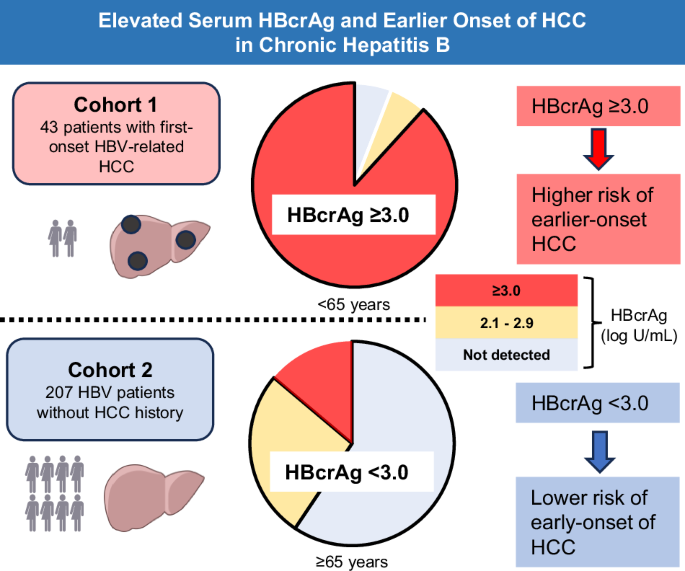
Figure 1a, b illustrate the distribution of HBcrAg positivity status among the 43 patients with initial onset of HBV-related HCC stratified by age group. The earlier-onset group (17 patients) harbored a significantly higher HBcrAg positivity rate compared with the late-onset group (26 patients). In the earlier-onset group, 88.2% of patients were HBcrAg-positive by the conventional method, with an additional 5.9% testing positive by the more sensitive iTACT method. HBcrAg negativity was observed in 5.9% of patients. In the late-onset group, 34.6% were positive by the conventional threshold, 34.6% were detectable only by the iTACT method, and 30.8% were undetectable.
Fig. 1: Distribution of HBcrAg positivity status by age group and HCC status.
a Patients with earlier-onset HCC (<65 years, n = 17) in Cohort 1. b Patients with late-onset HCC (≥65 years, n = 26) in Cohort 1. c HBV-infected patients without HCC aged <65 years (n = 106) in Cohort 2. d HBV-infectedpatients without HCC aged ≥65 years (n = 101) in Cohort 2. HBcrAg-positive by the conventional testing method (≥3.0 log U/mL), Yellow: HBcrAg-positive only by the iTACT method (2.1–2.9 log U/mL), Gay: HBcrAg not detected. HBcrAg, hepatitis B core-related antigen, HCC hepatocellular carcinoma, iTACT immunoassay technology for amplified chemiluminescence testing.
ROC curve analysis for discriminating earlier-onset from late-onset HBV-related HCC in Cohort 1
To further evaluate the ability of viral and fibrosis-related markers to distinguish earlier-onset from late-onset HCC within the diagnosed HCC cohort, we performed receiver operating characteristic (ROC) curve analysis in Cohort 1. Figure 2 displays ROC curves comparing the discriminatory ability of seven biomarkers: HBcrAg, HBsAg, HBV DNA, HBeAg, FIB-4 index, alpha-fetoprotein [AFP], and des-gamma-carboxy prothrombin [DCP]. HBcrAg demonstrated the highest discriminatory performance, with an area under the ROC curve (AUROC) of 0.80, optimal cut-off value of 3.0 log U/mL, sensitivity of 88.2%, and specificity of 69.2%, which corresponded to the detection threshold of the conventional HBcrAg assay. HBsAg had an AUROC of 0.67, cut-off value of 2.17 log IU/mL, sensitivity of 88.2%, and specificity of 52.2%. HBV DNA had an AUROC of 0.69, cut-off value of 1.80 log IU/mL, sensitivity of 94.1%, and specificity of 53.8%. HBeAg had an AUROC of 0.76, cut-off value of 0.20 COI, sensitivity of 62.5%, and specificity of 83.3%. FIB-4 index had an AUROC of 0.50, cut-off value of 5.24, sensitivity of 88.2%, and specificity of 26.9%. AFP had an AUROC of 0.52, cut-off value of 3.20 ng/mL, sensitivity of 82.4%, and specificity of 38.5%. DCP had an AUROC of 0.52, cut-off value of 83.0 mAU/mL, sensitivity of 41.2%, and specificity of 76.0%. These findings indicated that HBcrAg levels provided superior discriminatory ability for earlier-onset HCC compared with the other biomarkers. Using the optimal cut-off value of 3.0 log U/mL, earlier-onset HCC patients demonstrated a significantly higher HBcrAg positivity rate versus late-onset patients (88.2% vs 34.6%, p < 0.001), thereby confirming the discriminatory value of this biomarker for age-stratified characterization of HBV-related HCC (Fig. 1).
Fig. 2: ROC analysis of the discriminatory ability of HBcrAg, HBsAg, HBV DNA, HBeAg, FIB-4 index, AFP, and DCP for distinguishing earlier-onset from late-onset HBV-related HCC in Cohort 1.
AUROC area under the receiver operating characteristic curve, AFP alpha-fetoprotein, DCP des-gamma-carboxy prothrombin, FIB-4 fibrosis-4, HBcrAg hepatitis B core-related antigen, HBeAg hepatitis B e antigen, HBsAg hepatitis B surface antigen, HBV hepatitis B virus, HCC hepatocellular carcinoma, ROC receiver operating characteristic, Sen sensitivity, Spe specificity.
Patient characteristics in Cohort 2
The characteristics of the 207 HBV patients without a history of HCC in Cohort 2 are summarized in Table 3. This cohort served as a background population to investigate age-related differences in virological markers, particularly HBcrAg, among HBV-infected individuals who remained HCC-free. Median age was 63 years (IQR: 51–73), with 44.9% (93 patients) male. Median platelet count was 20.0 × 10⁴/μL (IQR: 16.4–23.6), and median FIB-4 index was 1.67 (IQR: 1.13–2.32). Regarding viral markers, median HBsAg level was 2.5 log IU/mL (IQR: −1.3 to 2.5), and iTACT-HBcrAg was 2.20 log U/mL (IQR: ND-3.10). Median HBV DNA level was ND (IQR: ND-2.20). HBeAg was positive in 16.4% (34 patients) of cases. NA therapy was noted in 30.0% (62 patients) of cases.
Table 3 Characteristics of HBV patients without HCC in Cohort 2
When stratified by age, 106 patients (51.2%) were classified into the younger group with a median age of 51 years, while 101 patients (48.8%) were classified as the older group with a median age of 73 years. The younger group demonstrated significantly higher HBsAg (3.0 vs 1.1 log IU/mL, p < 0.001), iTACT-HBcrAg (2.60 vs ND log U/mL, p < 0.001), and HBV DNA (ND vs ND log IU/mL, p = 0.001) than the older group. The frequency of HBeAg positivity (25.5% vs 6.9%, p < 0.001) and platelet count (21.3 vs 18.7 × 10⁴/μL, p < 0.001) were significantly greater as well. Although FIB-4 index increases with age due to its age-dependent formula, it remained significantly lower in the younger group (1.14 vs 2.20, p < 0.001).
Next, patients in Cohort 2 were categorized based on their HBcrAg levels into three groups: not detected (ND, 99 patients), <3.0 log U/mL (51 patients), and ≥3.0 log U/mL (57 patients), as summarized in Supplementary Table 1. Patients with higher HBcrAg levels (≥3.0 log U/mL) were significantly younger compared with those in the ND and <3.0 log U/mL groups (median age: 52 vs 67 and 67 years, respectively; both p < 0.005). There were no significant differences among the groups in sex distribution, platelet count, albumin, total bilirubin, AST, ALT, or HBV DNA levels. Serum HBsAg levels increased in parallel with HBcrAg levels. Patients in the ≥3.0 log U/mL group had significantly higher HBsAg levels than those in the ND and <3.0 log U/mL groups (median: 1804.0 IU/mL vs 13.9 and 226.0 IU/mL, respectively; both p < 0.001). The proportion of HBeAg-positive patients was markedly higher in the ≥3.0 log U/mL group (49.1%) compared with the ND (1.0%) and <3.0 log U/mL groups (9.8%) (both p < 0.001). Collectively, these findings suggest that an HBcrAg threshold of 3.0 log U/mL delineates two clinically distinct patient subsets, characterized by differences in viral antigen burden and virological activity.
Distribution of HBcrAg positivity and comparison with earlier-onset HCC
Figure 1c, d illustrate the distribution of HBcrAg positivity status in HBV patients without HCC (207 patients), focusing specifically on the older group (101 patients). The majority of older HBV patients without HCC (59.4%) were HBcrAg-negative. Among the remaining patients, 26.7% were HBcrAg-positive only in the more sensitive iTACT method, with only 13.9% positive by the conventional method. These findings demonstrate a pronounced age-associated decline in HBcrAg expression among HBV patients without HCC.
Notably, although younger patients in Cohort 2 exhibited higher HBcrAg levels than their older counterparts, their levels remained substantially lower than those observed in earlier-onset HCC patients from Cohort 1 (median: 2.60 vs 4.90 log U/mL) (Fig. 3). While these cohorts differ in clinical context, this finding suggests that markedly elevated HBcrAg may characterize the clinical phenotype of earlier-onset HBV-related HCC and may not be explained solely by age-related differences in viral activity.
Fig. 3: Serum HBcrAg levels (log U/mL, measured by iTACT-HBcrAg) are plotted across four groups stratified by age and presence of hepatocellular carcinoma (HCC): earlier-onset HCC (N = 17), late-onset HCC (N = 26), younger without HCC (N = 106), and older without HCC (N = 101).
The horizontal lines represent the median HBcrAg levels in each group. Undetectable values are plotted at the bottom of the graph. HBcrAg hepatitis B core-related antigen, HCC hepatocellular carcinoma.
Subgroup analysis of HBeAg-negative patients in Cohort 1
To further evaluate the potential interaction between HBcrAg and HBeAg status, we performed a subgroup analysis excluding HBeAg-positive patients. Clinical characteristics of HBeAg-negative patients are shown in Table 4. In the HBeAg-negative cohort (36 patients), patients were divided into earlier-onset (12 patients) and late-onset (24 patients) groups. ALT levels were significantly higher in the earlier-onset group (34 vs 20 U/L, p = 0.014), whereas other clinical parameters, including platelet count, albumin, total bilirubin, and FIB-4 index, did not differ significantly between the groups. Notably, iTACT-HBcrAg levels were significantly higher in the earlier-onset group than in the late-onset group (3.75 vs 2.50 log U/mL, p = 0.010) (Figs. 4 and 5). No significant differences were observed in HBsAg or HBV DNA levels. Even after excluding HBeAg-positive cases, HBcrAg levels remained significantly higher in the earlier-onset group than in the late-onset group, suggesting that the association is independent of HBeAg status.
Fig. 4: Serum HBcrAg levels (log U/mL, measured by iTACT-HBcrAg) in HBeAg-negative patients with hepatocellular carcinoma (HCC), stratified by age: earlier-onset HCC (N = 12) and late-onset HCC (N = 24).
The horizontal lines represent the median HBcrAg levels in each group. Undetectable values are plotted at the bottom of the graph. HBcrAg hepatitis B core-related antigen, HCC hepatocellular carcinoma.
Fig. 5: Distribution of HBcrAg positivity status by age group and HCC status in HBeAg-negative group.
a HBeAg-negative patients with earlier-onset HCC (<65 years, n = 12). b HBeAg-negative patients with late-onset HCC (≥65 years, n = 24). HBcrAg-positive by conventional the testing method (≥3.0 log U/mL); Yellow: HBcrAg-positive only by the iTACT method (2.1–2.9 log U/mL); Gray: HBcrAg not detected. HBcrAg hepatitis B core-related antigen, HCC hepatocellular carcinoma, iTACT immunoassay technology for amplified chemiluminescence testing.
Table 4 Characteristics of HBV patients who were HBeAg-negative in Cohort 1
