


In silico analysis
Validation of the molecular docking protocol
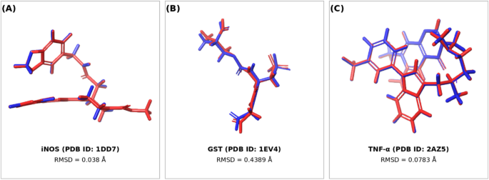
Re-docking of the corresponding co-crystallized ligands into the original binding sites of iNOS, GST, and TNF-α reproduced their experimentally determined binding orientations. The calculated heavy-atom RMSD values between the crystallographic and re-docked ligand poses were 0.0380 Å for iNOS, 0.4389 Å for GST, and 0.0783 Å for TNF-α. All RMSD values were markedly below the accepted threshold of 2.0 Å, indicating close agreement between the experimental and predicted ligand poses and supporting the reliability of the applied Glide XP docking protocol. Visual superposition of the crystallographic and re-docked poses further demonstrated close spatial overlap within the respective binding pockets (Fig. 1).
Fig. 1
Validation of the molecular docking protocol by re-docking analysis. Superposition of the crystallographic ligand poses (red) and their corresponding top-ranked re-docked poses (blue) within the binding sites of (A) iNOS, (B) GST, and (C) TNF-α. The calculated heavy-atom RMSD values were 0.038, 0.4389, and 0.0783 Å, respectively.
Docking of kaempferol with inducible nitric oxide synthase (iNOS)
Kaempferol demonstrated a high binding affinity toward inducible nitric oxide synthase (iNOS; PDB ID: 1DD7), as reflected by a favorable Glide docking score of − 9.199 kcal/mol. The compound was effectively positioned within the enzyme’s active site, where it established several stabilizing interactions. Notably, kaempferol acted as a hydrogen bond donor to TRP366 and as a hydrogen bond acceptor to MET368. The presence of these interactions supports the formation of a stable protein–ligand complex and suggests a potential inhibitory role of kaempferol against iNOS enzymatic activity (Fig. 2).
Fig. 2
Two-dimensional interaction diagram of Kaempferol docked into the active site of iNOS (PDB ID: 1DD7).
Docking of kaempferol with glutathione S-transferase (GST)
Docking simulations of kaempferol with glutathione S-transferase (GST; PDB ID: 1EV4) yielded a Glide docking score of − 5.691 kcal/mol, reflecting a moderate binding affinity. Within the GST binding pocket, kaempferol established hydrogen bond donor interactions with Val55 and Glu104, hydrogen bond acceptor interactions with Thr68, as well as π–cation interactions with Arg15. Although the predicted binding strength was lower than that observed for iNOS and TNF-α, the presence of these stabilizing contacts suggests that kaempferol may exert a modulatory influence on GST function, potentially impacting GST-mediated detoxification processes (Fig. 3).
Fig. 3
Two-dimensional interaction diagram of Kaempferol docked into the binding site of GST (PDB ID: 1EV4).
Docking of kaempferol with TNF-α
The most pronounced interaction was observed between kaempferol and TNF-α, as indicated by a favorable Glide docking score of − 6.820 kcal/mol. Kaempferol was positioned stably within the TNF-α binding region, forming two hydrogen bond donor interactions with key residues, namely Gln61 and Ser60, in addition to a π–π stacking interaction with Tyr119. Collectively, these interactions support the formation of a stable protein–ligand complex and suggest a potential inhibitory effect of kaempferol on TNF-α biological activity (Fig. 4).
Fig. 4
Two-dimensional interaction diagram of Kaempferol docked into the TNF-α binding site (PDB ID: 2AZ5).
Physicochemical characteristics of KMP-SeNPs
The dynamic light scattering analysis was used to determine the hydrodynamic size features of selenium nanoparticles synthesized with kaempferol (KMP-SeNPs). The nanoparticles had a Z-average diameter of 78.5 nm, which is indicative of a homogeneous size distribution in the nanoscale size range. The particle size profile as shown by the intensity proved to have a single, sharp peak at a value of around 80 nm, which suggests that the majority of the consistently scattered nanoparticles with a narrow distribution were present. The general quality of the measurement was good, and this proved the reliability of the size distribution data as in Fig. 5.
Fig. 5
DLS and zeta potential Description of SeNPs biosynthesized using kaempferol (KMP-SeNPs). (A) particle size distribution by intensity indicating nanoscale size and good dispersion, and (B) Zeta potential distribution showing a highly negative surface charge confirming colloidal stability of KMP-SeNPs.
Surfaces charge measurement also showed that KMP-SeNPs had a significantly negative zeta potential, having the mean value of −42.5 mV and a standard deviation of 4.10 mV. A high negative surface potential is evidence of strong electrostatic repulsive forces between nanoparticles which provide great colloidal stability and particle agglomeration resistance. Together with the other physicochemical characteristics, all of them indicate that the process of nanosized selenium particle green synthesis via kaempferol has been successful and that these particles can be used in further biological and therapeutic studies.
Transmission electron microscopy (TEM) analysis of KMP-SeNPs
TEM was used to explain the morphological and size properties of selenium nanoparticles that were biosynthesized using kaempferol (KMP-SeNPs). The TEM micrograph shows that the nanoparticles are mostly spherical with clear and smooth edges and dark appearance, which proves the successful development of elemental selenium in nanoscale. These particles are extensively dispersed across the field of vision with little aggregation, which constitutes a good stabilization of kaempferol in the process of synthesizing green as in Fig. 6.
Fig. 6
TEM image of SeNPs biosynthesized using kaempferol (KMP-SeNPs) (scale bar = 100 nm).
The majority of the nanoparticles are observed to have a narrow nanoscale size range, which can be well compared with the size of the particles observed in dynamic light scattering analysis. In another colloidal stability of KMP-SeNPs, the lack of big aggregates and the equal scattering of the sample also testify.
Cardiac enzymes
As presented in Figures 7, administration of 5-fluorouracil (5-FU) caused pronounced cardiovascular disease, as demonstrated by marked elevations in serum cardiac enzyme activities. Compared with the control group, 5-FU-treated rats exhibited substantial increases in CK-MB, troponin, and LDH by 77.17%, 106.25%, and 101.30%, respectively, indicating severe myocardial injury, disruption of cardiomyocyte membrane integrity, and enhanced leakage of intracellular enzymes into the circulation (p < 0.05).
Co-treatment with sodium selenite (Na₂SeO₃ & 5-FU) resulted in only partial amelioration of 5-FU-induced cardiac enzyme disturbances. Relative to the Normal Control group, CK-MB, troponin, and LDH levels remained elevated by 17.86%, 14.43%, and 35.27%, respectively. However, in contrast to the 5-FU group, these parameters showed moderate reductions of 33.50%, 44.53%, and 32.80%, reflecting a limited cardioprotective effect of inorganic selenium against 5-FU-induced myocardial damage (p < 0.05).
In contrast, kaempferol supplementation (KMP & 5-FU) produced a more pronounced protective effect on cardiac enzyme profiles. for the 5-FU group, serum CK-MB, troponin, and LDH activities were reduced by 38.43%, 37.04%, and 10.93%, respectively. Despite this improvement, enzyme levels remained greater than those observed in the Normal Control group, showing residual elevations of 9.07%, 29.82%, and 79.28%, suggesting partial but incomplete restoration of myocardial integrity (p < 0.05).
Notably, treatment with selenium nanoparticles biosynthesized using kaempferol (KMP-SeNPs & 5-FU) conferred the most substantial cardioprotective effect among all treated groups. In relation to the 5-FU group, CK-MB, troponin, and LDH concentrations were markedly reduced by 40.67%, 40.03%, and 26.56%, correspondingly. In contrast to the Normal Control group, enzyme activities exhibited only mild residual increases of 5.16%, 23.71%, and 47.81%, approaching near-physiological values and demonstrating superior preservation of myocardial structural and functional integrity (p < 0.05).
Fig. 7
Impact of kaempferol and kaempferol-mediated selenium nanoparticles on cardiac injury biomarkers in 5-fluorouracil–treated rats. (A) CK-MB, (B) cardiac troponin I (cTnI), and (C) LDH activities. The data, which includes statistical comparisons between groups, are presented as mean ± SEM (n = 5). *P < 0.05, **P < 0.01, ***P < 0.001, and ****P < 0.0001.
Oxidative stress and redox homeostasis markers
As presented in Figures 8, administration of 5-fluorouracil (5-FU) induced a profound oxidative stress state and severe disruption of redox homeostasis in cardiac tissue. Compared with the Normal Control group, 5-FU-treated rats exhibited marked elevations in Keap-1, malondialdehyde, and nitric oxide (NO) levels by 120.25%, 500.60%, and 170.67%, respectively. These changes were accompanied by pronounced suppression of endogenous antioxidant defenses, as evidenced by significant reductions in GSH, SOD, CAT, GR, GPx, and Nrf2 by 38.84%, 43.41%, 55.11%, 38.69%, 40.49%, and 41.82%, respectively, confirming excessive ROS and NOS production, antioxidant depletion, and inhibition of cytoprotective signaling pathways following 5-FU intoxication (p < 0.05).
Oral supplementation with inorganic selenium (Na₂SeO₃ & 5-FU) resulted in only partial mitigation of oxidative damage. Relative to the Normal Control group, Keap-1, MDA, and NO levels remained significantly elevated by 40.89%, 144.71%, and 41.29%, respectively, while antioxidant parameters including GSH, SOD, CAT, GR, GPx, and Nrf2 continued to show residual reductions of 20.34%, 20.06%, 27.56%, 16.89%, 14.11%, and 7.65%, respectively. When contrasted with the 5-FU group, these alterations reflected modest improvements in oxidative status, indicating a limited protective efficacy of inorganic selenium against 5-FU-induced redox imbalance (p < 0.05).
In contrast, kaempferol supplementation (KMP & 5-FU) exerted a more substantial antioxidative effect. Compared with the 5-FU group, Keap-1, MDA, and NO levels were significantly reduced by 30.75%, 52.18%, and 39.98%, respectively. Concurrently, GSH, SOD, CAT, GR, GPx, and Nrf2 levels were markedly restored by 45.16%, 44.53%, 66.23%, 31.42%, 28.60%, and 40.66%, respectively. Despite these improvements, most oxidative stress and antioxidant parameters did not fully return to Normal Control values, indicating partial but incomplete restoration of redox homeostasis (p < 0.05).
Notably treatment with selenium nanoparticles biosynthesized using kaempferol (KMP-SeNPs & 5-FU) produced the most pronounced normalization of oxidative stress biomarkers. Relative to the 5-FU group, Keap-1, MDA, and NO levels declined markedly by 50.05%, 77.60%, and 63.06%), respectively, approaching near-physiological values. Simultaneously, antioxidant defenses were robustly enhanced, with GSH, SOD, CAT, GR, GPx, and Nrf2 levels increasing by 75.28%, 76.12%, and 122.38%, 63.29%, 63.34%, and 62.02%), respectively. By comparing it with the Normal Control group.
Fig. 8
Effects of kaempferol and kaempferol-mediated selenium nanoparticles on oxidative stress and antioxidant defense in 5-fluorouracil–induced toxicity. (A) Keap-1, (B) MDA, (C) NO, (D) GSH, (E) SOD, (F) CAT, (G) GR, (I) GPx, and (J) Nrf2 expression. Data are presented as mean ± SEM (n = 5) with statistical comparisons among groups. *P < 0.05, **P < 0.01, ***P < 0.001, and ****P < 0.0001.
Inflammatory cytokines and immunoregulatory gene expression
As illustrated in Figures 9, administration of 5-fluorouracil (5-FU) induced a marked inflammatory reaction accompanied by profound disruption of immune regulatory signaling. Compared with the Normal Control group, TNF-α and IL-6 values in 5-FU-administered rats escalated significantly by 192.95%, and 333.54%, respectively, while IL-10 was significantly reduced by 70.31%.
At the molecular level, expression of NF-κB was strongly upregulated by 175.45%, whereas expression of the regulatory T-cell marker FOXP3 was markedly downregulated by 75.08%. These findings indicate excessive inflammatory activation, cytokine imbalance, and impairment of immunoregulatory mechanisms following 5-FU exposure (p < 0.05) (Figs. 10).
Co-treatment with sodium selenite (Na₂SeO₃ & 5-FU) resulted in partial improvement of inflammatory and transcriptional disturbances. Relative to control group, TNF-α and IL-6 values remained elevated by 39.90% and 99.17%, respectively, while IL-10 showed a modest reduction by 12.15%. NF-κB expression persisted above basal levels by 54.45%, and FOXP3 expression remained reduced by 18.07%. When evaluated against the 5-FU group, sodium selenite reduced TNF-α and IL-6 by 52.27% and 54.06%, respectively, increased IL-10 by 195.68%, suppressed NF-κB expression by 43.92%, and enhanced FOXP3 expression by 228.58%, indicating a moderate but incomplete anti-inflammatory and immunoregulatory effect (p < 0.05).
In contrast, kaempferol supplementation (KMP & 5-FU) produced a more pronounced attenuation of inflammatory signaling. In contrast to 5-FU group, TNF-α and IL-6 levels declined by 33.53%, and 50.17%, respectively, while IL-10 increased substantially by 173.23%. Concurrently, NF-κB expression was reduced by 36.82%, and FOXP3 expression was restored by 213.86%. However, relative to the control group, TNF-α, IL-6, and NF-κB remained elevated by 94.78%, 116.09%, and 73.99%, respectively, and FOXP3 expression remained reduced by 21.75%, reflecting partial recovery of inflammatory and immunoregulatory balance (p < 0.05).
Notably, treatment with selenium nanoparticles biosynthesized using kaempferol (KMP-SeNPs & 5-FU) exerted the most robust protective effect. Notably, the 5-FU group, TNF-α and IL-6 were markedly reduced by 56.45%, and 71.36%, respectively, while IL-10 increased by 213.27%. In parallel, NF-κB expression was strongly suppressed by 58.55%, and FOXP3 expression was markedly enhanced by 287.48%. Compared with the control group, TNF-α, IL-6, and NF-κB exhibited only minimal residual elevations by 27.59%, 24.13%, and 14.18%, respectively, while FOXP3 expression was nearly normalized by 3.41%, demonstrating near-complete restoration of inflammatory and immunoregulatory homeostasis (p < 0.05) (Figs. 9 and 10).
Fig. 9
Effects of kaempferol and kaempferol-mediated selenium nanoparticles on inflammatory cytokines in 5-fluorouracil–induced toxicity. (A) TNF-α, (B) IL-6, and (C) IL-10 protein values. Data are presented as mean ± SEM (n = 5) with statistical comparisons among groups. *P < 0.05, **P < 0.01, ***P < 0.001, and ****P < 0.0001.
Fig. 10
Effects of kaempferol and kaempferol-mediated selenium nanoparticles on gene expression in 5-fluorouracil–induced toxicity. mRNA expression of (A) NF-κB and (B) FOXP3. Data are presented as mean ± SEM (n = 5) with statistical comparisons among groups. *P < 0.05, **P < 0.01, ***P < 0.001, and ****P < 0.0001.
Apoptotic biomarkers
As illustrated in Figures (11), administration of 5-FU triggered a pronounced apoptotic response in cardiac tissue, as reflected by marked alterations in key pro- and anti-apoptotic markers. In contrast to the control group, 5-FU-injected rats exhibited significant increases in Bax and Caspase-3 by 83.38%, and 215.22%, respectively, accompanied by a substantial reduction in Bcl-2 by 49.60%. These findings indicate excessive activation of apoptotic signaling pathways and loss of cellular survival mechanisms following 5-FU exposure (p < 0.05).
Co-treatment with sodium selenite (Na₂SeO₃ & 5-FU) resulted in partial attenuation of apoptosis-related disturbances. Relative to the control group, Bax and Caspase-3 levels remained elevated by 32.19%, and 45.33%, respectively, while Bcl-2 remained reduced by 15.57%. When compared with the 5-FU group, sodium selenite administration reduced Bax and Caspase-3 by 27.93%, and 53.90%, respectively, and increased Bcl-2 by 67.45%, indicating a moderate anti-apoptotic effect that was insufficient to fully restore apoptotic balance (p < 0.05).
In contrast, kaempferol supplementation (KMP & 5-FU) exerted a more pronounced modulatory effect on apoptotic signaling. Compared with the 5-FU group, Bax and Caspase-3 levels declined by 32.49%, and 49.38%, respectively, while Bcl-2 levels increased by 44.68%. However, relative to the control group, Bax and Caspase-3 remained elevated by 23.87%, and 59.54%, respectively, and Bcl-2 remained reduced by 27.04%, indicating partial but incomplete suppression of apoptosis (p < 0.05).
Notably, treatment with selenium nanoparticles biosynthesized using kaempferol (KMP-SeNPs & 5-FU) produced the most substantial anti-apoptotic effect. Relative to the 5-FU group, Bax and Caspase-3 levels were markedly reduced by 42.86%, and 66.59%, respectively, while Bcl-2 expression was significantly enhanced by 68.13%. Compared with the control group, Bax and Caspase-3 exhibited only minimal residual increases of 4.77%, and 5.27%, respectively, and Bcl-2 levels approached near-physiological values 15.23%, indicating near-complete restoration of apoptosis-regulatory balance and highlighting the superior cytoprotective efficacy of KMP-SeNPs against 5-FU-caused cardiac injury (p < 0.05).
Fig. 11
Effects of kaempferol and kaempferol-mediated selenium nanoparticles on apoptotic markers in 5-fluorouracil–induced toxicity. (A) Bax, (B) Caspase-3, and (C) Bcl-2 values in experimental groups. Data are presented as mean ± SEM (n = 5) with statistical comparisons among groups. *P < 0.05, **P < 0.01, ***P < 0.001, and ****P < 0.0001.
Immunohistochemical examination of kaempferol-mediated selenium nanoparticles on Nrf-2 and Keap-1
IHC analysis revealed a marked expression of the NRF-2 in cardiac tissue of control, KMP-SeNPs&5FU, and KMP-5FU treated groups without prominent variance (p > 0.05), whereas the Na2SeO3&5FU and 5FU groups had weak expression. Both KMP&5FU group and KMP-SeNPs&5FU groups differed significantly with 5FU regarding Nrf-2 IHC immunoreactivity (P < 0.001) (Fig. 12).
Fig. 12
Protective effect of KMP-SeNPs on cardiac immunoreactivity of Nrf-2 in 5FU-induced cardiotoxicity in rats. Photomicrographs of tissue of the heart from all cohorts. The reactivity of Nrf-2 was evident in the tissues as a brown tone produced by DAB chromogen. (DAB, ×400). (A) The control group showed normal cardiac muscle structure with moderate Nrf-2 staining. (B) 5FU and (D) Na2SeO3&5FU treated groups revealed weak Nrf-2 immunostaining. In contrast, (C) KMP&5FU group revealed moderate expression, while (E) KMP-SeNP&5FU-treated group displayed moderate Nrf-2 staining. (F) Quantitative assessment of immunostaining region % for Nrf-2 was expressed as mean ± SEM (n = 5). Statistical evaluation utilizing one-way ANOVA accompanied by Duncan’s post hoc analysis. ns: not significant; *P < 0.05, **P < 0.01, ***P < 0.001, and ****P < 0.0001.
Moreover, Keap1 immunohistochemical staining was mild in the cardiac tissues of the control, KMP-SeNPs&5FU, and KMP&5FU groups. Although both treatment groups exhibited slightly higher Keap1 expression than the control group, the difference was statistically significant. In contrast, the Na2SeO3&5-FU showed moderate expression and 5-FU groups showed moderate Keap1 immunoreactivity (Fig. 13). Furthermore, the KMP-SeNPs&5FU group exhibited significantly lower Keap1 immunoreactivity than the 5FU group (P < 0.001).
Fig. 13
Protective effect of KMP-SeNPs on cardiac immunoreactivity of Keap-1 in 5FU-induced cardiotoxicity in rats. Photomicrographs of cardiac tissues from all groups. The eactivity of Keap-1 was observed in the tissues as a brown hue generated by the DAB chromogen. (DAB, ×400). (A) The control group showed normal cardiac muscle structure with weak Keap-1 staining. (B) 5FU and treated groups revealed marked Keap-1 immunostaining. In contrast, (D) Na2SeO3&5FU group revealed moderate expression, while (C) KMP&5FU and (E) KMP-SeNP&5FU-treated group displayed mild Keap-1 staining. (F) Quantitative assessment of immunostaining region % for Keap-1 was expressed as mean ± SEM (n = 5). Statistical analysis by one-way ANOVA with Duncan’s post hoc test. ns: non-significant; *P < 0.05, **P < 0.01, ***P < 0.001, and ****P < 0.0001.
Histopathological examination
The histological analysis of the control group demonstrated typical heart tissue architecture, including branched muscle fibers with central oval vesicular nuclei, acidophilic sarcoplasm, and extracellular gaps with blood capillaries Figure (14 A). The 5FU group revealed symptoms of cardiac necrosis, including substantial fragmentation, hypereosinophilia, perimysial edema, and nuclear pyknosis. The interstitial oedema in the tissue gaps increased significantly Figure (14B). Na2SeO3−5FU treated group showed limited improvement compared to 5FU group with pyknotic myocytes and perimysial edema Figure (14 C). The morphological appearance in the KMP&5FU-treated group was significantly improved, with myocytic morphology preserved and interstitial capillaries eliminated Figure (14D). The KMP-SeNPs&5FU-treated group displayed an approximately normal histology pattern of cardiac myocytes Figure (14E).
Fig. 14
Histopathological analysis of cardiac tissues. (A) Photomicrograph of the cardiac tissue of control rat showing normal histological structure cardiac myocytes with acidophilic myocardial fibers, oval vesicular nuclei (N). (B) Section from 5FU-treated group showed necrotic muscle fibers (*) with pyknotic cardiomyocytes (short arrow), widely separated myocytes (double arrow) and inflammatory cellular infiltration (long arrow). (D) Na2SeO3&5FU group showed perimysial edema (double arrow) and inflammatory cellular infiltration (long arrow), and apoptotic cell (short arrow). Both (C) KMP&5FU and (E) KMP-SeNPs&5FU-treated group showed preserved cardiomyocyte morphology with central oval vesicular nuclei (N), exhibiting approximately normal structure. (H&E, ×400).
