


Details on key resource are shown in Table 1.
Table 1 Material information.
Cell culture
All cell lines were maintained at 37 °C in a humidified incubator with 5% CO2. Human AML cell lines including U937 (RRID: CVCL_0007), HL-60 (RRID: CVCL_0002) and THP1 (RRID: CVCL_0006) were obtained from the Cell Bank of Type Culture Collection, Chinese Academy of Sciences (CAS; Shanghai, China), and cultured in RPMI-1640 medium (Gibco, Grand Island, NY, USA) supplemented with 10% fetal bovine serum (FBS; ABM). Human BMSCs (HS-5; RRID: CVCL_3720; ATCC, Manassas, VA, USA) and HEK293T cells (CAS; RRID: CVCL_0063) were cultured in high-glucose Dulbecco’s Modified Eagle’s Medium (DMEM; Gibco) supplemented with 10% FBS. All cell lines underwent short tandem repeat (STR) profiling for authentication and were routinely tested and confirmed to be free of mycoplasma contamination.
Extraction of acute myeloid leukaemia blasts and bone marrow mesenchymal stem cells
This study was approved by the Committee for the Ethical Review of Research, Fujian Medical University Union Hospital (2023KJCX044). Human bone marrow aspirates were collected from patients with AML prior to treatment at Fujian Medical University Union Hospital, Fuzhou, China. Details on samples from patients with AML are shown in Table 2. AML blasts were purified using CD34 microbeads (Miltenyi Biotec, Bergish-Gladbach, Germany). The extraction of bone marrow mesenchymal stromal cells were carried out in accordance with previous reports23. Briefly, mononuclear cells (MNCs) were isolated using Ficoll Paque density centrifugation (TBD, Tianjin, China). MNCs were seeded with a density of 5 million cells/6-cm dish in RPMI1640 (Gibco) supplemented with 20% FBS (ABM). Non-adherent cells were removed and adherent bone marrow mesenchymal stem cells were passaged with a confluence of 80% and expanded up.
Table 2 Patient’s information.
In vitro co-culture of acute myeloid leukaemia cells with stromal cells
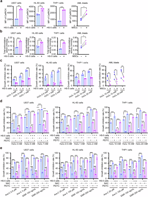
HS-5 cells or bone mesenchymal stem cells were seeded at a density of 1.0 × 10⁵ cells per well in six-well plates. Upon reaching ~70% confluence, the medium was replaced with 2 ml of complete RPMI-1640 medium containing 10% FBS and 5.0 × 10⁵ AML cells or 10.0 × 10⁵ AML blasts. After 24 h of co-culture, AML cells expressing human CD45 or CD34 were isolated using anti-human CD45/CD34 immunomagnetic beads (Miltenyi Biotec).
CCK-8 cell viability assay
AML cells (2 × 10⁴/well) were seeded in 96-well plates and treated with cytarabine (Ara-C, Pharmacia, Italy), daunorubicin (DNR, Shandong New Era Pharmaceutical Co. Ltd, Linyi, China) or H2O2 (Sigma-Aldrich, St. Louis, Missouri, USA) for 24 h. Cell viability was assessed using the Cell Counting Kit-8 (Dojindo, Tokyo, Japan) as per the manufacturer’s protocol. After 4-h incubation at 37 °C, absorbance was measured at 450 nm and 630 nm using a SpectraMax i3X microplate reader (Molecular Devices, Silicon Valley, CA, USA). Growth inhibition rate was calculated as (mean OD450–630 of negative control cells − mean OD450–630 of drug-treated cells) / (mean OD450–630 of negative control cells − mean OD450–630 of blank control) × 100%. Each condition was tested in triplicate and repeated in three independent experiments.
Apoptosis assay
AML cells from co-culture or monoculture were harvested, and 5.0 × 10⁵ cells were stained with 5 μl Annexin V-APC and 5 μl 7-AAD (KeyGen, Jiangsu, China) for 15 min at room temperature in the dark. Apoptosis was analysed by flow cytometry (Celesta, BD Biosciences, Franklin Lake, NJ, USA) with 30,000 events per sample.
Live/dead cell staining
Cells (5.0 × 10⁵) were stained with 0.5 μl Fixable Viability Dye eFluor 450 (eBioscience, San Diego, CA, USA) in 500 μL for 30 min on ice in the dark. Live (FVD–) and dead (FVD+) populations were analysed by flow cytometry (30,000 events/sample).
Total antioxidant activity assay
The total levels of various antioxidant macromolecules, antioxidant small molecules and enzymes within a system represent the overall antioxidant activity of that system. AML cells were lysed by ultrasonication in PBS, and total antioxidant capacity was quantified using the Total Antioxidant Capacity Assay Kit (Beyotime, Shanghai, China). Absorbance at 414 nm was measured using a microplate reader. Each assay included two technical replicates and was independently repeated three times.
8-hydroxy-2’-deoxyguanosine (8-OHdG) ELISA assay
Following 24-h Ara-C exposure, genomic DNA was extracted using the Wizard Genomic DNA Purification Kit (Promega, Madison, WI, USA), and DNA content was quantified by NanoDrop (Thermo Fisher Scientific, Waltham, MA, USA). 8-OHdG levels were determined from 2 μg DNA using an ELISA kit (Cusabio, Wuhan, China) as per the manufacturer’s instructions, and absorbance at 450 nm was measured. Data are expressed as relative 8-OHdG levels, with untreated cells or cells infected with the empty vector are normalized to 1. Values are shown as mean ± standard error of the mean from technical triplicates.
Intracellular and mitochondrial reactive oxygen species detection
AML cells (5.0 × 10⁵) were stained with CellROX Deep Red (500 nM, Thermo Fisher Scientific) or MitoSOX Red (3 μM, Thermo Fisher Scientific) in Hank’s buffer for 45 or 30 min, respectively, at 37 °C. After two washes, fluorescence was analysed by flow cytometry. Viable cells (FVD–) were gated, and 30,000 events were collected per sample.
Intracellular and mitochondrial Ca2+ measurement
For cytosolic Ca2+, cells were incubated with Calbryte 630 AM (5 μM, AAT Bioquest, Sunnyvale, CA, USA) in Hank’s buffer containing 0.04% Pluronic F-127 (Thermo Fisher Scientific) and 1 mM probenecid (MCE, Monmouth Junction, NJ, USA) for 45 min. Intracellular Ca2+ was detected by flow cytometry. For calcium flux dynamics of AML cells, AML cells stained with Calbryte 630 AM were stimulated by BzATP and AZ10606120, and the changes of intracellular Ca2+ in the cells were recorded using a fluorescent microplate reader. For mitochondrial Ca2+, cells were stained with 5 μM Rhod-2 AM (Thermo Fisher Scientific) under similar conditions, then incubated for 6 h in complete medium to clear cytoplasmic dye. Mitochondrial Ca2+ levels were measured by a microplate reader. Data are presented as relative fluorescence intensity, with untreated cells or cells infected with the empty vector normalized to 1.
Western blot analysis
Whole-cell lysates were prepared in RIPA buffer containing PMSF, phosphatase and protease inhibitors, then denatured in 5× SDS buffer. Proteins were separated by SDS-PAGE, transferred to 0.45-μm PVDF membranes (Millipore, Boston, MA, USA), blocked with 8%–10% milk, and incubated with primary antibodies overnight (details on antibody information are shown in Table 1). HRP-conjugated secondary antibodies were applied, and signals detected using a Bio-Rad chemiluminescence system (Hercules, CA, USA). Band intensity was quantified using ImageJ.
Generation of stable cell lines
The human P2RX7 overexpression plasmid (Ubi-MCS-SV40-puromycin) was obtained from GeneChem (Shanghai, China). sgRNAs targeting P2RX7, Nrf2, Keap1 and PGAM5 (all exon 2) were synthesized by Sangon Biotech (Shanghai, China) and cloned into the LentiCRISPRv2 vector (Addgene, Cambridge, MA, USA). sgRNA sequences were as follows:
(1) P2RX7: sg1: 5’-TGATGACAGGCTCTTTCCGC-3’; sg2: 5’-GTTGTGTCCCGAGGTAAGGA-3’; (2) Nrf2: sg1: 5’-GCGACGGAAAGAGTATGAGC -3’; (3) Keap1: sg1: 5’-GGTCAAGTACCAGGATGCAC-3’, sg2: 5’-TGTGTCCTCCACGTCATGAA-3’); (4) PGAM5: sg1: 5’-GAAGAGGAACGTGGAATCTG-3’.
HEK293T cells (CAS) were co-transfected with the target plasmid, psPAX2, and pMD2.G (ratio 4:3:1) using Lipofectamine 3000 (Thermo Fisher Scientific). Viral supernatants were harvested at 24 and 52 h, filtered (0.45 μm) and concentrated using 25% PEG 8000 in 0.75 M NaCl (1∶4 ratio) overnight at 4 °C, then centrifuged at 4000 × g for 20 min. Pellets were resuspended in growth media and stored at −80 °C. AML cells were transduced with the lentiviral particles and selected with puromycin (2.5 μg/ml; Solarbio, Shanghai, China) for 10 days. Knockout efficiency was confirmed by Western blot; overexpression was validated by real-time quantitative PCR (RT-qPCR) and Western blot.
Real-time quantitative PCR
Total RNA was extracted with TRIzol (Thermo Fisher Scientific), and cDNA was synthesized using a Reverse Transcription Kit (Thermo Fisher Scientific). RT-qPCR was performed using Blastaq 2× qPCR MasterMix (ABM, Vancouver, Canada) on a 7500 system (Applied Biosystems, New York, USA). ACTB was used for normalization, and the 2–ΔΔCt method was applied. Primer sequences were as follows:
(1) P2RX7:
forward: 5’- GGGCGGAATAATGGGCATTGAG -3’;
reverse: 5’- AAGGCGACGGAAACTGTATTTGG -3’.
(2) Nrf2:
forward: 5’- CACATCCAGTCAGAAACCAGTGG -3’;
reverse: 5’- GGAATGTCTGCGCCAAAAGCTG -3’.
(3) ACTB:
forward: 5’- CCAACCGCGAGAAGATGA -3’;
reverse: 5’- TCCATCACGATGCCAGTG -3’.
Luciferase reporter assay
Three tandem copies of the antioxidant response element (ARE) sequence (5’-GTGACAAAGCAATCCCGTGACAAAGCAATCCCGTGACAAAGCAATAAAGCTTAGACACTAGAGGGTATATAATGGAAGCTCGACTTCCAG-3’) were cloned into a firefly luciferase reporter plasmid (F-luc, GeneChem). AML cells were co-transfected with equal amounts of the ARE-F-luc construct and a renilla luciferase plasmid (R-luc, GeneChem) to control for transfection efficiency. After 48 h, cells were co-cultured with HS-5 stromal cells for 24 h. Luciferase activity was measured using the Dual-Luciferase Reporter Assay System (Promega), and relative transcriptional activity was calculated as the ratio of firefly to renilla luciferase (F-luc/R-luc), normalized to a value of 1.
Immunoprecipitation
Cells were lysed in NP-40 lysis buffer supplemented with PMSF and protease inhibitor cocktail for 30 min at 4 °C. Lysates were pre-cleared with 25 μl of protein A/G magnetic beads (Millipore) at room temperature, and then incubated with primary antibodies overnight at 4 °C with rotation. Subsequently, 50 μl of fresh beads were added and incubated for 30 min at room temperature. Bead-bound immunocomplexes were washed three times with 0.05% PBST and eluted in 5× SDS loading buffer. Eluates were analysed by Western blot.
Ubiquitination assay
To assess Nrf2 ubiquitination, AML cells were lysed in NP-40 buffer containing PMSF and protease inhibitors. Endogenous Nrf2 was immunoprecipitated as described above, and ubiquitinated Nrf2 was detected by Western blot using an anti-ubiquitin antibody (Abcam, Cambridge, UK).
Mice
All animal procedures were approved by the Animal Ethics Committee of Fujian Medical University (Protocol No. IACUC FJMU 2024-0279) and performed in accordance with institutional guidelines. 4-week-old male NOD/ShiLtJGpt-Prkdcem26Cd52Il2rg em26Cd22/Gpt (NCG) mice and 8-week-old male C57BL/6 J mice were purchased from Guangdong Yakult Biotech Co., Ltd (China) and housed in a specific pathogen-free facility with ventilated cages. Mice were maintained under standard 12-h light/dark cycles with controlled temperature and humidity, and provided with autoclaved food and water ad libitum. Mice were randomly assigned to experimental groups. Each group comprised mice housed 5 per cage, with animal numbers indicated in the corresponding figure legends. Mice were monitored daily for signs of distress, and humane endpoints included ≥20% weight loss, hindlimb paralysis, severe hunching or limited mobility. All in vivo studies were conducted in the Animal Resources Center of Fujian Medical University.
Acute myeloid leukaemia xenotransplantation in NOD/ShiLtJGpt-Prkdc
em26Cd52
Il2rg
em26Cd22 /Gpt mice
AML xenografts were established by injecting 4.0 × 10⁵ AML cells suspended in 200 μl PBS into NCG mice via the tail vein. Two weeks post-injection, mice were randomly assigned to four treatment groups: vehicle control, Ara-C (20 mg/kg/day), AZ10606120 (10 mg/kg/day), or a combination of both agents. Treatments were administered intravenously once daily for 5 consecutive days. Mice were euthanized upon signs of terminal illness (e.g. hindlimb paralysis, coma). AML burden was quantified by flow cytometry using APC-conjugated anti-human CD45 antibody (Biolegend, San Diego, CA, USA) following mechanical dissociation and erythrocyte lysis of liver, spleen and bone marrow tissues. To assess antioxidant capacity in residual AML cells from bone marrow, human CD45+ cells were isolated using magnetic beads (Miltenyi Biotec), and total antioxidant activity and 8-OHdG levels were measured ex vivo.
MLL-AF9 driven murine acute myeloid leukaemia model
MLL-AF9 AML cells were generated as previously described24,25. Briefly, the retroviral constructs MSCV-MLL-AF9-IRES-YFP (30 μg) along with the packaging plasmid pCL-ECO (15 μg) were co-transfected into HEK293T cells using Lipofectamine 3000 to produce the MLL-AF9 retrovirus. Lineage-negative foetal liver cells isolated from C57BL/6 J embryos were transduced with MLL-AF9 retrovirus and then intravenously injected into sublethal irradiation (6.5 Gy) C57BL/6 J mice (8–10 weeks old). YFP+ bone marrow cells were sorted from primary recipient mice by flow cytometry and transplanted into non-irradiated secondary recipients at a dose of 2.0 × 10⁵ cells per mouse. MLL-AF9 leukaemic mice of secondary recipients were administered vehicle, Ara-C (100 mg/kg/day), AZ10606120 (10 mg/kg/day), or a combination of both agents for 5 days via intraperitoneal injection. AML burden was analysed by flow cytometry and then YFP+ bone marrow cells were sorted to assess the antioxidant capacity.
Statistical analysis
All statistical analyses were performed using SPSS version 25. Data distribution was tested for normality using the Shapiro–Wilk test. For normally distributed data, data are presented as mean ± standard error of the mean or standard deviation and compared using Student’s t test (for two groups) or one-way analysis of variance followed by Dunnett’s post hoc test (for multiple groups). For non-normally distributed data, data are presented as median with interquartile range and compared using the Mann–Whitney U test or Kruskal–Wallis test. Survival analysis was performed using Kaplan–Meier survival curves for the mice, and statistical significance was determined using the log-rank (Mantel–Cox) test. A P value < 0.05 was considered statistically significant.
