


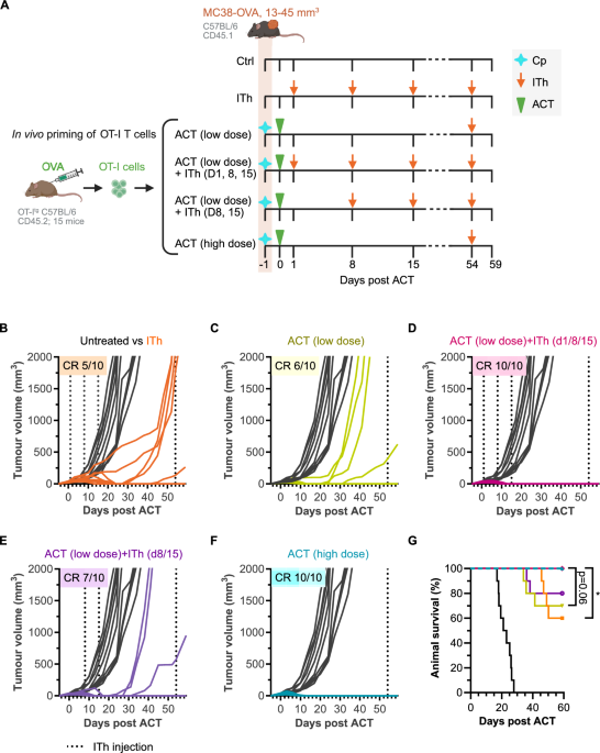
Our results demonstrate that combining mRNA immunotherapy with TCRtg T cell transfer more effectively suppressed tumour growth and improved survival in MC38‑OVA–bearing mice compared with either treatment alone. mRNA immunotherapy promoted expansion, reactivation, tumour infiltration and an effector phenotype of the transferred OT-I T cells. It also induced endogenous, antigen-specific CD8+ T cells, enhancing the OVA-specific immune response. Together, these data suggest that the addition of mRNA immunotherapy has the potential to overcome some of the main challenges of ACT therapy against solid tumours – even without prior lymphodepletion. Notably, full therapeutic efficacy required early initiation of mRNA immunotherapy, starting one day after low-dose ACT. While a delayed start at Day 8 led to comparable OT-I T cell expansion at later time points (Day 40 & Day 54), fast expansion of the transferred CD8+ T cells at early time points (e.g., Day 11) was likely critical for controlling the rapidly growing tumours.
The lack of durable efficacy represents one of the major challenges of ACT, reflected by frequent therapy failure after initial clinical responses15. Limited persistence of transferred T cells is one of the major factors contributing to relapse. In addition, conventional ex vivo expansion protocols, often relying on IL-2, may drive differentiation toward effector phenotypes and reduce the proportion of less differentiated, stem-like T cells, which have been associated with improved persistence and long-term responses26. Our findings suggest that these limitations may be mitigated by intermittent administration of LNP-encapsulated mRNA encoding the target antigen in the recipient in vivo, even at a late time point when memory T cells are present in very low numbers.
The tumour relapse observed following ITh or low-dose ACT monotherapy might be caused by limited persistence and/or progressive functional impairment of T cells, including exhaustion or anergy. In addition, recognition by antigen-specific CD8⁺ T cells could be impaired by the emergence of antigen-loss or MHC class I–deficient tumour variants.
Patients often receive IL-2 after ACT to expand the transferred T cells, which can be associated with severe systemic side effects caused by the activation of bystander host cells12. Prolonged IL-2 administration can also result in the expansion and activation of CD4+ Foxp3+ regulatory T cells, restricting administration to short term usage27. In our study, administration of mRNA immunotherapy led to a strong in vivo expansion of transferred T cells, even at later timepoints where minimal numbers of remaining TCRtg T cells were observed, suggesting that the addition of mRNA immunotherapy has the potential to enhance survival and longevity of transferred T cells. Importantly, our results demonstrate that TCRtg T cells derived from OT-I mice do not require IL-2 administration for mRNA immunotherapy-mediated expansion after adoptive transfer. If this finding extends to ex vivo engineered and expanded T cells currently used for ACT in the clinic, it could potentially reduce the risk of IL-2-associated toxicities.
The long-term clinical efficacy of ACT is frequently hampered by a loss of effector functions, as chronic stimulation during ex vivo expansion can lead to increased frequencies of dysfunctional or exhausted T cells15. mRNA immunotherapy can reactivate TCRtg T cell effector function in vivo. Strong reactivation of expanded TCRtg T cells was confirmed by the expression of PD-1 and TIM3, with additional SLAMF6 expression indicating that the cells had retained their self-renewal capacity. Interestingly, SLAMF6 expression in T cells has been found to correlate positively with checkpoint inhibitor response28, elevated immune activity and better prognosis in both breast cancer and melanoma29. In addition, an induction of a heterogenous pool of memory T cells, including persistent effector memory CD8+ T cells with the capability for fast expansion to antigen re-exposure and memory precursor cells was shown.
Effective T cell infiltration into solid tumours is crucial for ACT success30. In our study, we have presented evidence that mRNA immunotherapies can significantly increase the number of tumour-infiltrating TCRtg T cells correlating with the enhanced number of TCRtg T cells in the periphery. Importantly, the tumour-infiltrating TCRtg T cells featured improved cytotoxic potential, as shown by increased Granzyme B expression, indicating that administration of mRNA immunotherapy may improve not only the quantity, but also the quality of the T cell response.
Tumours often escape immunotherapy by genetic or epigenetic suppression of antigen expression and presentation pathways31. As TCRtg T cell therapy relies on presentation of a single target epitope, induction of intrinsic tumour escape mechanisms including emergence of antigen-loss variants or loss of epitope presentation by loss or down-regulation of the respective HLA class I allele expression represent formidable risks31,32. Addition of an mRNA immunotherapeutic encoding additional tumour antigens could hypothetically broaden the immune response beyond the antigen targeted by the TCRtg T cells and potentially decrease the risk of immune evasion. Such additional tumour antigens might even be more efficient in priming endogenous T cells than the antigen recognized by the transgenic TCR because they avoid competition between naïve endogenous and transferred TCRtg T cells, an effect which likely explains the lower induction of endogenous SIINFEKL-specific CD8+ T cells by ITh in the presence of ACT (Fig. 4C, D). In the current work we observed induction of endogenous CD8+ T cell responses against the immunodominant CD8 epitope of the mRNA-encoded antigen OVA in mice without lymphodepletion, while potentially increased breadth of the response remains to be shown in future studies.
Before receiving ACT, patients undergo lymphodepleting chemotherapy, potentially causing long-term cytopenia33. Prolonged systemic side effects such as grade 3 or 4 anaemia, thrombocytopenia, neutropenia, leukopenia, or lymphopenia, and an increased infection risk are common consequences of lymphodepleting chemotherapy3. The main aim of lymphodepletion is the reduction of endogenous lymphocytes to improve engraftment, homing and survival of transferred T cells13. Indeed, we observed prominent expansion of transferred T cells only when ACT was combined with mRNA immunotherapy in lymphodepleted hosts. We also observed that lymphodepletion was strictly required for tumour growth inhibition when mice were treated with ACT monotherapy. These observations are consistent with recent work34 demonstrating that lymphodepleting preconditioning enhances expansion of transferred T cells and promotes elimination of primary tumours, but at the same time compromises host antitumor immunity and reduces protection against antigen-loss variants that are resistant to ACT. Accordingly, while mRNA immunotherapy induced endogenous T cell responses against the encoded antigen in our study, these responses were markedly reduced following lymphodepletion prior to ACT. Interestingly, our data demonstrates that robust anti-tumour responses against large tumours can be achieved by combining TCRtg T cell therapy and mRNA immunotherapy without lymphodepletion. As such, our data suggests that addition of mRNA immunotherapy to ACT may eliminate the need for lymphodepletion, thereby reducing severe ACT-induced side effects, as well as the observed reduction of mRNA-induced endogenous T cell responses in mice receiving combination therapy with lymphodepletion.
Finally, the timely generation of sufficient numbers of TCRtg T cells to achieve effective treatment remains a major challenge14. In this study, combining a low dose of TCRtg T cells with mRNA immunotherapy resulted in sustained therapeutic efficacy, suggesting that mRNA immunotherapy may help overcome this limitation by enabling the infusion of lower T‑cell numbers followed by in vivo expansion of the transferred cells.
Our preclinical observations in a murine model highlight the potential of combining TCRtg T cell therapy with mRNA-based ITh. Further studies are required to validate these findings in the human setting and to assess their applicability to clinically established TCRtg T cell therapy protocols. To this end the current study has several limitations. First, the model employed TCRtg CD8⁺ T cells derived from transgenic mice following in vivo activation, in contrast to the ex vivo engineered, expanded and polyclonal human T cells used in clinical practice. Second, the engineered MC38‑OVA model may not fully capture the heterogeneity and antigen amounts observed in clinical settings. Nonetheless, as the tumour cells express only moderate amounts of OVA that alone do not induce measurable SIINFEKL-specific CD8+ T cell responses, they can serve as a valuable model. Third, the SIINFEKL peptide targeted in this system exhibits high affinity for both the presenting MHC molecule and the transgenic OT‑I TCR; therefore, additional target antigens and tumour models should be evaluated to confirm generalizability. Fourth, while the capacity of ITh to induce T cell responses specific to the encoded tumour antigens has been well established35, further studies using mRNAs encoding antigens not recognized by the transferred TCRtg would help to substantiate this effect in the context of TCRtg T cell therapy. Finally, the pronounced expansion of antigen-specific OT-I T cells following mRNA immunotherapy supports a dominant role for antigen-driven stimulation. However, as an irrelevant mRNA control was not included in the experimental design it cannot be excluded that innate immune activation induced by the mRNA–LNP formulation contributes to the observed therapeutic effects. Future studies incorporating control mRNAs will be required to delineate the relative contributions of antigen-specific versus antigen-independent mechanisms.
