


Biological insights from spatial omics into tissue architecture and disease states in pancreatic ductal adenocarcinoma and its precursor lesions
Spatial organization and heterogeneity in pancreatic ductal adenocarcinoma
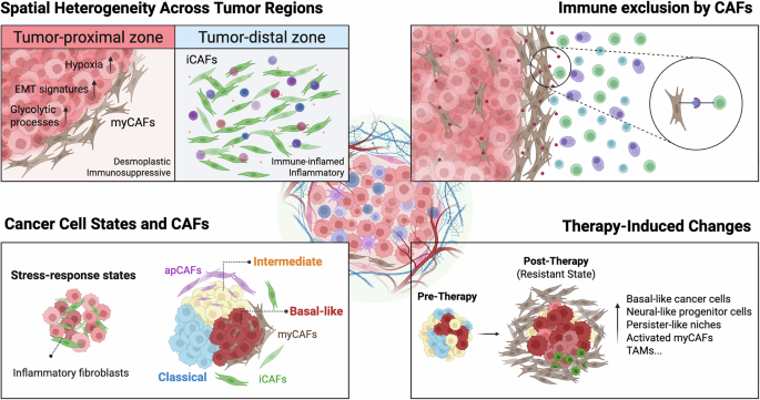
Spatial profiling studies have recently redefined PDAC as a highly compartmentalized ecosystem rather than a disordered mixture of malignant cells and stromal cells. Early spatial transcriptomic work, performed prior to the commercial release of the 10x Genomics Visium platform, generated the first region-resolved atlas of PDAC and delineated distinct cancer, immune and fibroblast niches within intact tumour sections31. By integrating spatial data with single-cell RNA sequencing, this study demonstrated distinct transcriptomic programmes across ductal epithelium, malignant cells, and stromal compartments that are spatially constrained in a manner consistent with histological architecture. Intriguingly, cancer cells with divergent transcriptomic states preferentially co-localize with specific stromal compartments. For example, inflammatory fibroblasts are often associated with tumour cells enriched for stress–response signatures. Subsequent studies comparing different tumour regions further revealed that the tumour periphery is enriched for hypoxia-associated programmes, glycolytic processes and epithelial–mesenchymal transition signatures compared with tumour centres32,33. These peripheral zones are also characterized by increased abundance of myofibroblastic cancer-associated fibroblasts (myCAFs), reinforcing the concept that cancer-cell states and fibroblast subtypes are spatially co-patterned, rather than randomly distributed32.
Molecular subtyping efforts based on bulk and single-cell transcriptomics have classified PDAC into classical, basal-like and intermediate/hybrid states with distinct clinical implications9,10,12,34,35. Although dissociated single-cell approaches established that multiple tumour lineages can coexist within individual tumours, they could not resolve how these programmes are spatially arranged. Spatially resolved analyses have demonstrated that lineage coexistence maps onto defined architectural contexts and exhibits non-random associations with fibroblast subtypes, particularly in tumour-proximal regions. Laser capture microdissection-based spatial profiling first identified spatially confined stromal states, including “reactive” and “desert” niches defined by distinct fibroblast programmes with divergent immune activity and therapeutic responsiveness3. Desert niches appeared to support tumour differentiation and confer chemoprotective properties, whereas reactive niches promoted tumour progression and emergence of basal-like phenotypes. More recent spatial transcriptomic studies have shown that inflammatory CAFs are preferentially enriched in tumour-distal regions, whereas myCAFs localize adjacent to malignant glands36,37. In larger cohorts, interspersion of classical, basal-like, and intermediate tumour states has been observed within single tumours, accompanied by structured myCAF enrichment at tumour–stroma interfaces and in basal-like regions30. Notably, intermediate tumour states — positioned transcriptionally between classical and basal-like programmes and linked to poor prognosis — exhibit preferential spatial proximity to antigen-presenting CAFs and immunomodulatory niches30,37. Whether CAF localization actively drives tumour phenotypic transitions or reflects adaptation to tumour-derived cues remains unresolved. Nonetheless, spatial analyses unequivocally demonstrate that fibroblast heterogeneity is architecturally structured and tightly aligned with malignant cell states.
Beyond tumour–fibroblast coupling, CAF organization has emerged as a central determinant of immune architecture in PDAC. Single-cell analyses previously suggested that fibroblasts can remodel immune landscapes14,38, and recent spatial profiling studies have directly visualized immune exclusion within defined tissue niches. Enrichment of myCAFs in tumour-adjacent regions is consistently associated with reduced T cell and plasma-cell infiltration, forming physical and chemokine-mediated barriers at the tumor–stroma interface, and is linked to poor clinical outcomes30,36,39,40. These findings position myCAF-rich zones as spatial mediators of immune evasion in PDAC. In addition to the CAF-driven immune exclusion, immune infiltration in PDAC often segregates into immunologically “hot” and “cold” sub-environments. Multiple immunosuppressive niches have been identified and linked to clinical outcomes. There are regions enriched for exhausted CD8+ T cells and NKT cells adjacent to immunosuppressive myeloid niches41. Regionally localized macrophage populations influence tumour-intrinsic transcriptional dichotomies and contribute to CD8+ T cell exclusion, thereby reinforcing spatially confined immunosuppressive ecosystems that can be modulated by immunotherapy and chemotherapy4. Finally, close proximity of M2-like macrophages to tumour cells correlates with poor disease-free survival, underscoring the clinical relevance of immune spatial profiling42.
Unfortunately, these spatial patterns are not fixed but instead undergo significant remodelling in response to therapy. Comparisons between treatment-naïve and treated tumors have demonstrated that treatment can alter the distribution of surrounding stromal populations, leading to shifts in tumour–CAF interactions and downstream pathways that are rarely observed in untreated specimens43,44,45,46. In addition, distinct niches enriched for persister-like tumour cells, activated stromal subsets and altered immune infiltration have been identified, suggesting that treatment can also influence localized adaptive responses to therapeutic intervention47. These spatially structured changes may contribute to therapeutic resistance by creating a protective microenvironment that can support tumour-cell survival and limit immune-mediated tumour clearance. Together, these findings suggest that therapy drives coordinated changes in cell behaviour, cell–cell interactions and stromal organization within the TME, underscoring the importance of spatial context in understanding treatment response and therapeutic efficacy.
Collectively, these spatial studies (primarily leveraging transcriptomics) characterize PDAC as a structured disease with evolving tissue ecologies (Fig. 1). Such microenvironmental architectures are dynamically remodelled across tumour regions and under therapeutic pressure, influencing immune exclusion, stromal reprogramming, lineage plasticity and adaptive resistance. To date, most spatial datasets have focused on established tumours, leaving a critical gap in understanding when these architectural rules first emerge. Studying the earliest spatial rewiring events that occur before overt malignancy is necessary for understanding the microenvironmental conditions that permit tumour initiation. In this regard, extending spatial profiling to pancreatic precursor lesions provides an invaluable opportunity to capture the initial steps of epithelial–stromal–immune reorganization that ultimately shape the trajectory towards invasive disease. In the following section, we review current knowledge of pancreatic precursor lesions from a spatial perspective and discuss how early architecture reprogramming may influence PDAC evolution.
Fig. 1: Spatial organization and functional heterogeneity of the pancreatic ductal adenocarcinoma microenvironment.
Pancreatic ductal adenocarcinoma (PDAC) stroma exhibits a distinct spatial architecture; tumour-proximal zones are characterized by hypoxia, epithelial-to-mesenchymal transition (EMT) signatures, glycolytic processes and myofibroblastic cancer-associated fibroblast (myCAF) enrichment, whereas tumour-distal zones harbour inflammatory cancer-associated fibroblasts (iCAFs) and immune-inflamed profiles. This heterogeneity facilitates selective crosstalk between specific CAF subtypes, including myCAFs, iCAFs and antigen-presenting cancer-associated fibroblasts (apCAFs), and diverse malignant transcriptional states, such as classical, intermediate and basal-like lineages. CAFs actively drive immune evasion by establishing physical and biochemical barriers that promote immune exclusion from the tumour parenchyma. Following therapeutic intervention, the microenvironment undergoes extensive remodelling towards a resistant state, marked by shifts in cancer-cell plasticity and the expansion of activated myCAFs and tumour-associated macrophages (TAMs).
Early epithelial and microenvironmental remodelling in precursor lesions
Increasing evidence indicates that pancreatic precursor lesions — including pancreatic intraepithelial neoplasia (PanIN) and intraductal papillary mucinous neoplasms (IPMNs) — form spatially differentiate ecosystems in which epithelial plasticity and microenvironmental remodelling develop prior to overt invasion (Fig. 2). Spatially resolved analyses have uncovered molecular divergence across PanIN stages, from low-grade to high-grade lesions43. Progression from low-grade to high-grade PanIN is associated with a transition from inflammatory signalling towards proliferative and invasion-related programmes in the epithelium48. In addition, fibroblasts surrounding PanIN lesions display CAF-like transcriptional features observed in invasive PDAC, supporting a model in which epithelial–stromal co-evolution begins before malignant transformation48. Spatial analyses have also revealed early immune reorganization during the progression. For example, immature tertiary lymphoid structures (TLSs) assemble adjacent to PanIN lesions and are enriched for CD4+ T cells, with comparatively lower densities of regulatory T cells and exhausted phenotypes than observed in established PDAC49. Together, these findings indicate that PanIN-associated immune architecture is spatially organized yet remains qualitatively distinct from the immune landscapes of invasive disease.
Fig. 2: Spatial studies-based molecular landscape during pancreatic ductal adenocarcinoma progression from precursor lesions.
The transition to pancreatic ductal adenocarcinoma (PDAC) through distinct precursor pathways — pancreatic intraepithelial neoplasia (PanIN) and intraductal papillary mucinous neoplasm (IPMN) — is marked by progressive stromal activation and immune remodelling. In the PanIN-to-PDAC axis (top), early engagement of cancer-associated fibroblast (CAF)-like cells and inflammatory signalling coincides with changes in tertiary lymphoid structures (TLS), which evolve from immature aggregates to mature structures. Similarly, IPMN progression (bottom) involves a shift from a low-risk state towards a high-risk phenotype, supported by intensified CAF-like signatures, an immunosuppressive shift toward M2-like macrophage polarization and the recruitment of checkpoint-expressing T cells.
Spatial transcriptomic profiling of IPMN has further refined our understanding of early epithelial diversification. Multiple studies demonstrate coexistence of transcriptionally distinct epithelial programmes within individual lesions, including “low-risk” and “high-risk” states that often parallel classical and basal-like lineages observed in PDAC, even when lesions appear microscopically similar. Notably, high-risk molecular signatures can be detected within histologically low-grade regions50,51, underscoring discordance between morphology and molecular risk. Lineage-specifying transcription factors appear to govern these early epithelial trajectories. For example, NKX6‑2 has been identified as a master regulator of an indolent gastric-like programme, and its loss marks progression towards high-grade dysplasia52. Acquisition of basal-like programmes and concomitant loss of exocrine differentiation markers correlate strongly with invasive potential, mirroring lineage transitions in established PDAC50,51,52. Microenvironmental remodelling accompanies these epithelial shifts. High-grade non-invasive IPMNs can already display carcinoma-like epithelial programmes alongside stromal activation and emergence of fibroblasts adopting CAF-like signatures53,54. In parallel, immune niches evolve from plasma-cell- and mast-cell-enriched environments in low-grade lesions towards immunosuppressive states marked by M2-like macrophages and checkpoint-associated T cell phenotypes during progression50,52,54,55. These findings position IPMN as an evolving spatial ecosystem in which epithelial plasticity and stromal and immune restructuring unfold prior to invasion.
A critical limitation of many precursor-focused studies is their reliance on pancreata harbouring invasive PDAC, raising the possibility of tumour-associated field effects. Establishing a molecular and architectural baseline for the normal exocrine pancreas is therefore essential for distinguishing early oncogenic reprogramming from background tissue variability. Spatial profiling of pancreata from healthy organ donors provides such a reference framework. Multimodel analyses combining multiplex immunohistochemistry, single-cell RNA-seq and GeoMx spatial transcriptomics profiling have revealed that microscopic PanIN lesions are surprisingly common in ostensibly healthy organs across a wide age range56. Although donor-derived sporadic PanINs share core transcriptional features with those from PDAC-bearing pancreata, tumour-associated lesions exhibit enhanced basal-like gene expression and evidence of early molecular divergence. Moreover, the surrounding stroma in donor pancreata differs substantially from peritumoral PDAC stroma, with distinct fibroblast and macrophage signatures, suggesting that elements of microenvironmental activation can arise in isolation but remain spatially constrained in the absence of invasive disease56. Complementary spatial proteomic analyses further indicate that molecular reprogramming may precede overt histological alteration. PanINs arising within tumour-bearing pancreata exhibit a pronounced field effect characterized by activation of stress adaptation, immune engagement and metabolic rewiring programmes, whereas PanINs from healthy donors largely retain a quiescent molecular state57. These data define early spatially patterned molecular states that may prime tissue regions for subsequent invasion.
Not only do organ-donor-derived spatial maps provide a reference for sporadic preneoplastic lesions but they also enable rigorous benchmarking against normal pancreatic tissue architecture. As summarized in Table 1, this area is growing, with multiple donor-derived spatial datasets emerging across diverse platforms, including GeoMx, CosMx, Visium, Xenium and spatially resolved proteomics. Although comprehensive mapping of the normal human pancreas remains in its early stages and available resources are still limited in number, these studies already capture regional anatomical heterogeneity and microenvironmental variation across different cell compartments or states. Importantly, they also document disease-relevant perturbations, such as diabetes or cystic fibrosis, thereby establishing an essential baseline against which cancer-associated spatial programmes can be interpreted. Integrating organ-donor atlases with datasets from precursor lesions and invasive PDAC will be critical for disentangling early pathogenic reprogramming from normal tissue variability and for reconstructing the initial spatial trajectories that culminate in pancreatic tumorigenesis.
Table 1 Publicly available spatial omics datasets generated from human organ donor pancreas.
In summary, spatial profiling studies across PDAC and its precursor lesions show that cancer development and progression are closely associated with progressive remodelling of tissue architecture, driven by dynamic changes among diverse cell populations. These datasets outline a spatial continuum that links normal pancreatic structure to early neoplastic alterations and, ultimately, to the complex tumour ecosystems in invasive PDAC. Although current spatial omics technologies provide fundamental biological insights, their broader application remains limited by resources, technical requirements, and challenges in standardization across multi-cohort studies and platforms. Thus, most investigations are still restricted to relatively small sample sizes and specialized sample preparation. There is a critical need for approaches that can translate detailed spatial atlases into clinically useful interpretations applicable to larger and more diverse patient populations. Digital pathology and AI-driven image analysis have emerged as practical and highly complementary strategies. Using routine histological slides, computational models trained on whole-slide images (WSIs) establish scalable and quantitative assessment of tissue architecture and spatial patterning across large clinical samples. These approaches can capture morphological features correlated with epithelial states, stromal structure and immune distribution, thereby connecting molecularly defined spatial atlases with large-scale studies of pancreatic cancer progression. Integrating AI-driven histomorphology analysis with spatial omics frameworks represents a promising research direction for translating spatial biology into clinically scalable diagnostics and prognostic tools. In the sections below, we discuss several compelling developments in digital pathology and AI in this field and outline potential strategies for their integration with spatial biology approaches.
Digital pathology and computational analysis of pancreatic ductal adenocarcinoma histology
Digital pathology converts conventional histology slides into computationally accessible WSIs, typically from H&E-stained tissue sections, now routinely acquired at gigapixel scale58. Conventional histopathological assessment, while remaining the clinical gold standard, is constrained by inter-observer variability and limited scalability for capturing the complex spatial features characteristic of PDAC, including the heterogeneous admixture of tumour glands, desmoplastic stroma and variable immune infiltrates59,60,61. Digital pathology addresses these limitations through standardized visualization, large-scale data sharing and computational analysis of tissue morphology, and has become the primary substrate for AI-based methods. Quantitative characterization of tumour architecture and microenvironmental composition across whole-tissue sections can capture features that are difficult to assess by manual review alone. WSIs with gigapixel-scale size present substantial computational challenges and are therefore typically processed using patch-based representations at predefined magnifications62,63. This enables scalable computation while preserving fine-grained histological detail.
Workflows based on patch extraction can facilitate multi-scale analysis by enabling models to integrate information across spatial resolutions, ranging from local cellular and nuclear patterns captured at higher magnifications to tissue-level architecture at lower magnifications62. Multi-scale representations are well suited to PDAC, where tumour–stroma boundaries, immune infiltration density, and glandular architecture can differ substantially between adjacent tissue regions. Furthermore, pipelines commonly include tissue detection/segmentation, artefact filtering, and stain or colour normalization (or augmentation) to mitigate technical variability from staining and scanning, which can improve robustness and generalizability across cohorts and institutions64.
The section below describes AI methodologies that operate on these images, and their specific applications to PDAC.
Supervised learning approaches
In early stages of AI applications in digital pathology, supervised learning with annotated WSIs was utilized for tasks such as tumour detection, histological grading and classification. Landmark studies in detection of breast-cancer metastasis65, Gleason grading of prostate cancer66 and classification of lung-cancer subtype67 evaluated that deep-learning models can match or exceed pathologist-level performance when trained on large annotated datasets. Supervised approaches in PDAC have been applied to tumour detection in endoscopic ultrasound-guided fine-needle biopsy specimens, where low tumour cellularity and tissue fragmentation pose diagnostic challenges68. Deep-learning-based segmentation of tumour epithelium and stromal compartments has further enabled quantitative characterization of the PDAC TME from routine H&E slides, including automated estimation of tumor–stroma ratios with prognostic relevance69 and identification of stromal and lymphocyte features associated with survival70. However, supervised learning is constrained by the need for extensive expert annotations for training. In PDAC, this is a bottleneck, where region-level labelling of extreme histological heterogeneity marked by pathologists is extensively laborious and potential inter-observer disagreement exists.
Weakly supervised and multiple-instance learning
The reliance of supervised learning on detailed spatial annotations has motivated weakly supervised approaches that leverage slide-level labels63. Multiple-instance learning (MIL) treats a WSI as a “bag” of image patches and trains models using slide-level labels such as diagnosis, survival or molecular status, without requiring explicit annotation of informative regions. This is well suited to histopathology, where slide-level clinical and molecular annotations are often available at scale, whereas pixel- or region-level delineations are labour intensive. MIL-based approaches have demonstrated clinically relevant predictions across cancer types, including survival prediction in mesothelioma without region-level annotations71 and attention-based frameworks that both classify slides and localize diagnostically informative regions62. Weakly supervised models have also shown that mutation status, gene-expression patterns and transcriptomic subtypes can be inferred from H&E WSIs without matched, detailed spatial annotations72, a practical advantage for scaling to large retrospective cohorts.
In PDAC, Saillard et al. applied a weakly supervised framework to predict transcriptomic subtypes and stratify prognosis from routine H&E slides28. As large annotated PDAC image datasets remain scarce, slide-level learning strategies are particularly attractive for this disease. Notably, this approach revealed image-derived features not captured by standard pathology review, including tile-level subtype maps that expose intratumoural heterogeneity, minor aggressive subtype components and spatial tumor–stroma mixing patterns that are associated with prognosis. These findings illustrate that weakly supervised models can not only classify PDAC tumours but also resolve morphological heterogeneity at a spatial resolution finer than conventional assessment.
Representation learning and foundation models
Recent work has shifted towards representation learning that can learn generalizable feature embeddings from histopathology images that transfer across downstream tasks22,73. In practice, pretrained representations are commonly used as fixed-feature extractors with lightweight downstream models, as initialization for task-specific fine-tuning, or for embedding-based retrieval and clustering22,74. Patch-level embeddings typically require aggregation for whole-slide applications, often implemented using attention-based pooling or MIL approaches, to produce slide-level representations62,73. Rather than training task-specific features from scratch, these models reuse pretrained visual embeddings adaptable across tissue types and disease contexts. Building on these advances, the alignment of histology embeddings and spatial omics embeddings is opening new possibilities for predicting spatial molecular states and cell-type composition directly from H&E images, a direction with promise for mapping the complex PDAC microenvironment from routine slides at scale75. More recently, multimodal vision-language models have aligned histology image embeddings with pathology text, including report-derived supervision, to support retrieval, report generation, and other cross-modal applications23,76.
A paramount concern of pathology foundation models is to improve robustness in domain shift arising from differences in staining, tissue processing and scanning protocols across institutions21,77. For PDAC, where multi-institutional cohorts are essential but tissue processing and staining vary considerably across centres, foundation-model representations may help to mitigate the domain shift that has limited the generalizability of earlier models.
Prognostic, predictive and biomarker applications in pancreatic ductal adenocarcinoma
A major translational objective is to derive prognostic and treatment-relevant predictions from routine histology. In PDAC, weakly supervised whole-slide models have stratified patients by survival from H&E images alone28,78. As noted in the supervised approach section above, a deep-learning model based on stromal and immune features extracted from histology have been associated with survival70, suggesting that both learned representations and interpretable morphological characteristics carry prognostic information.
Beyond prognosis, earlier studies have linked image-derived signatures to treatment outcomes in clinically treated cohorts. An AI-derived histological signature was associated with disease-specific survival among patients receiving adjuvant gemcitabine79, and separate efforts have reported prediction of postoperative recurrence from digitized histology in resected PDAC cohorts that received adjuvant therapy80. These treatment-associated biomarkers represent a class of image-derived markers that complement molecular profiling. A notable direction is histology-to-transcriptomics inference, linking local morphology to molecular profiles. Ahmadvand et al. explored H&E-based molecular subtyping in PDAC using subtype labels calculated by gene expressions, motivating prospective evaluation in clinical settings27. These signatures in PDAC provide a natural interface with spatial profiling technologies, enabling cross-modal validation and spatially resolved interpretation of morphological heterogeneity.
Integration with spatial biology
Spatial transcriptomic and proteomic technologies provide a molecular reference for validating AI-derived morphological signals from routine histology. Spatial profiling can anchor and contextualize image model readouts, whereas histology can extend spatial insights as a scalable companion modality to larger cohorts where spatial profiling remains impractical18,81.
We consider three strategies for integrating spatial omics with histology-based image analysis (Fig. 3). The first integration strategy is cross-modal validation, in which image-derived tissue states or spatial patterns inferred from H&E images are compared against spatially resolved molecular programmes measured either on matched tissue sections or across independently profiled cohorts. Second, histology-to-omics translation is one of possible scenarios, where spatial omics labels or spatially defined niches serve as supervision to train image models that predict molecular programmes from H&E images at scale. Recent tools have advanced this direction through histology-guided high-resolution spatial transcriptomics82,83 and large-scale biomarker discovery from pathology archives84. Continued development of benchmarking resources such as paired spatial transcriptomics and WSI datasets will be important for maturing this integration framework85. The third strategy, which has emerged more recently, is multimodal embedding alignment. Histology and gene-expression embeddings from co-registered tissue sections are projected into a shared representation space through contrastive learning, rather than comparing predictions or transferring labels across modalities75. The resulting aligned embeddings can then be used for tasks such as cross-modal retrieval, tissue annotation from bulk expression references, or cell-type decomposition guided by both image and transcriptomic features.
Fig. 3: Integration of spatial omics with artificial intelligence-based histology analysis in pancreatic ductal adenocarcinoma.
A representative haematoxylin and eosin (H&E) section from a pancreatic ductal adenocarcinoma (PDAC) resection specimen illustrates the two dominant tissue compartments used as morphological inputs: neoplastic glands (red) and desmoplastic stroma (blue). In Strategy 1 (cross-modal validation), image-derived tissue states are compared against molecular programmes measured by spatial omics, anchoring AI predictions to a molecular reference. In Strategy 2 (histology-to-omics translation), spatial omics measurements from a paired cohort provide supervised labels to train an image model that predicts molecular programmes from routine H&E at scale. In Strategy 3 (multimodal embedding alignment), histology-patch embeddings from foundation models and gene-expression embeddings from spatial profiling are mapped into a shared representation space through contrastive learning, enabling direct assessment of morphology–transcriptome correspondence and supporting downstream applications such as cross-modal retrieval, tissue annotation and cell-type decomposition.
In PDAC, these integration strategies are particularly relevant given the spatially confined nature of tumour-stromal niches. As noted above, spatial transcriptomic analyses of treatment-naive and therapy-exposed PDAC have revealed multicellular dynamics and treatment-induced remodelling of tumour and stromal compartments, including spatially restricted driver programmes and transitional cell populations that cooperate with specific microenvironmental states43,44,45. These spatially resolved treatment-response or resistance phenotypes are one of the natural targets for cross-modal validation with AI-based histological models, as treatment-associated morphological changes are often visible on H&E but lack standardized quantitative descriptors. A practical workflow for PDAC could therefore use spatial omics to discover and validate spatially localized biology in smaller deeply profiled cohorts, then deploy AI-enabled pathology to apply these findings as quantitative readouts across larger multi-institutional cohorts. The pronounced desmoplastic stroma of PDAC, whose glandular and stromal patterns on H&E encode distinct microenvironmental states, may be particularly amenable to such histology-to-spatial-programme prediction, although this remains to be systematically tested.
Current limitations and practical considerations
Although the field of AI for digital pathology has moved quickly, several practical bottlenecks continue to limit the reliable application of AI to PDAC histology and its translation into clinical use. One primary obstacle is inter-institutional generalization. Differences in tissue processing, fixation and staining protocols, scanner hardware, image resolution and patient demographics introduce substantial domain shift, often degrading model performance. Pathology foundation models have been proposed as ways to address this86, but none of these is guaranteed to hold up when applied to new datasets. For example, training cohorts cannot fully capture subtle morphological variance, particularly for cancers with tremendous heterogeneity, such as PDAC. Also, these generalization challenges extend beyond algorithmic refinement. Deploying models in a clinical setting requires standardized quality assurance, multi-institutional validation and alignment with regulatory and accreditation standards. Current guidance has accordingly shifted emphasis from standalone performance metrics towards end-to-end workflow validation87,88,89.
Pathology foundation models have improved feature extraction and cross-task transferability, but PDAC remains underrepresented in most pretraining datasets22,73. It remains untested whether their embeddings adequately capture the morphological distinctions that matter in this disease, such as classical versus basal-like glandular patterns, activated versus quiescent stroma, or the spatial mixing of tumour and stromal compartments. This gap is compounded at the slide level, where predictions still rely on clinical labels such as survival or recurrence that conflate tumour biology with adjuvant therapy regimens, surgical margin status and patient-level variables90,91. Cohort curation, confounder modelling and transparent reporting of cohort composition are still needed to make sure that predictions reflect actual biology rather than artefacts of study design. On the interpretability side, attention-based heatmaps are commonly used for patch attribution but can end up reflecting shortcut learning rather than biologically meaningful signals. Counterfactual analysis and structured stress testing offer more rigorous ways to check whether model outputs actually correspond to meaningful tissue patterns such as tumour–stroma interfaces or stromal activation92,93.
Finally, data governance and reproducibility are practical barriers that should not be underestimated. The computational cost of WSI analysis, combined with privacy constraints and institutional reluctance around data sharing, makes cross-institutional benchmarking difficult in practice. Moving forward, standardized evaluation protocols, prospective endpoint preregistration and transparent documentation of dataset provenance and domain characteristics will all be needed. The scarcity of paired histology–spatial transcriptomics datasets for PDAC in existing benchmarks94 is another bottleneck, as it limits the ability to perform cross-modal validation of image-derived predictions.
